. 2002 Jun;16(6):1280-98.
doi: 10.1210/mend.16.6.0841.

An Otx-related homeodomain protein binds an LHbeta promoter element important for activation during gonadotrope maturation

Affiliations

An Otx-related homeodomain protein binds an LHbeta promoter element important for activation during gonadotrope maturation

Suzanne B Rosenberg et al. Mol Endocrinol. 2002 Jun.

Abstract

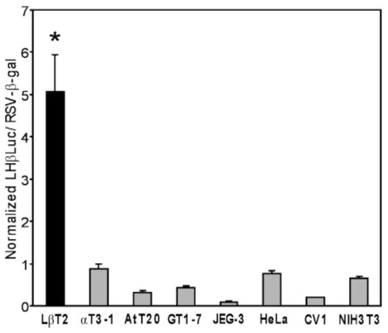
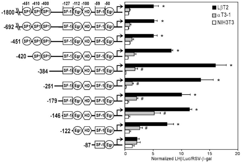
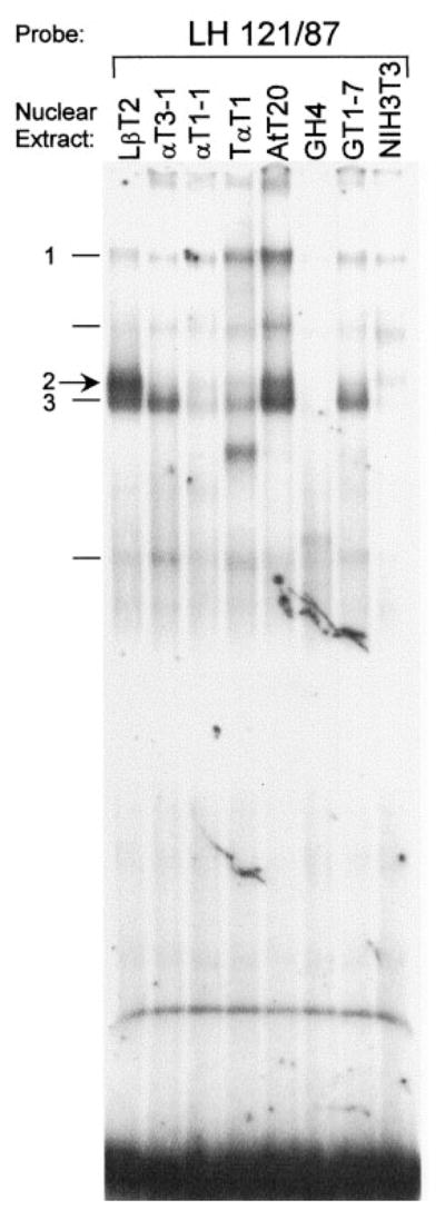
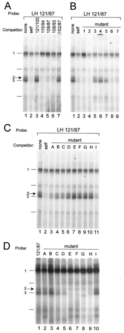
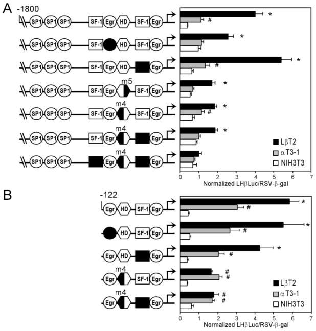
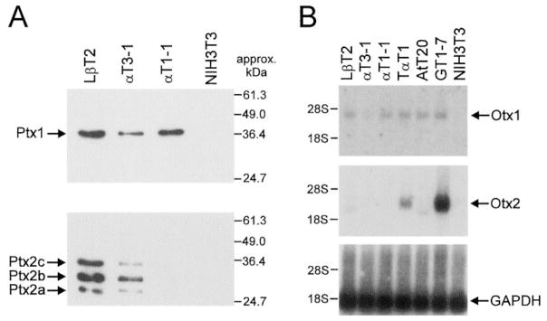
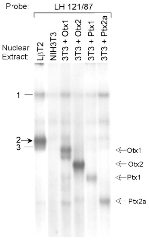
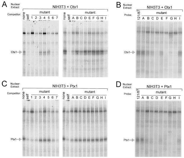
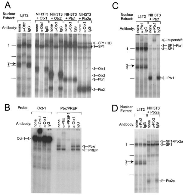
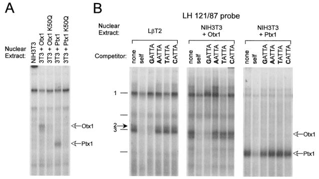
The hormone-secreting cell types of the anterior pituitary differentiate in a specific spatial and temporal manner. The alpha-subunit of the glycoprotein hormones appears at embryonic d 11.5 in the mouse, followed by steroidogenic factor-1, which distinguishes the gonadotrope progenitor cells, around embryonic d 14. Gonadotrope maturation is marked by the onset of LHbeta-gene expression 2 d later. The alphaT3-1 and LbetaT2 immortalized mouse pituitary cell lines correspond to these later sequential stages of gonadotrope differentiation. In addition to the early markers of the gonadotrope lineage present in alphaT3-1 cells, LbetaT2 cells also express markers of a mature gonadotrope, including LHbeta and FSHbeta. Using transient transfections to compare expression among gonadotrope and nongonadotrope-derived cell types, we show that the rat 1.8-kb LHbeta promoter directs reporter gene expression specifically to the mature gonadotrope LbetaT2 cell line. Promoter truncation and mutagenesis analyses indicate that the homeodomain (HD) element located at approximately -100 bp relative to the transcriptional start site is essential for this selectivity to LbetaT2 cells when compared with alphaT3-1 cells. In EMSAs, this HD site binds a protein present in LbetaT2 but not other gonadotrope-derived cells. Antibody supershift and competition experiments indicate that this LbetaT2 nuclear protein is a K50 HD protein related to the Otx family, though it is not a known pituitary homeobox transcription factor protein. These studies indicate a role for a novel Otx-related HD protein in gonadotrope maturation during development.


Figures

Fig. 1
Fig. 1. The Rat LH β-Subunit Promoter Is Specifically Active in LβT2 Cells
The 1.8-kb rat LHβ promoter was linked to the Luc reporter gene (LHβLuc) and transiently transfected into pituitary-derived LβT2 (differentiated gonadotrope), αT3-1 (precursor gonadotrope), and AtT20 (corticotrope) cells and nonpituitary-derived GT1-7 (hypothalamic), JEG-3 (placental), HeLa (cervical fibroblast), CV1 (kidney), and NIH3T3 (fibroblast) cells. RSV-β-gal was cotransfected as an internal control. To control for differences in transfection efficiencies and transcription rates between distinct cell lines, a parallel control, RSV-Luc was transfected into each cell line with the RSV-β-gal internal control. The ratio of RSV-Luc divided by RSV-β-gal was set at 100 for each cell line (not shown). Results represent the mean ± SEM of at least three independent experiments (n ≥ 3). The bar marked with an asterisk (*) (LβT2 cells) is statistically significant from all other bars (P < 0.05). Significant differences between other cell lines are not shown.
Fig. 2
Fig. 2. The Region Between −122 and −87 of the Rat LHβ Promoter is Necessary for LβT2 Cell Specificity
Truncations of the LHβ promoter linked to Luc were transiently transfected into LβT2, αT3-1, and NIH3T3 cells (identified by black, gray, and white bars, respectively). Diagrams at left depict the 5′ deletions used as well as the numerous regulatory elements identified in the rat LHβ promoter, including HD and binding sites for SP1, SF-1, and Egr. Results represent the mean ± SEM of at least three independent experiments (n ≥ 3). Asterisks (*) designate a significant difference from αT3-1 and NIH3T3 cells (P < 0.05); # indicates a significant difference from NIH3T3 cells (P < 0.05).
Fig. 3
Fig. 3. A Complex Present in LβT2 Cells, but Absent from Other Gonadotrope-Derived Cell Lines, Binds the LHβ Promoter
EMSA was conducted using the LH 121/87 probe and nuclear extracts from pituitary and nonpituitary-derived cell lines, indicated above each lane: LβT2 (gonadotrope), αT3-1 (precursor gonadotrope), αT1-1 (uncommitted gonadotrope/thyrotrope), TαT1 (thyrotrope), AtT20 (corticotrope), GH4 (so-matotrope), GT1-7 (hypothalamic neuroendocrine), and NIH3T3 (fibroblast). Complexes in LβT2 cells are indicated at left. Complex 2 exhibits strong binding in the mature gonadotrope LβT2 nuclear extracts, but not the αT3-1 or αT1-1 precursor nuclear extracts.
Fig. 4
Fig. 4. Complex 2 Binds the HD Element of the LHβ Promoter
A–C, EMSA was conducted using the LH 121/87 probe and nuclear extract from LβT2 cells. Competitions were performed using 100-fold excess unlabeled oligonucleotide as indicated above each lane: None (no competitor), self (wild-type LH 121/87 competitor). A, Competitions using truncated LH 121/87 oligonucleotide competitors indicate that complex 2 binds in the region from −108 to −94. B and C, Competitions using mutated LH 121/87 oligonucleotides indicate that complex 2 binds the HD element. D, EMSA was conducted using oligonucleotides containing single base-pair mutations as probes and nuclear extract from LβT2 cells. The closed arrow at left indicates complex 2; complexes 1 and 3 are also indicated. The results are summarized and sequences of all oligonucleotide competitors and probes are depicted in Table 1.
Fig. 5
Fig. 5. Mutation of the HD Element Abrogates LβT2 Cell Specificity
Site-directed mutagenesis was performed to mutate the 5′ Egr-1 binding site, the 3′ SF-1 binding site, the 5′ SF-1 binding site, and the HD in both the −1800LHβLuc and −122LHβLuc reporter genes. Two different HD mutations were examined (m4 and m5) as indicated in the diagram at left. HDm4 and HDm5 correspond to mutant 4 and mutant 5 used in Figs. 4 and 8 and shown in Table 1. Transient transfections of mutant LHβLuc plasmids were performed in LβT2, αT3-1, and NIH3T3 cells (identified by black, gray, and white bars, respectively). A, Mutations in the context of −1800LHβLuc. B, Mutations in the context of −122LHβLuc. Results represent the mean ± SEM of at least three independent experiments, each performed in triplicate (n ≥ 9). Asterisks (*) designate a significant difference from αT3-1 and NIH3T3 cells (P < 0.05); # indicates a significant difference from NIH3T3 cells (P < 0.05).
Fig. 6
Fig. 6. Expression of Otx and Ptx HD Proteins in Pituitary-Derived Cell Lines
A, Ptx1 and Ptx2 are expressed in both LβT2 and αT3-1 cells. A, Western blot with approximately 15 μg of nuclear extract from LβT2, αT3-1, αT1-1, and NIH3T3 cells was incubated with antibodies specific to Ptx1 or Ptx2. For Ptx1, a single band is observed in LβT2, αT3-1, and αT1-1 cells. The three bands observed with the Ptx2 antibody represent the three isoforms of Ptx2 (21), as indicated. B, Otx1, but not Otx2, is expressed in gonadotrope-derived cell lines. A Northern blot with approximately 2 μg poly(A) mRNA from LβT2, αT3-1, αT1-1, TαT1, AtT20, GT1-7, and NIH3T3 cells was probed with radiolabeled mouse cDNA fragments coding for Otx1 or Otx2. The GAPDH cDNA was used to allow visualization of the quantity of RNA loaded in each lane. Otx1 is detected in all cell types except NIH3T3, although at low levels in αT3-1 cells. Otx2 is detected in TαT1 and GT1-7 cells but not any of the gonadotrope-derived cell lines.
Fig. 7
Fig. 7. Known Members of the Ptx and Otx Families of HD Proteins Do Not Comigrate with Complex 2
EMSA was performed using the LH 121/87 probe and nuclear extracts from LβT2 cells, NIH3T3 cells, and NIH3T3 cells transiently transfected with an expression vector coding for Otx1 (3T3 + Otx1), Otx2 (3T3 + Otx2), Ptx1 (3T3 + Ptx1), or Ptx2a (3T3 + Ptx2a), as indicated above each lane. Open arrows at right indicate the migrations of the Otx and Ptx family members as labeled. The closed arrow at left indicates complex 2.
Fig. 8
Fig. 8. Binding of Otx1 and Ptx1 to the LHβ Promoter HD Element
A and C, EMSA was conducted using the LH 121/87 probe, nuclear extracts from NIH3T3 cells transiently transfected with an expression vector coding for either Otx1 (NIH3T3 + Otx1) (A) or Ptx1 (NIH3T3 + Ptx1) (C), and oligonucleotide competitors containing either 3 bp or single base-pair point mutations spanning the HD element (Table 1). Competitions were performed using 100-fold excess unlabeled oligonucleotide as indicated above each lane: None (no competitor), self (wild-type LH 121/87 competitor). B and D, EMSA was conducted using oligonucleotides containing single base-pair mutations as probes with NIH3T3 + Otx1 (B) or NIH3T3 + Ptx1 (D) nuclear extracts. The results are summarized and the sequences of all oligonucleotide competitors are depicted in Table 1. A and B, Otx1 binding to the LHβ promoter relies mainly on the core HD recognition sequence (GATTA). Migration of Otx1 is indicated by an open arrow. C and D, Ptx1 binding to the LHβ promoter is more dependent on several nucleotides outside the core HD sequence. Migration of Ptx1 is indicated by an open arrow.
Fig. 9
Fig. 9. Complex 2 Contains an Otx-Related HD Protein
A, Complex 2 contains a factor that is related to Otx HD transcription factors. EMSA was performed using nuclear extracts from LβT2 cells and NIH3T3 cells transiently transfected with an expression vector coding for Otx1 (NIH3T3 + Otx1), Otx2 (NIH3T3 + Otx2), Ptx1 (NIH3T3 + Ptx1), or Ptx2a (NIH3T3 + Ptx2a). Antibodies directed against Otx1 (α-Otx1) or normal rabbit IgG (IgG) were included in the reactions as indicated. The closed arrow at left indicates complex 2; open arrows at right indicate the migrations of the Otx and Ptx family members, SP1, and bands representing the concurrent binding of proteins to both the SP1 and HD sites (SP1 + HD), as labeled. The LβT2 lanes shown were run on a separate gel from the transfected NIH3T3 lanes shown. B, The Otx1 antibody does not cross-react with TALE or POU-domain containing HD proteins. EMSA was performed using nuclear extracts from LβT2 cells and Oct-1 (5′-TGTCGAATGCAAATCACTAGAA-3′ (top strand)] (51) or Pbx/PREP (5′-AGCGCGGGGCGCATCAATCAATTTCG-3′ (top strand)] (52) consensus probes. Antibodies directed against Oct-1, Pbx 1/2/3 (Pbx), PREP-1, or Otx1 were included in the reactions as indicated. C, Ptx1 from LβT2 cells does bind the LH 121/87 probe, but is not present in complex 2. EMSA was conducted using nuclear extracts from LβT2 cells (left) and NIH3T3 cells transiently transfected with an expression vector coding for Ptx1 (NIH3T3 + Ptx1, right). Antibodies directed against Ptx1 (α-Ptx1) or normal rabbit IgG (IgG) were included in the reactions as indicated. The closed arrow at left indicates complex 2; open arrows at right indicate Ptx1, SP1, SP1+Ptx1, and the Ptx1 supershift as labeled. D, Ptx2 is not present in complex 2. EMSA was conducted using nuclear extracts from LβT2 cells (left) and NIH3T3 cells transiently transfected with an expression vector coding for Ptx2a (NIH3T3 + Ptx2a, right). Antibodies directed against Ptx2 (α-Ptx2) or normal rabbit IgG (IgG) were included in the reactions as indicated. The closed arrow at left indicates complex 2; open arrows at right indicate Ptx2a, SP1, and SP1+Ptx2a as labeled.
Fig. 10
Fig. 10. Complex 2 Contains a K50 HD Transcription Factor with Binding Specificity Similar to that of Otx1
A, Lysine at position 50 of the HD is essential for Otx1 and Ptx1 interaction with the LHβ promoter HD element. Site-directed mutagenesis was performed to mutate the coding sequence of both Ptx1 and Otx1 such that the lysine at the 50th position of the HD (K50) was replaced by a glutamine (Q50). EMSA was performed using the LH 121/87 probe and nuclear extracts from NIH3T3 cells transiently transfected with an expression vector coding for wild-type Otx1 (NIH3T3 + Otx1), mutant Otx1 (NIH3T3 + Otx1 K50Q), wild-type Ptx1 (NIH3T3 + Ptx1), or mutant Ptx1 (NIH3T3 + Ptx1 K50Q), as indicated above each lane. Open arrows at right indicate the migrations of Otx1 and Ptx1. B, Complex 2 shares binding specificity with the Otx family of HD transcription factors but not the Ptx family of HD transcription factors. EMSA was performed using LβT2 (left), NIH3T3 + Otx1 (middle), and NIH3T3 + Ptx1 (right) nuclear extracts. Results using NIH3T3 + Otx2 and NIH3T3 + Ptx2a nuclear extracts are not shown, but Otx2 is competed similarly to Otx1 and Ptx2a is competed similarly to Ptx1. Oligonucleotide competitors containing a core HD sequence (5′-TGTACAGTCCTCANATTATTCTCAGG-3′ where N is either G (GATTA), A (AATTA), T (TATTA), or C (CATTA)] (33) were included in the reactions as indicated above each lane. The closed arrow at left indicates complex 2; open arrows at right indicate the migrations of the Otx1 and Ptx1.

Similar articles

Cited by

References

    1. Pierce JG, Parsons TF. Glycoprotein hormones: structure and function. Annu Rev Biochem. 1981;50:465–495. - PubMed
    1. Alarid ET, Windle JJ, Whyte DB, Mellon PL. Immortalization of pituitary cells at discrete stages of development by directed oncogenesis in transgenic mice. Development. 1996;122:3319–3329. - PubMed
    1. Graham KE, Nusser KD, Low MJ. LβT2 gonadotroph cells secrete follicle stimulating hormone (FSH) in response to activin A. J Endocrinol. 1999;162:R1–R5. - PubMed
    1. Pernasetti F, Vasilyev VV, Rosenberg SB, Bailey JS, Huang H-J, Miller WL, Mellon PL. Cell-specific transcriptional regulation of FSHβ by activin and GnRH in the LβT2 pituitary gonadotrope cell model. Endocrinology. 2001;142:2284–2295. - PubMed
    1. Halvorson LM, Ito M, Jameson JL, Chin WW. Steroidogenic factor-1 and early growth response protein 1 act through two composite DNA binding sites to regulate luteinizing hormone β-subunit gene expression. J Biol Chem. 1998;273:14712–14720. - PubMed

Publication types

Substances