. 2002 Jun 1;22(11):4264-73.
doi: 10.1523/JNEUROSCI.22-11-04264.2002.

CASK participates in alternative tripartite complexes in which Mint 1 competes for binding with caskin 1, a novel CASK-binding protein

Affiliations

CASK participates in alternative tripartite complexes in which Mint 1 competes for binding with caskin 1, a novel CASK-binding protein

Katsuhiko Tabuchi et al. J Neurosci. .

Abstract

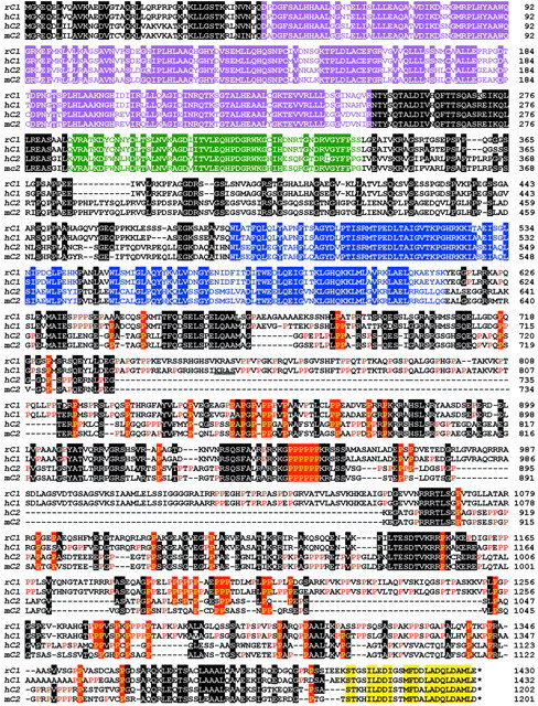
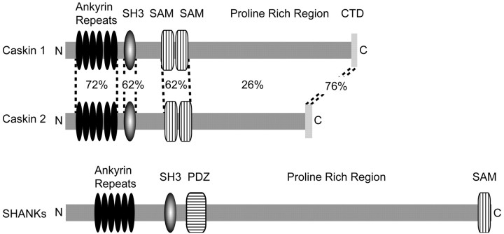
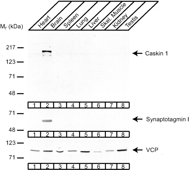
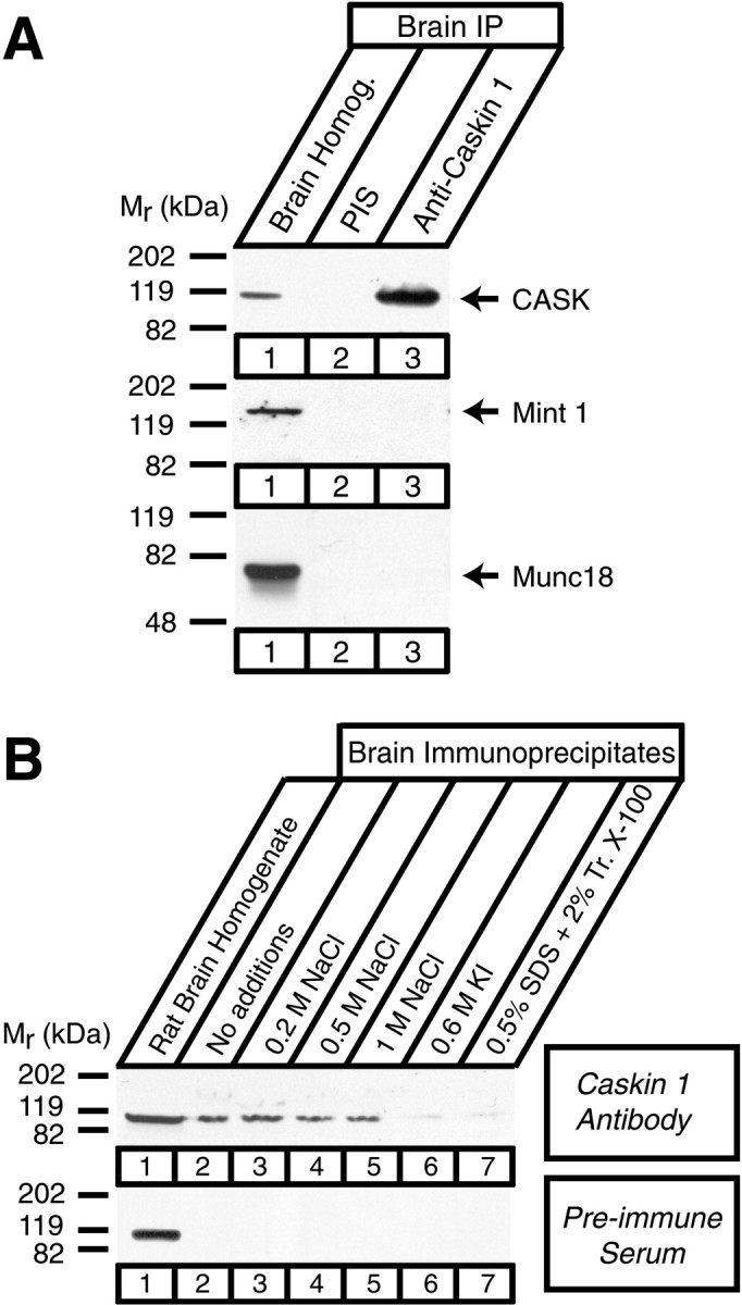
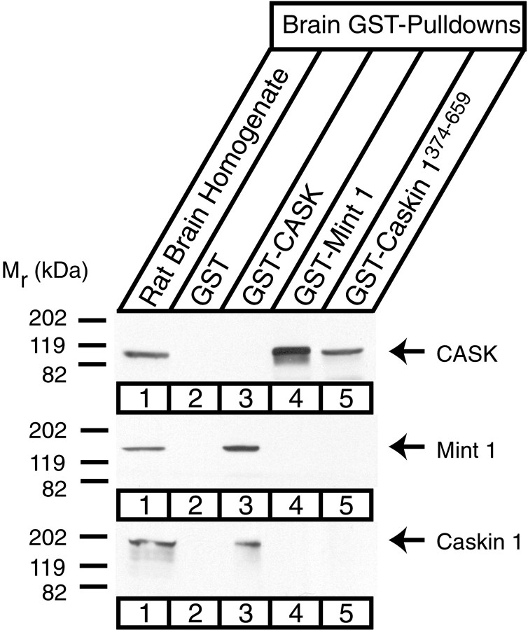
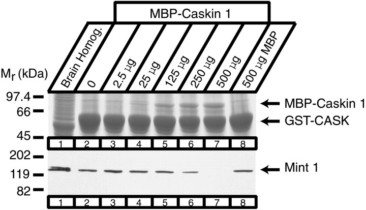
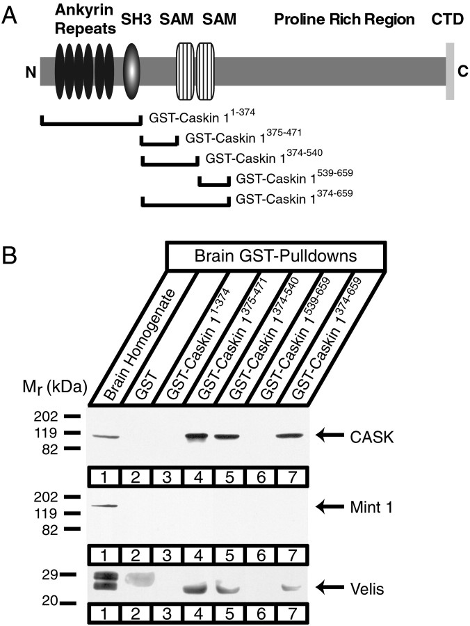
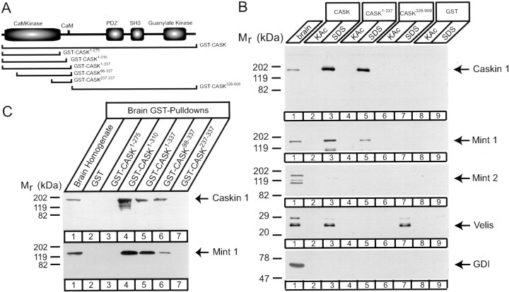
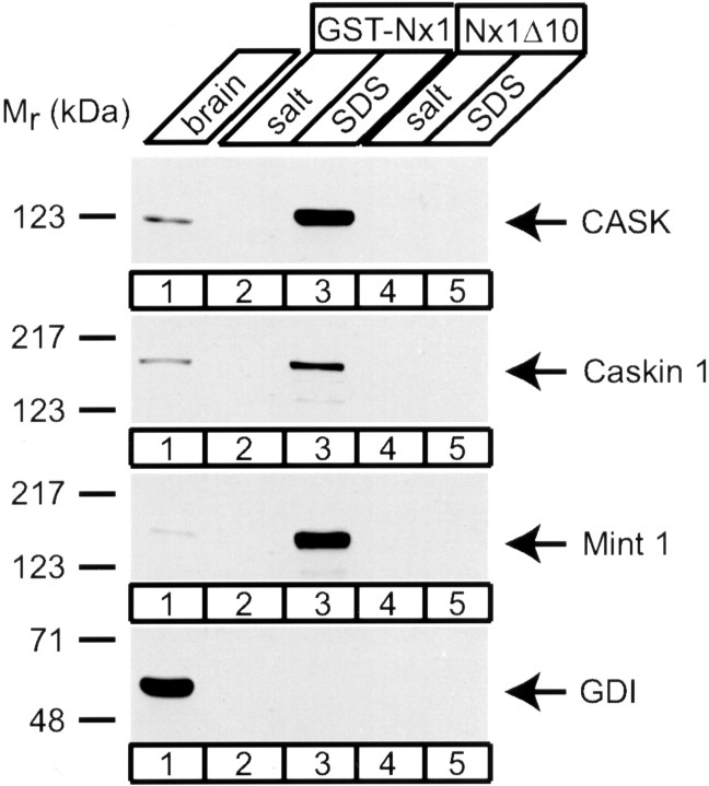
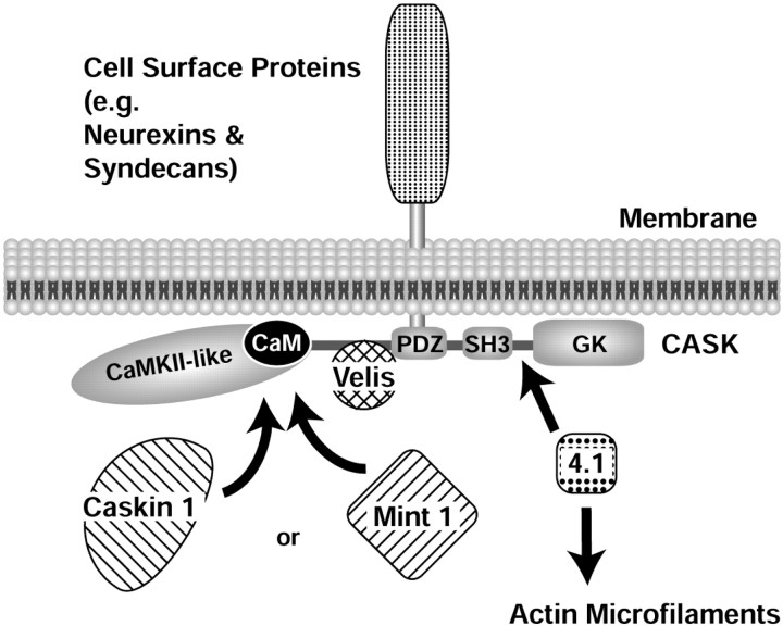
CASK, an adaptor protein of the plasma membrane, is composed of an N-terminal calcium/calmodulin-dependent protein (CaM) kinase domain, central PSD-95, Dlg, and ZO-1/2 domain (PDZ) and Src homology 3 (SH3) domains, and a C-terminal guanylate kinase sequence. The CaM kinase domain of CASK binds to Mint 1, and the region between the CaM kinase and PDZ domains interacts with Velis, resulting in a tight tripartite complex. CASK, Velis, and Mint 1 are evolutionarily conserved in Caenorhabditis elegans, in which homologous genes (called lin-2, lin-7, and lin-10) are required for vulva development. We now demonstrate that the N-terminal CaM kinase domain of CASK binds to a novel brain-specific adaptor protein called Caskin 1. Caskin 1 and a closely related isoform, Caskin 2, are multidomain proteins containing six N-terminal ankyrin repeats, a single SH3 domain, and two sterile alpha motif domains followed by a long proline-rich sequence and a short conserved C-terminal domain. Unlike CASK and Mint 1, no Caskin homolog was detected in C. elegans. Immunoprecipitations showed that Caskin 1, like Mint 1, is stably bound to CASK in the brain. Affinity chromatography experiments demonstrated that Caskin 1 coassembles with CASK on the immobilized cytoplasmic tail of neurexin 1, suggesting that CASK and Caskin 1 coat the cytoplasmic tails of neurexins and other cell-surface proteins. Detailed mapping studies revealed that Caskin 1 and Mint 1 bind to the same site on the N-terminal CaM kinase domain of CASK and compete with each other for CASK binding. Our data suggest that in the vertebrate brain, CASK and Velis form alternative tripartite complexes with either Mint 1 or Caskin 1 that may couple CASK to distinct downstream effectors.


Figures

Fig. 1.
Fig. 1.
Sequence analysis of Caskins. Alignment of the primary sequences of rat and human Caskin 1 (rC1 andhC1, respectively) and human and mouse Caskin 2 (hC2 and mC2, respectively). Sequences are identified on the left and numbered on theright. Residues that are identical between Caskins 1 and 2 in at least two of the sequences shown are highlighted by a domain-specific color code: The N-terminal ankyrin repeats are shown inpurple, the SH3 domain is in green, the SAM domains are in blue, and the Caskin-specific C-terminal domain is in yellow. Outside of these defined domains, shared sequences are highlighted in blackexcept for prolines, which are shown in the C-terminal half of the proteins on a red background if conserved among different Caskins and in red typeface if specific for a given Caskin isoform.
Fig. 2.
Fig. 2.
Domain structures of Caskins. The domain structures of Caskins 1 and 2 are shown schematically and compared with the domain structure of Shanks. Numbers between the Caskin 1 and 2 structures indicate percentage identity between the various domains. The color code used corresponds to that of Figure1.
Fig. 3.
Fig. 3.
RNA blotting analysis of Caskin 1 expression. A blot containing poly(A)-enriched RNA from the indicated rat tissues was probed at high stringency with a probe from the Caskin 1 cDNA.Numbers at left indicate positions of size markers.
Fig. 4.
Fig. 4.
Characterization of Caskin 1 antibodies. Proteins from total rat brain homogenate and from COS cells transfected with a full-length Caskin 1 expression vector were analyzed by immunoblotting with antibodies raised against a synthetic peptide corresponding to the C-terminal 15 residues of Caskin 1. Signals were visualized by ECL.Numbers at left indicate positions of molecular weight markers. Preimmune serum did not cause a signal (data not shown).
Fig. 5.
Fig. 5.
Immunoblotting analysis of Caskin 1 expression in adult rat tissues. Total proteins from the indicated tissues (50 μg/lane) were analyzed by immunoblotting with antibodies to Caskin 1 (top), the synaptic vesicle protein synaptotagmin 1 as a control for a brain-specific protein (middle), and vasolin-containing protein (VCP) as a control for a widely expressed protein (bottom). Signals were visualized by ECL. Numbers at leftindicate positions of molecular weight markers.
Fig. 6.
Fig. 6.
Localization of Caskin 1 analyzed by immunocytochemistry. A–C, Adjacent cryostat sections from rat cerebellum stained with antibodies to Rab3A (A) or Caskin 1 (B) or with Caskin 1 preimmune serum (C). The molecular layer (ml), granule cell layer (gl), and deep cerebellar nuclei (dcn) are identified. Scale bar in C, 0.1 mm (applies to A–C). D,E, Cultured hippocampal neurons double-labeled with a monoclonal antibody to synaptophysin (D) and a polyclonal antibody to Caskin 1 (E). Scale bar inE, 40 μm; (applies to D,E).
Fig. 7.
Fig. 7.
Coimmunoprecipitation of CASK and Caskin 1 from rat brain homogenates. A, Proteins solubilized from rat brains in Triton X-100 (lane 1) were immunoprecipitated with preimmune serum (PIS) (lane 2) and polyclonal antibodies to Caskin 1 (lane 3). Precipitated proteins were analyzed by immunoblotting with monoclonal antibodies to CASK, Mint 1, and Munc18-1 as indicated. IP, immunoprecipitates. B, Immunoprecipitates from rat brain homogenates (lane 1) with Caskin 1 antiserum or preimmune serum were washed in the presence of increasing concentrations of NaCl (lanes 3–5) or of the denaturing agents 0.6 m KI and 0.5% SDS as indicated (lanes 6 and 7) and analyzed by immunoblotting with monoclonal antibodies to CASK. Signals were visualized by ECL.Numbers at left indicate positions of molecular weight markers.
Fig. 8.
Fig. 8.
Confirmation of the CASK–Caskin 1 interaction by GST pulldowns. Proteins from rat brain homogenates (lane 1) were bound to GST alone (lane 2) or to GST fusion proteins of the N-terminal CaM kinase domain of CASK (lane 3) or of the N-terminal sequences of Mint 1 (lane 4) or of Caskin 1 (lane 5). Bound proteins were analyzed by immunoblotting with antibodies to CASK, Mint 1, and Caskin 1 as indicated. Numbers on theleft indicate positions of molecular weight markers.
Fig. 9.
Fig. 9.
Caskin 1 competes with Mint 1 for CASK binding. Proteins from rat brain homogenates (Homog.) (lane 1) were bound to immobilized GST–CASK1-337 in the presence of increasing concentrations of recombinant MBP–Caskin 1 fusion protein up to 500 μg (lanes 2–7) or of MBP alone at 500 μg (lane 8). Bound proteins were visualized by Coomassie blue staining (top) or immunoblotting with Mint 1 antibodies (bottom). Numbers atleft indicate positions of molecular weight markers.
Fig. 10.
Fig. 10.
Mapping of the CASK-binding site on Caskin 1 by GST pulldowns. A, Position of GST–Caskin 1 fusion proteins used for pulldowns in B.B, GST pulldowns of rat brain proteins with the indicated Caskin fusion proteins were analyzed by immunoblotting for CASK, Mint 1, and Velis as indicated. Signals were visualized by ECL.Numbers at left indicate positions of molecular weight markers.
Fig. 11.
Fig. 11.
Mapping of the Caskin 1 binding site on CASK.A, Domain structure of CASK and positions of GST–CASK fusion proteins used for pulldowns. B, C, GST pulldowns of proteins in rat brain homogenates with the indicated CASK fusion proteins analyzed by immunoblotting for the proteins identified atright. B, Proteins were eluted with 0.8m K-acetate (KAc) followed by SDS sample buffer, whereas in C, proteins bound to the beads were examined. Note that in CASK, not only the CaM kinase domain but also the region homologous to the autoregulatory sequence of CaM kinase II are required for binding. Also note the separation between the common Mint 1–Caskin 1 and the Veli binding sites on CASK. Signals were visualized by ECL. Numbers at leftindicate positions of molecular weight markers.
Fig. 12.
Fig. 12.
Assembly of CASK–Caskin 1 and CASK–Mint 1 complexes on the immobilized cytoplasmic domain of neurexin. Immobilized GST fusion protein of the wild-type cytoplasmic tail of neurexin 1 (GST–NxI) or of mutant cytoplasmic tail lacking the final 10 residues (NxIΔ10) was used for binding reactions with rat brain homogenates and eluted with 0.8 m K-acetate buffer (salt) followed by SDS sample buffer (SDS). Samples were analyzed by immunoblotting for CASK, Mint 1, Caskin 1, and GDI (as a negative control). Note the tight binding of CASK and Caskin 1 to the neurexin tail that requires the C-terminal residues of neurexin.Numbers at left indicate positions of molecular weight markers.
Fig. 13.
Fig. 13.
Model of the protein–protein interactions mediated by CASK. CASK is recruited to the plasma membrane via interactions of its PDZ domain with the C-terminal cytoplasmic tails of cell-surface proteins, for example neurexins, syndecans, and SynCaM. As a multidomain protein, CASK binds to multiple potential downstream effectors, many of which are also multidomain adaptor proteins containing domains similar to those of CASK. The N-terminal CaM kinase II homology region binds to calmodulin as a function of Ca2+ and to Caskin 1 and Mint 1 independently of Ca2+ (data not shown). The central sequence N-terminal to the PDZ domain binds to Velis, and the C-terminal SH3 and guanylate kinase (GK) domains and connecting sequences bind to protein 4.1, thereby recruiting actin filaments, and to Ca2+ channels (data not shown); in addition, the C-terminal regions may be involved in intramolecular and intermolecular interactions of CASK.

Similar articles

Cited by

References

    1. Biederer T, Südhof TC. Mints as adaptors: direct binding to neurexins and recruitment of munc18. J Biol Chem. 2000;275:39803–39806. - PubMed
    1. Biederer T, Südhof TC. Cask and protein 4.1 support F-actin nucleation on neurexins. J Biol Chem. 2001;276:47869–47876. - PubMed
    1. Borg JP, Yang Y, De Taddeo-Borg M, Margolis B, Turner RS. The X11alpha protein slows cellular amyloid precursor protein processing and reduces Abeta40 and Abeta42 secretion. J Biol Chem. 1998a;273:14761–14766. - PubMed
    1. Borg JP, Straight SW, Kaech SM, de Taddeo-Borg M, Kroon DE, Karnak D, Turner RS, Kim SK, Margolis B. Identification of an evolutionarily conserved heterotrimeric protein complex involved in protein targeting. J Biol Chem. 1998b;273:31633–31636. - PubMed
    1. Butz S, Okamoto M, Südhof TC. A tripartite protein complex with the potential to couple synaptic vesicle exocytosis to cell adhesion in brain. Cell. 1998;94:773–782. - PubMed

Publication types

MeSH terms

Associated data