. 2002 Jun 1;22(11):4335-45.
doi: 10.1523/JNEUROSCI.22-11-04335.2002.

c-Jun N-terminal protein kinase (JNK) 2/3 is specifically activated by stress, mediating c-Jun activation, in the presence of constitutive JNK1 activity in cerebellar neurons

Affiliations

c-Jun N-terminal protein kinase (JNK) 2/3 is specifically activated by stress, mediating c-Jun activation, in the presence of constitutive JNK1 activity in cerebellar neurons

Eleanor T Coffey et al. J Neurosci. .

Abstract

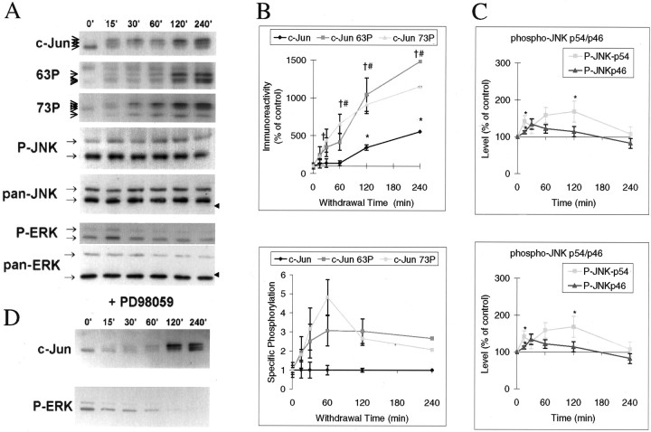
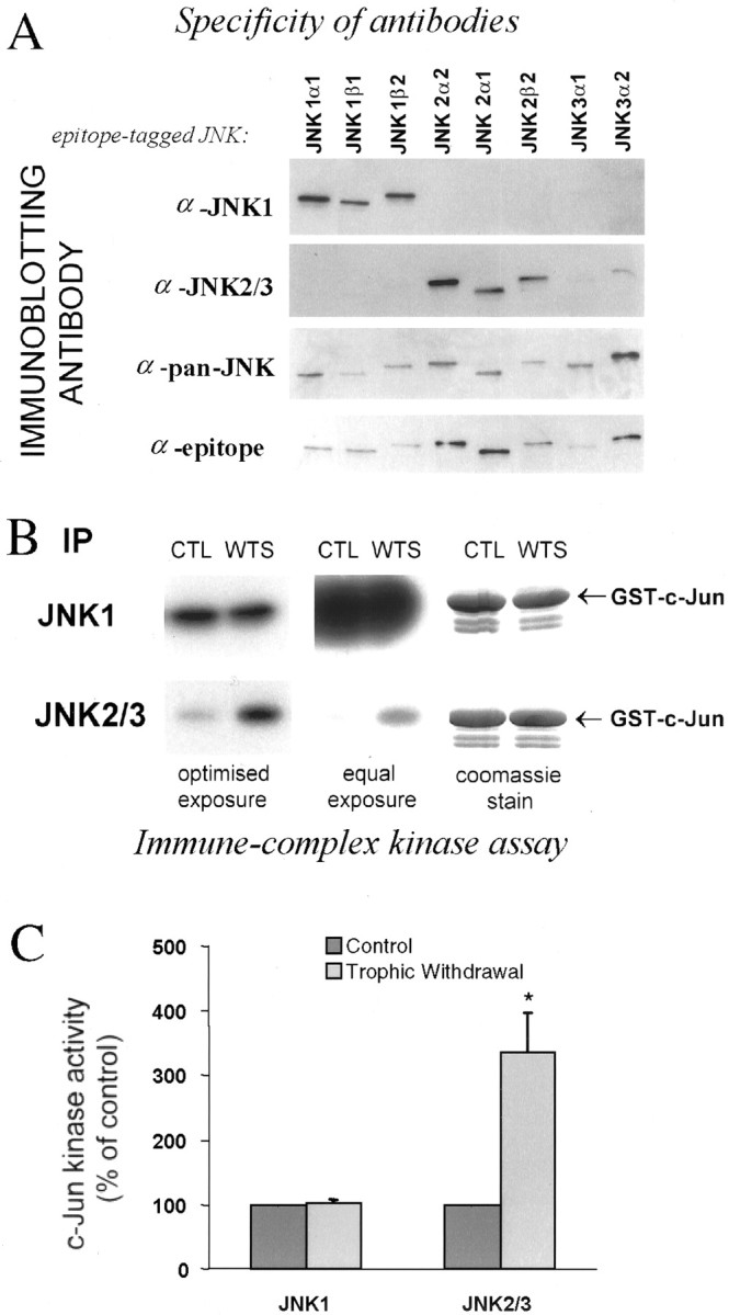
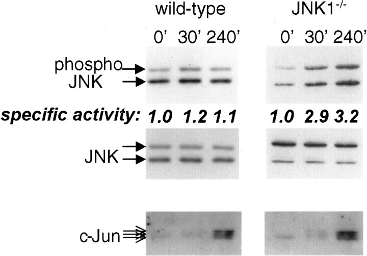
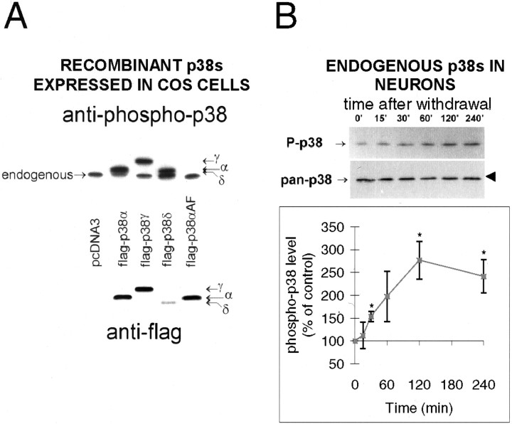
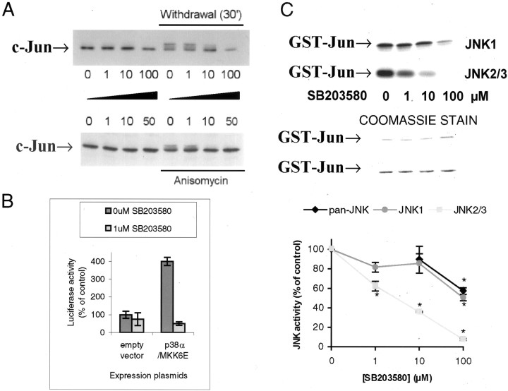
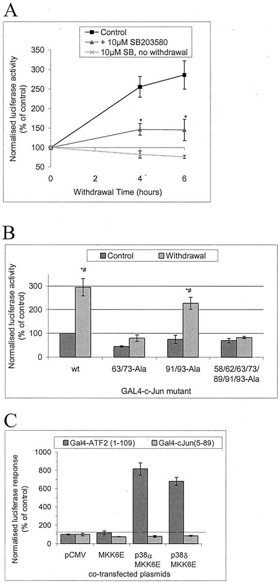
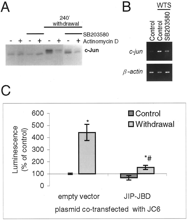
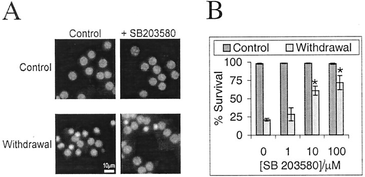
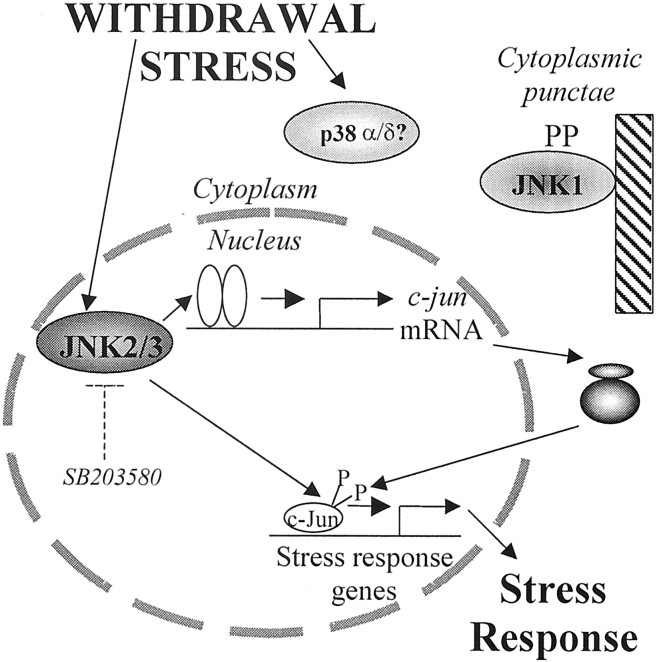
c-Jun is considered a major regulator of both neuronal death and regeneration. Stress in primary cultured CNS neurons induces phosphorylation of c-Jun serines 63 and 73 and increased c-Jun protein. However, total c-Jun N-terminal protein kinase (JNK) activity does not increase, and no satisfactory explanation for this paradox has been available. Here we demonstrate that neuronal stress induces strong activation of JNK2/3 in the presence of constitutively and highly active JNK1. Correspondingly, neurons from JNK1(-/-) mice show lower constitutive activity and considerably higher responsiveness to stress. p38 activity can be completely inhibited without effect on c-Jun phosphorylation, whereas 10 micrometer SB203580 strongly inhibits neuronal JNK2/3, stress-induced c-Jun phosphorylation, induced c-Jun activity, and neuronal death in response to trophic withdrawal stress. Neither constitutive JNK1 activity nor total neuronal JNK activity were significantly affected by this concentration of drug. Thus, neuronal stress selectively activates JNK2/3 in the presence of mechanisms maintaining constitutive JNK1 activity, and this JNK2/3 activity selectively targets c-Jun, which is isolated from constitutive JNK1 activity.


Figures

Fig. 1.
Fig. 1.
Withdrawal of trophic support rapidly induces c-Jun phosphorylation without corresponding activation of total JNK.A, Cerebellar granule neurons were deprived of trophic support by incubating the cells with medium free of serum and without additional KCl for the times indicated, and lysates were immunoblotted for total c-Jun, phosphoserine 63 c-Jun (“63P”), phosphoserine 73 c-Jun (“73P”), activated, dually phosphorylated JNK (“P-JNK”) and ERK (“P-ERK”), pan-ERK, and pan-JNK, as indicated by thearrows. Representative data are shown. Thetopmost band detected by anti-phosphoserine 63 c-Jun is nonspecific; it is constitutively present and does not co-migrate with c-Jun immunoreactivity. Arrowheads on theright of the blots indicate molecular mass marker (42 kDa). B, Top panel, Blots from replicate experiments as shown in A were scanned, and intensities of bands corresponding to those indicated by the arrows to theleft of the blots were quantitated from unsaturated films from replicate experiments, normalized to the maximum values, and scaled to percentage of initial (control) values. Multiple exposures were taken with preflashed films to avoid nonlinear response, saturated, or undetectably faint bands that may occur in any individual exposure. Means ± SEM are shown (n = 3–6). Values significantly different from 0′ control levels (pairedt test; p < 0.05 or better) are indicated by * (for c-Jun), # (c-Jun73P) or † (c-Jun63P).Bottom panel, The data were normalized to the levels of c-Jun at each time point, demonstrating that regulation of specific c-Jun phosphorylation rises to a plateau (the 60′ JunP73 peak being not significantly above the plateau) before total c-Jun levels change.C, P-JNK and P-ERK levels were quantitated as inB. P-JNK isoforms are resolved as top (54 kDa) and bottom (46 kDa) bands, and ERK1 and 2 are resolved as top (44 kDa) and bottom bands (42 kDa), respectively, as indicated by the arrows. The P-JNK exposure shown is a long exposure saturated in the 46 kDa bands to demonstrate the slight regulation of the fainter 54 kDa bands. Values significantly different from 0′ control levels (pairedt test; p < 0.05 or better) are indicated by *. D, Cerebellar granule neurons were deprived of trophic support for the times indicated above the figure as in A, but in the presence of 50 μm of the selective MEK1/2 inhibitor PD98059 to prevent ERK activation, added one hour before withdrawal of trophic support in each case. No inhibitor was added to the control (0′). Lysates were immunoblotted with antibodies recognizing c-Jun and the activated dually phosphorylated form of ERK.
Fig. 2.
Fig. 2.
Selective activation of JNK2/3 by withdrawal of trophic support in the persistent presence of high constitutive JNK1 activity. A, JNK isoforms were expressed in COS-7 cells, and selective JNK1 and JNK2/3 antibodies were identified by immunoblotting as indicated. The bottom paneldemonstrates JNK isoform loading by blotting for the epitope tag of the JNK constructs. B, Trophic support was withdrawn from cerebellar neurons for 2 hr, JNK1 and JNK2/3 were immunoprecipitated from the cells with antibodies used in A, and activity was measured by in vitro kinase assay with c-Jun substrate. The center panel shows equal exposure time to the gels, the left panel shows nonsaturated film, and the right panel shows the Coomassie-stained gel, indicating equal substrate loading. C, Quantitated activity is displayed as a percentage of control activity (n = 4). An * in the histogram indicates significant difference from control by paired t test (p < 0.05).
Fig. 3.
Fig. 3.
Regulation of total JNK revealed in neurons from JNK1−/− mouse. Cerebellar granule neurons were prepared from JNK1−/− or wild-type mice and treated as in Figure 1, trophic support being withdrawn for 0–240 min, as indicated above the corresponding lanes. Phospho-JNK, total JNK, and c-Jun, detected by immunoblotting, are indicated by the arrows. An increase in phospho-JNK level was observed in JNK1−/−lysates. Mean values of specific JNK activation (phosphoJNK/JNK quantitated from blots), normalized to the initial levels, are indicated below the corresponding phospho-JNK lanes. Data shown is representative of two or three replicates (wild-types) and three replicates (JNK1−/−).
Fig. 4.
Fig. 4.
Compartmentalization and granularity of JNK isoform localization. A, Cerebellar granule neurons were fixed and stained with JNK isoform-specific antibodies or without primary antibody as shown. Confocal scans are shown. The fluorescence intensity profiles along the white linesare shown in the inset line graphs (arbitrary scale), demonstrating the extranuclear localization of JNK1 and the relative abundance of JNK2/3 in the nucleus. Note also the granularity of JNK1 staining along processes and the relatively diffuse JNK2/3 staining.B, Immunoblot detection of JNK1 and JNK2/3 from cytoplasmic and nuclear fractions indicate no change in levels in either compartment in response to 2 hr withdrawal of trophic support. Representative blots from three replicates are shown. The nuclear JNK1 blots required longer exposure time than the corresponding cytoplasmic blot, in accordance with the relative distribution shown by immunofluorescent staining in Figure 4A.
Fig. 5.
Fig. 5.
Activation of p38 is detected by a phospho-specific p38 antibody that recognizes active forms of all p38 species. COS-7 cells were transfected with pcDNA3 vectors containing flag-tagged p38α, γ, δ, or as controls, p38αAF, which cannot be phosphorylated or no insert (“pcDNA3”), and anisomycin-treated to activate p38. The ability of anti-phospho-p38 to recognize the activated isoforms was determined by immunoblotting (top panel), and expression levels verified by flag immunoblotting (bottom panel). The flag-tag of the constructs caused retarded mobility, allowing them to be distinguished from endogenous anisomycin-activated p38 as shown.B, Lysates of cerebellar granule neurons stimulated as in Figure 1 were immunoblotted with phospho-p38 antibody and pan-p38 antibody as shown. The arrows on the leftof the blots indicate the proteins, and the arrowhead on the right indicates the 42 kDa Mr marker. Quantitated data (means ± SEM; n = 3–4) were normalized and scaled to percentage of initial (control) values. The * represents a significant difference from control (p < 0.05 or better; pairedt test).
Fig. 6.
Fig. 6.
SB203580 inhibits c-Jun phosphorylation and JNK2/3 immunoprecipitated from cerebellar granule neurons. A,SB203580 was added to neurons 1 hr before either no treatment (left lanes) or 30′ withdrawal stress (top panel) or anisomycin (10 μg/ml) treatment (bottom panel). c-Jun phosphorylation was detected by mobility shift, demonstrating that ≥10 μmSB203580 inhibits the mobility shift in both cases. B,HeLa cells were transfected with empty vector or plasmids encoding constitutively active MKK6 and p38α, together with plasmids encoding GAL4-ATF2 and the GAL4-driven luciferase reporter pGL3-G5E4Δ38 and pRL-CMV as internal control. Cells were grown for 24 hr after transfection with DMSO carrier with or without SB203580 as indicated, cells were lysed and normalized luciferase expression activity data assayed by dual luciferase assay is shown (mean ± SEM;n = 3). C, JNK1 and JNK2/3 were immunoprecipitated with specific antibodies (compare Fig.2A) from cells withdrawal-stressed for 2 hr, and immune-complex kinase assay performed in the presence of concentrations of SB203580 as shown (top panel). Center panel shows Coomassie-stained gels, indicating equal substrate loading. Bottom panel shows meaned data (±SEM) from replicates, demonstrating high sensitivity of JNK2/3 to SB203580 (IC50 ≈ 3 μm). Results with a pan-JNK antibody are also shown, demonstrating that total JNK has properties consistent with JNK1 (compare Fig. 2B, right panel) An asterisk denotes significant difference from control (p < 0.05 or better; paired t test).
Fig. 7.
Fig. 7.
Withdrawal of trophic support induces transcriptional activation of GAL4-c-Jun in a manner sensitive to 10 μm SB203580 and dependent on phosphorylation sites serines 63/73; p38 isoforms cannot activate it. A,Cerebellar granule neurons were transfected with GAL4-luciferase reporter, GAL4-c-Jun(6–89) fusion construct, Renilla luciferase internal control, and empty vector (pCMV) to bring total DNA to 4 μg/well of a 12 well plate. Cells were deprived of trophic support in the presence or absence of 10 μm SB203580 (added 1 hr before withdrawal of trophic support) for the times shown or treated with SB203580 without withdrawal of trophic support alone for an equivalent time. This was achieved by transfecting all samples at the same time, withdrawing trophic support at different times, and lysing all samples at the same time, thus that all samples had the same amount of time to express the transfected plasmids. Firefly luciferase activity was normalized to the Renilla luciferase internal standard. Normalized activity levels were expressed as a percentage of values from samples with continued trophic support. Means ± SEMs (n = 3–5) are shown. An * indicates that the values in the presence of SB203580 are significantly different from in the absence of the drug (p < 0.05; pairedt test). B, Cerebellar granule neurons were transfected as in A but with GAL4 fused to c-Jun (5–105) wild-type “wt” or Ser/Thr→Ala point mutants as shown. Neither GAL4-c-Jun(5–105) construct in which Ser63/73 is mutated to Ala is significantly activated by withdrawal of trophic support; both wt and Thr91/93Ala are activated to similar extents (2.9- and 3.0-fold, respectively). Means ± SEM (n = 4) are shown. An * indicates a significant increase in comparison with control value with wt, a # indicates significant difference from control using the same GAL4 fusion protein (p ≤ 0.05; pairedt test). C, The possibility of c-Jun activation in A by p38 was investigated by transfecting neurons inA but with GAL4 fusions of either c-Jun(6–89) as inA or ATF2(1–109) as a control p38 substrate, and cotransfected with plasmids as shown to express and activate specific isoforms of p38. Data shown represents mean ± range (n = 2).
Fig. 8.
Fig. 8.
Withdrawal-stress induced c-Jun mRNA and protein are inhibited by SB203580, and activation of c-Jun promoter is prevented by the JNK inhibitor JIP-JBD in cerebellar granule neurons. A, Cerebellar granule neurons were withdrawal-stressed in the presence or absence of SB203580 (10 μm) and/or actinomycin D (5 μm) added 1 hr before withdrawal of trophic support as indicated. Actinomycin D, which targets RNA synthesis, inhibits the withdrawal-induced accumulation of c-Jun, whereas SB203580 inhibits both the accumulation and the phosphorylation-associated mobility shift of c-Jun. B,SB203580 (10 μm) or carrier was added 1 hr before withdrawal of trophic support as indicated. After 3 hr, RNA was isolated, and actin and c-jun mRNA were amplified by RT-PCR and detected on agarose gels with ethidium bromide. A representative experiment is shown. C, Neurons were cotransfected with c-Jun promoter-driven luciferase reporter plasmid (pGL3-JC6), Renilla luciferase internal standard, and 1 μg of either empty vector (pCMV) or JIP-JBD to inhibit JNK signaling as shown. After 20 hr, neurons were withdrawal-stressed for 4 hr where indicated. Reporter induction was calculated as described in the legend to Figure 7(n = 3). The JNK inhibitor prevented induction of c-jun promoter reporter expression by withdrawal of trophic support. * and # indicate significant differences from empty vector-transfected, control, and withdrawal-stressed values, respectively (p < 0.05 or better; pairedt test).
Fig. 9.
Fig. 9.
Neuronal death induced by withdrawal of trophic support is reduced by SB203580. Cerebellar granule neurons were cultured in medium containing serum and 20 mm additional KCl. Death induced by withdrawal of trophic support was evoked by incubating the cells with medium free of serum and without additional KCl, in the presence or absence of SB203580 added 1 hr before withdrawal of trophic support. DNA was stained with Hoechst 33342 24 hr later. A, Fluorescence images of Hoechst 33342: DNA complexes show healthy nuclei in controls. Withdrawal of trophic support induced pyknosis of over half the nuclei in a manner largely prevented by 10 μm SB203580. B, Percentage of neurons present scored as alive (means ± SEMs;n = 4) is shown. An * indicates that survival with SB203580 is significantly higher than without (p < 0.001 by paired ttest).
Fig. 10.
Fig. 10.
Scheme depicting proposed JNK and p38 regulation in stressed cerebellar neurons. Stresses, e.g., withdrawal of trophic support, selectively activate JNK2/3. This leads to c-Jun phosphorylation and activation of promoters of stress responsive genes such as c-jun itself, ultimately leading to the response of the neuron to stress. A p38 isoform is also activated, but its role is unknown. In contrast, JNK1 is constitutively active; in spite of this, it is predominantly associated with cytoplasmic structures and unable to phosphorylate c-Jun in the nucleus.

References

    1. Alessi DR, Cuenda A, Cohen P, Dudley DT, Saltiel AR. PD098059 is a specific inhibitor of the activation of MAPKK in vitro and in vivo. J Biol Chem. 1995;270:27489–27494. - PubMed
    1. Barila D, Mangano R, Gonfloni S, Kretzschmar J, Moro M, Bohmann D, Superti-Furga G. A nuclear tyrosine phosphorylation circuit: c-Jun as an activator and substrate of c-Abl and JNK. EMBO J. 2000;19:273–281. - PMC - PubMed
    1. Behrens A, Sibilia M, Wagner EF. Amino-terminal phosphorylation of c-Jun regulates stress-induced apoptosis and cellular proliferation. Nat Genet. 1999;21:326–329. - PubMed
    1. Buchdunger E, Zimmermann J, Mett H, Meyer T, Muller M, Druker BJ, Lydon NB. Inhibition of the Abl protein-tyrosine kinase in vitro and in vivo by a 2-phenylaminopyrimidine derivative. Cancer Res. 1996;56:100–104. - PubMed
    1. Clarke N, Arenzana N, Hai T, Minden A, Prywes R. EGF induction of the c-jun promoter by a Rac pathway. Mol Cell Biol. 1998;18:1065–1073. - PMC - PubMed

Publication types

MeSH terms