. 2002 Jun 1;22(11):4364-71.
doi: 10.1523/JNEUROSCI.22-11-04364.2002.

Domain 2 of Drosophila para voltage-gated sodium channel confers insect properties to a rat brain channel

Affiliations

Domain 2 of Drosophila para voltage-gated sodium channel confers insect properties to a rat brain channel

Iris Shichor et al. J Neurosci. .

Abstract

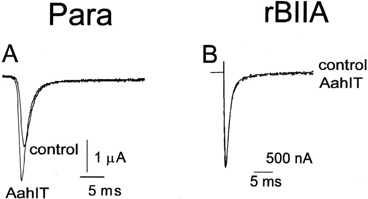
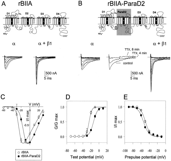
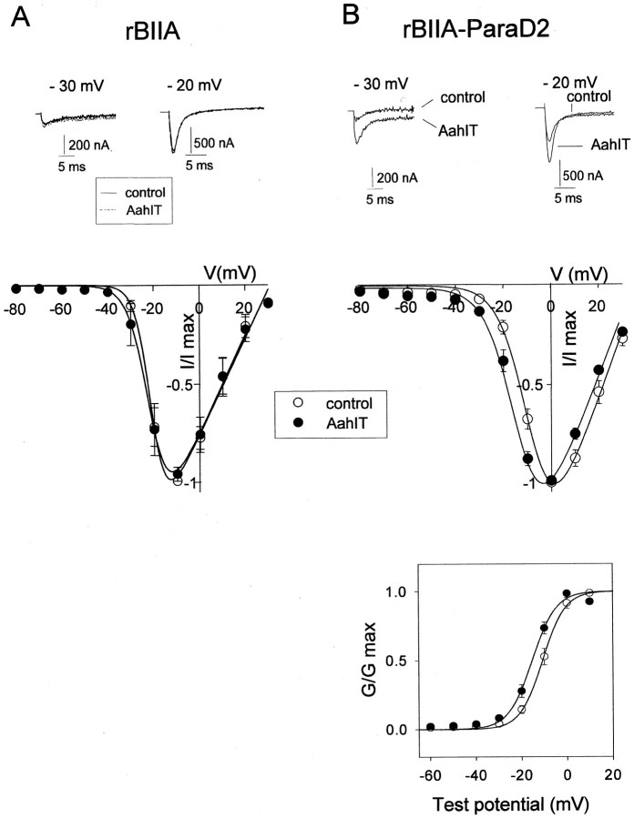
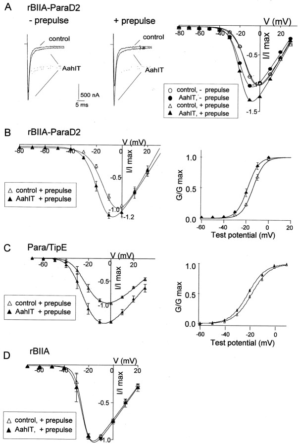
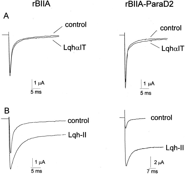
The ability of the excitatory anti-insect-selective scorpion toxin AahIT (Androctonus australis hector) to exclusively bind to and modify the insect voltage-gated sodium channel (NaCh) makes it a unique tool to unravel the structural differences between mammalian and insect channels, a prerequisite in the design of selective pesticides. To localize the insect NaCh domain that binds AahIT, we constructed a chimeric channel composed of rat brain NaCh alpha-subunit (rBIIA) in which domain-2 (D2) was replaced by that of Drosophila Para (paralytic temperature-sensitive). The choice of D2 was dictated by the similarity between AahIT and scorpion beta-toxins pertaining to both their binding and action and the essential role of D2 in the beta-toxins binding site on mammalian channels. Expression of the chimera rBIIA-ParaD2 in Xenopus oocytes gave rise to voltage-gated and TTX-sensitive NaChs that, like rBIIA, were sensitive to scorpion alpha-toxins and regulated by the auxiliary subunit beta(1) but not by the insect TipE. Notably, like Drosophila Para/TipE, but unlike rBIIA/beta(1), the chimera gained sensitivity to AahIT, indicating that the phyletic selectivity of AahIT is conferred by the insect NaCh D2. Furthermore, the chimera acquired additional insect channel properties; its activation was shifted to more positive potentials, and the effect of alpha-toxins was potentiated. Our results highlight the key role of D2 in the selective recognition of anti-insect excitatory toxins and in the modulation of NaCh gating. We also provide a methodological approach to the study of ion channels that are difficult to express in model expression systems.


Figures

Fig. 1.
Fig. 1.
rBIIA is not sensitive to the excitatory insect selective toxin AahIT, in contrast to Drosophila Para. Oocytes expressing the Drosophila Para channel (together with the TipE subunit; A) or the rBIIA channel (together with the β1-subunit; B) were voltage clamped at a holding potential of −80 mV, and currents were elicited by a depolarizing pulse to −10 mV. Na+ currents in single oocytes were recorded before (control) and 13 min after application of 1 μm(A) or 3 μm(B) AahIT.
Fig. 2.
Fig. 2.
Comparison between the chimera, rBIIA-ParaD2, and the rBIIA currents. A,B, Top, A schematic presentation of rBIIA (A) and rBIIA-ParaD2 (B) channel domains (D1–D4), the latter having rBIIA D1, D3, and D4 and the Para Drosophila D2 (ParaD2; gray box). Bottom, Na+ currents elicited in oocytes expressing α-subunits of rBIIA (A) or rBIIA-ParaD2 (B) alone (α) or together with β1- subunit (α+β1; 1:1) as denoted. Oocytes were held at −100 mV, followed by 40 msec depolarizations to voltages varying from −80 to +30 mV in 10 mV increments. Middle trace in B shows rBIIA-ParaD2 currents elicited by stepping to 0 mV from −80 mV holding potential, before and 4 or 8 min after 1 μm TTX application. C, Normalized peak Na+ currents (I/Imax) of rBIIA (open circles) and rBIIA-ParaD2 (filled circles) plotted as a function of membrane voltage (V). Each value is mean ± SEM from seven oocytes. The experimental results were fitted using a modified Bolzmann equation (Eq. 1 in Materials and Methods). D, Normalized conductance (G/Gmax)–voltage relationships derived from the data in C. Data were fitted using Equation 1 (see Materials and Methods).Symbols are as in C. Values for half-activation potential (V1/2) and for the slope factor (k) were as follows: rBIIA,V1/2 = −24.7 ± 1.4 mV,k = 2.3 ± 0.24 mV; rBIIA-ParaD2,V1/2 = −13.6 ± 0.8 mV,k = 5.1 ± 0.56 mV; showing a significant difference between rBIIA and rBIIA-ParaD2 (paired two-tailedt test; p < 0.001).E, Steady-state inactivation of the rBIIA (open circles) and rBIIA-ParaD2 (filled circles). Oocytes were held at −80 mV, and 200 msec steps to prepulse potentials (Vprepulse) from −90 to −10 mV in 10 mV increments were given before eliciting currents by 50 msec steps to −10 or 0 mV (as detailed in Materials and Methods). Fractional current (I/Imax) was plotted as a function of Vprepulse and fitted to Equation 2 (see Materials and Methods). Each pointrepresents mean ± SEM values from four oocytes. Values for half-inactivation potential (V1/2) and for the slope factor (k) were as follows: rBIIA, V1/2 = −49 ± 0.75 mV,k = 5.7 ± 0.12 mV; rBIIA-ParaD2,V1/2 = −54 ± 0.7 mV,k = 6.4 ± 0.13 mV; showing a significant difference between rBIIA and rBIIA-ParaD2 (using paired two-tailedt test; p < 0.005 forV1/2; p < 0.01 for k).
Fig. 3.
Fig. 3.
AahIT affects the rBIIA-ParaD2 but not the rBIIA channels. A, B, Top, rBIIA (A) and rBIIA-ParaD2 (B) currents elicited after step depolarizations to −30 or −20 mV (as denoted above traces) before and 10 min after application of 3 μm (A) or 1.4 μm (B) AahIT. Concentrations up to 10 μm were checked to give the same results.Middle, Normalized peak currents (I/Imax) of each oocyte plotted as a function of membrane voltage (V), before (open circles,control) and 10 min after (filled circles) application of AahIT. Each pointrepresents the mean ± SEM values from three oocytes treated with 3 μm (A) or of six oocytes treated with 1.4–2.8 μm (B) AahIT. Data were fitted using Equation 1 (see Materials and Methods). No significant difference between maximal currents of control and AahIT-treated oocytes was observed. Bottom, Normalized conductance (G/Gmax)–voltage relationships derived from the data in the middle panel, fitted to Equation 1. Values for V1/2 andk were as follows: control,V1/2 = −10 ± 1.2 mV,k = 4.7 ± 0.15 mV; AahIT-treated oocytes,V1/2 = −15.5 ± 1 mV,k = 4.99 ± 0.07 mV; showing a significant difference (p ≤ 0.003 using two-tailed paired t test) in V1/2between the control and AahIT.
Fig. 4.
Fig. 4.
The effect of AahIT is potentiated by a conditioning depolarizing pulse. A, rBIIA-ParaD2 currents elicited by step depolarization to −10 mV without (−prepulse, left) or after (+prepulse, middle) a 2 msec prepulse to +50 mV, before (control) and after (AahIT) application of 1.4 μmAahIT. Right, Normalized rBIIA-ParaD2 current–voltage relationships measured in a single oocyte, before (open triangles, control) and after (filled triangles) application of 1.4 μm AahIT using the prepulse protocol. Data showing the effect of the toxin in the same oocyte without using the prepulse protocol (as in Fig. 3B) was superimposed (open and filled circles for control and AahIT, respectively). Currents were normalized to maximal current of control. Data were fitted using Equation 1 (see Materials and Methods).Gmax values without prepulse were as follows: 53.2 and 52.4 μS for control and AahIT-treated oocytes, respectively. Gmax values with prepulse were as follows: 43.5 and 50.6 μS for control and AahIT-treated oocytes, respectively. B, C, Left, Normalized peak currents (I/Imax of control) of rBIIA-ParaD2 (B) or Para/TipE (C) plotted as a function of membrane voltage (V), before (open triangles) and 10 min after (filled triangles) application of 1–2 μm AahIT, using a prepulse protocol. Each point represents the mean ± SEM values from four oocytes. Right, Voltage dependence of activation derived from the current–voltage relationships (left) presented as an activation curve of control (open triangles) and AahIT-treated (filled triangles) oocytes. NormalizedG–V relationships were fitted using Equation 1 (see Materials and Methods). Values for rBIIA-ParaD2 (B) were as follows: control,V1/2 = −14 ± 0.67 mV,k = 4.7 ± 0.09 mV,Gmax = 31.48 ± 3; AahIT-treated oocytes, V1/2 = −19.5 ± 1 mV,k = 4.6 ± 0.1 mV,Gmax= 38.6 ± 2.6; showing significant differences (paired two-tailed t test) inV1/2 (p < 0.005) and Gmax (p < 0.033) between the control and AahIT-treated oocytes. Values for Para/TipE (C) were as follows:V1/2 = −19 ± 2.23 ,k = 6.5 ± 0.3,Gmax = 19.42 ± 1.37; AahIT-treated oocytes, V1/2 = 22.2 ± 2.7, k = 6.2 ± 0.2,Gmax = 26.31 ± 2.84; showing significant differences (paired two-tailed t test) inV1/2 (p < 0.007) and Gmax (p < 0.019) between the control and AahIT-treated oocytes. D, Normalized currents (I/Imax), elicited using the prepulse protocol, plotted as a function of membrane voltage (V) in either control (open triangles; n = 7) or 10 μmAahIT treated (filled triangles;n = 4) in rBIIA-expressing oocytes.
Fig. 5.
Fig. 5.
The effect of scorpion α-toxins on rBIIA-ParaD2 and rBIIA. Oocytes were held at −80 mV, and maximal rBIIA or rBIIA-ParaD2 currents, recorded every 1 min, were elicited by a depolarizing pulse to −10 or 0 mV, respectively. A, rBIIA-ParaD2 (right) and rBIIA (left) currents, before (control) and 13 min after application of 1 μm LqhαIT. Same results were obtained with the toxin at 5 μm. B, rBIIA-ParaD2 (right) and rBIIA (left) currents, before (control) and 13 min after application of 200 nm Lqh-II.

Similar articles

Cited by

References

    1. Auld VJ, Goldin AL, Krafte DS, Marshall J, Dunn J, Catterall WA, Lester HA, Davidson N, Dunn RJ. A rat brain Na+ channel α subunit with novel gating properties. Neuron. 1988;1:449–461. - PubMed
    1. Auld VJ, Goldin AL, Krafte DS, Catterall WA, Lester HA, Davidson N, Dunn RJ. A neutral amino acid change in segment IIS4 dramatically alters the gating properties of the voltage-dependent sodium channel. Proc Natl Acad Sci USA. 1990;87:323–327. - PMC - PubMed
    1. Cahalan MD. Modification of sodium channel gating in frog myelinated nerve fibres by Centruroides sculpturatus scorpion venom. J Physiol (Lond) 1975;244:511–534. - PMC - PubMed
    1. Catterall WA. Cellular and molecular biology of voltage-gated sodium channels. Physiol Rev. 1992;72:S15–S48. - PubMed
    1. Catterall WA. Structure and function of voltage-gated ion channels. Annu Rev Biochem. 1995;64:493–531. - PubMed

Publication types

MeSH terms

LinkOut - more resources