. 2002 Jun 1;22(11):4456-67.
doi: 10.1523/JNEUROSCI.22-11-04456.2002.

Developmental regulation of small-conductance Ca2+-activated K+ channel expression and function in rat Purkinje neurons

Affiliations

Developmental regulation of small-conductance Ca2+-activated K+ channel expression and function in rat Purkinje neurons

Lorenzo A Cingolani et al. J Neurosci. .

Abstract

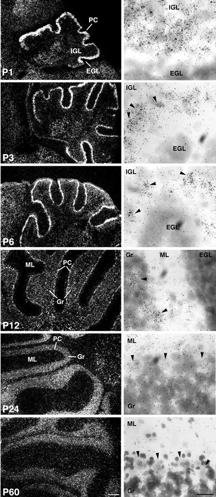
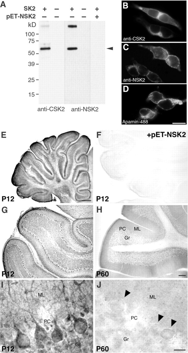
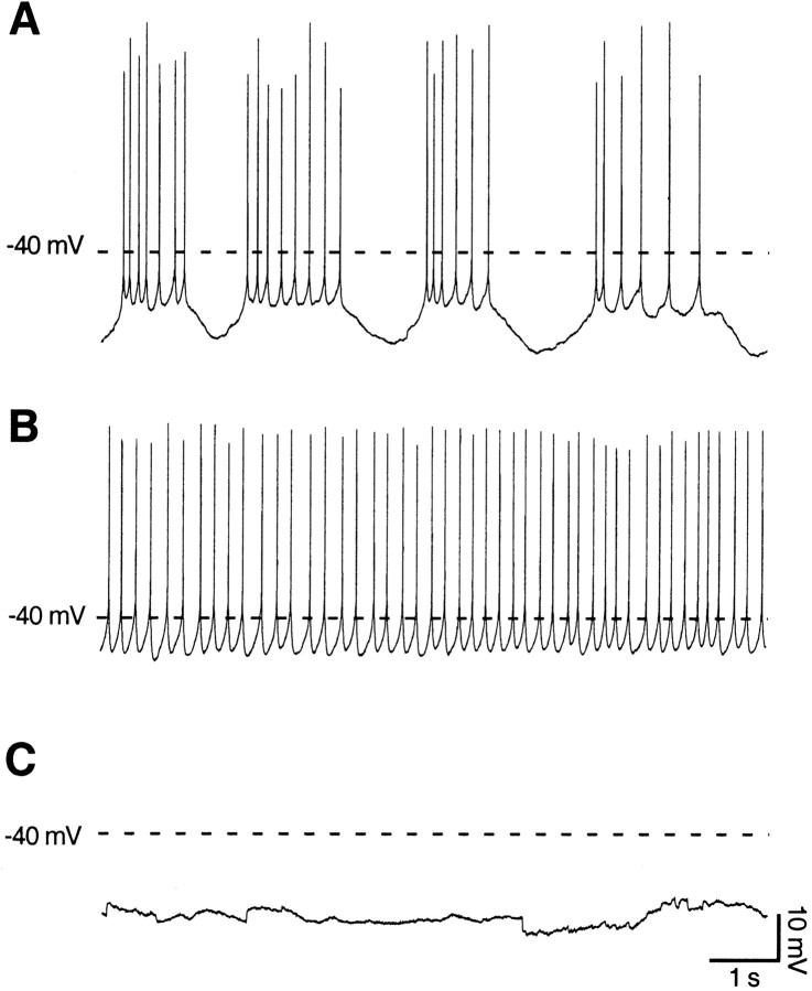
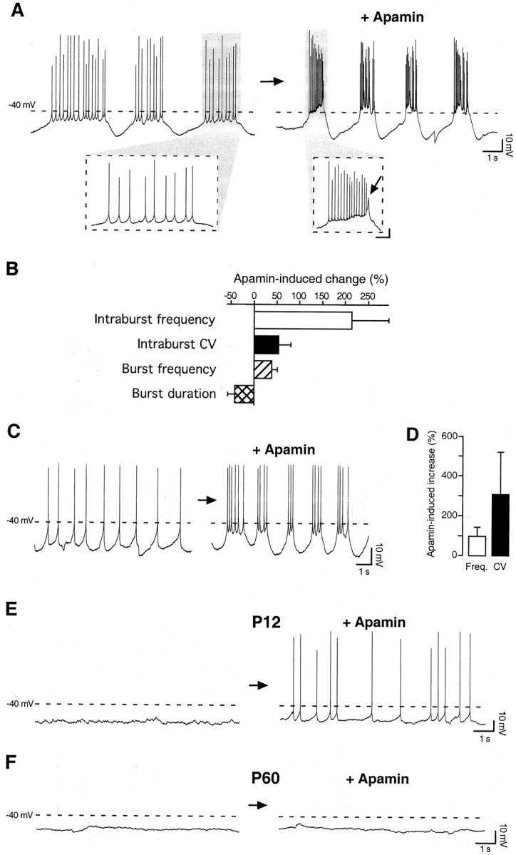
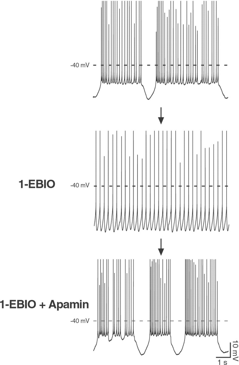
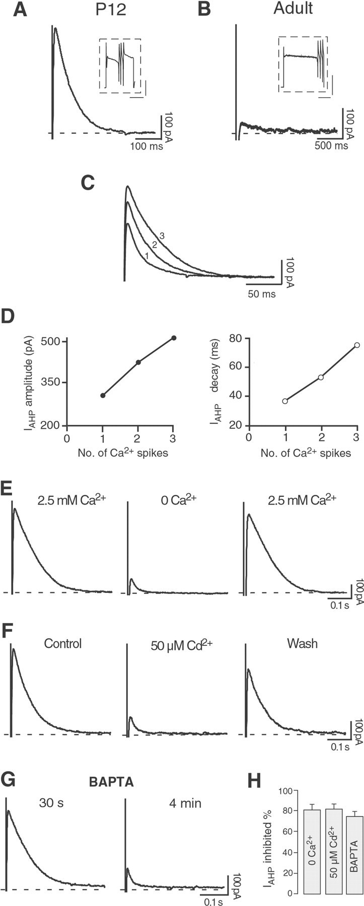
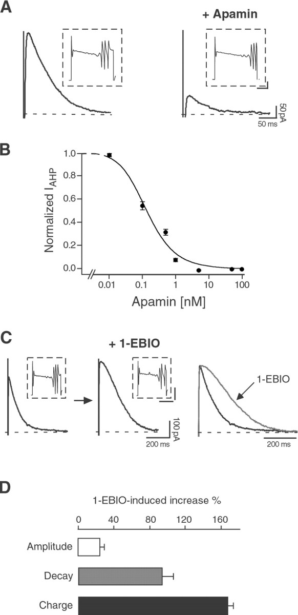
Calcium transients play an important role in the early and later phases of differentiation and maturation of single neurons and neuronal networks. Small-conductance calcium-activated potassium channels of the SK type modulate membrane excitability and are important determinants of the firing properties of central neurons. Increases in the intracellular calcium concentration activate SK channels, leading to a hyperpolarization of the membrane potential, which in turn reduces the calcium inflow into the cell. This feedback mechanism is ideally suited to regulate the spatiotemporal occurrence of calcium transients. However, the role of SK channels in neuronal development has not been addressed so far. We have concentrated on the ontogenesis and function of SK channels in the developing rat cerebellum, focusing particularly on Purkinje neurons. Electrophysiological recordings combined with specific pharmacological tools have revealed for the first time the presence of an afterhyperpolarizing current (I(AHP)) in immature Purkinje cells in rat cerebellar slices. The channel subunits underlying this current were identified as SK2 and localized by in situ hybridization and subunit-specific antibodies. Their expression level was shown to be high at birth and subsequently to decline during the first 3 weeks of postnatal life, both at the mRNA and protein levels. This developmental regulation was tightly correlated with the expression of I(AHP) and the prominent role of SK2 channels in shaping the spontaneous firing pattern in young, but not in adult, Purkinje neurons. These results provide the first evidence of the developmental regulation and function of SK channels in central neurons.


Figures

Fig. 1.
Fig. 1.
The expression level of SK2 mRNA is developmentally regulated in the rat cerebellum. In situhybridization was performed on sagittal sections at different developmental stages (P1, P3,P6, P12, P24, andP60) with SK2-specific oligonucleotides. Sections were coated with photoemulsion and exposed for 3 months. The probe for SK2 strongly labeled Purkinje cells (PC) from P1 to P12. The silver grains can be identified as white signals in the layer labeled as PC in the dark-field photomicrographs on the left. Light-field photomicrographs show cresyl violet-counterstained Purkinje cell neurons at high magnification (right panels), where the SK2 signal is visible as clusters of black dots over the cell nuclei. At P1, Purkinje neurons are organized in a multilayered band located between the internal and external granule layer and already display a high level of SK2 mRNA. At P3, P6, and P12, Purkinje cells are organized in a discrete monolayer, and arrowheads point to some of the silver grain clusters over Purkinje cell nuclei. Conversely, at P24 and P60 the signal observed in the dark-field photomicrographs (left panels) is attributable to a moderate level of expression of SK2 transcript in granule cells. The SK2 signal on single Purkinje cells (arrowheads in P24 andP60, right panels) is below the threshold limit of detection. EGL, External granule layer;Gr, granule cells; IGL, internal granule layer; ML, molecular layer; PC, Purkinje cells. Scale bars: dark-field, left panels, 250 μm; light-field, right panels, 25 μm.
Fig. 2.
Fig. 2.
The expression of SK2 protein is developmentally regulated in Purkinje neurons, as shown by immunohistochemistry with SK2-specific antibodies. A, Western blot on SK2 channel subunits expressed in HEK-293 cells and characterized with anti-CSK2 and affinity-purified anti-NSK2 antibody.Arrowheads indicate the bands corresponding to the SK2 channel subunit (lanes 1, 3). There are no bands visible in fractions from cells that do not express SK2 channel subunits (lanes 2, 4). No bands are detected after preadsorption of the primary antibody with the antigen (lane 5), demonstrating that the high molecular band corresponds to an aggregate of SK2 channel subunits.B, C, Immunofluorescence of HEK-293 cells transfected with SK2, showing a specific staining with both the anti-CSK2 and the anti-NSK2 antibodies. D, Labeling of membrane standing SK2 channels in transfected HEK-293 cells with a fluorescent derivative of apamin (apamin-Alexa488).EJ, Light-microscopy micrographs showing the immunohistochemical reactions of the affinity-purified anti-NSK2 antibody in the cerebellum at P12 and P60. InF, no staining is visible with preadsorbed antibodies (+pET-NSK2). In G and H, low-magnification views of the cerebellum show strong SK2 signals on the majority of Purkinje neurons at P12 (G), but only very weak ones on a few scattered Purkinje neurons on sections from adult rats (H, P60). InI and J, at a cellular level of resolution, Purkinje neurons show a strong SK2 signal on the somata and the dendritic stem at P12 (I), but not at P60 (J). In J, three Purkinje cells are indicated by arrowheads. One of them presents a light stain by anti-NSK2. This was observed in sporadic cases, but the level of staining at P60 was always lower than the one observed at earlier developmental stages (I).Gr, Granule cells; ML, molecular layer;PC, Purkinje cells. Scale bars: B, C, D, 20 μm; E, 0.5 mm; F, G, H, 150 μm;I, J, 20 μm.
Fig. 3.
Fig. 3.
Purkinje cells present different spontaneous firing patterns at P12. A, Fifty percent of the Purkinje cells presented a pattern of spontaneous activity characterized by rhythmic bursts of action potentials at P12. The mean burst frequency was 0.36 ± 0.03 Hz, the burst duration was 1.6 ± 0.2 sec, and the interburst intervals were 1.8 ± 0.2 sec (n = 32). B, Approximately 30% of the Purkinje cells showed a sustained tonic firing of single spikes with an average frequency of 4.4 ± 0.7 Hz and a CV of 0.24 ± 0.03 (n = 20). C, The remaining 20% of the cells did not present any spontaneous firing (silent cells) and displayed an average membrane resting potential of −57 ± 2 mV (n = 12). All traces displayed were recorded in the whole-cell configuration, with no steady current injected. Thedashed line corresponds to a membrane potential of −40 mV. Calibration is the same for all three traces.
Fig. 4.
Fig. 4.
The SK channel blocker apamin modifies the spontaneous firing behavior of Purkinje neurons at P12 but not at adult age. A, When applied on spontaneously bursting Purkinje cells, apamin (100 nm) increased both the frequency of the action potentials within each burst (intraburst frequency) and the frequency of the bursts (burst frequency). Moreover, apamin shortened the duration of each burst and enhanced the Ca2+spike terminating each burst (inset,arrow). Calibration in inset: 10 mV, 250 msec. B, Bar diagram summarizing the effects of apamin (100 nm) on spontaneously bursting Purkinje cells. The intraburst frequency was increased by 213 ± 81% (n = 3), and the burst frequency was increased by 37 ± 12% (n = 4), whereas the burst duration was reduced by 40 ± 15% (n = 4). Besides increasing the intraburst firing frequency, apamin decreased the regularity of the firing within each burst, as quantified by the increase in the CV by 54 ± 27% (n = 3).C, When applied to spontaneously single-spiking Purkinje cells, apamin (100 nm) favored the transition to a bursting firing pattern. D, Bar diagram summarizing the effect of apamin on single-spiking Purkinje cells. The overall firing frequency was increased by 97 ± 44%, and the CV was increased by 305 ± 213% (n = 5). E, At P12, apamin (100 nm) induced spontaneous firing of single spikes, without affecting the resting membrane potential (−52 ± 2 mV before and −50 ± 2 mV after apamin application;p = 0.65; n = 4).F, By contrast, apamin (100 nm) did not change the spontaneous firing behavior of silent Purkinje cells at adult age (P60). The dashed line inA, C, E, andF represents a membrane potential of −40 mV. No steady current was injected during these whole-cell current-clamp recordings.
Fig. 5.
Fig. 5.
The SK channel enhancer 1-EBIO exerts an opposite effect to the blocker apamin on the spontaneous firing pattern of Purkinje neurons at P12. When applied to a spontaneously bursting Purkinje cell (top panel), 1-EBIO (100 μm) promoted the transition to a single-spiking pattern, with action potentials fired at a low frequency (3.5 ± 0.6 Hz) and in a very regular manner (CV = 0.17 ± 0.05;middle panel). Additionally, every action potential was followed by a pronounced afterhyperpolarization in the presence of 1-EBIO (middle panel), a clear indication of an increased SK channel activity. The application of apamin (100 nm) completely reversed the effect on the firing pattern induced by 1-EBIO and demonstrates unequivocally that 1-EBIO specifically enhanced SK channel activity (bottom panel). Similar results were obtained in four cells. All traces were recorded in the whole-cell configuration. Calibration is the same for all traces. Spike height was truncated for better resolution of the afterhyperpolarizations after single or bursts of action potentials.
Fig. 6.
Fig. 6.
Purkinje neurons present a Ca2+-activated K+ current after depolarization-induced Ca2+ spikes at P12, but not at adult age under the same conditions. A, Tail current after a depolarizing pulse (200 msec to +10 mV) sufficient to elicit three Ca2+ spikes in a Purkinje cell at P12 in the presence of the Na+ channel blocker TTX (0.5 μm) and of the BK channel blocker penitrem A (10 μm). A similar current was observed in nine cells. Calibration ininset: 1 nA, 100 msec. B, In an adult Purkinje cell, a longer depolarizing pulse (600 msec to −10 mV) was needed to elicit three Ca2+ spikes as inA, in the presence of identical concentrations of TTX and penitrem A. The Ca2+ influx did not induce a tail current as observed at P12. Similar results were obtained in four cells. Calibration in inset: 2 nA, 200 msec.C, D, The tail current amplitude and duration increased proportionally to the number of all-or-none Ca2+ spikes triggered by the depolarizing pulses. The numbers beside the current traces (1,2, 3) indicate the number of Ca2+ spikes preceding each tail current.E, In P12 Purkinje cells, the tail current disappeared in Ca2+-free medium (0 Ca2+; 5 mm Mg2+). This effect was fully reversible. F, The Ca2+ channel blocker Cd2+ (50 μm) strongly and reversibly suppressed the tail current. G, When the Ca2+ chelator BAPTA (10 mm) was enclosed in the pipette solution, the tail current was progressively reduced during the first minutes in the whole-cell configuration.H, Bar diagram summarizing the results presented inEG. In Ca2+-free medium the tail current was reduced by 81 ± 5% (n = 3); 50 μmCd2+ suppressed the tail current by 82 ± 5% (n = 5). In the presence of BAPTA, the tail current was inhibited by 75 ± 5% (n = 4).
Fig. 7.
Fig. 7.
The Ca2+-dependent tail current in P12 Purkinje neurons is an SK2-mediatedIAHP, sensitive to apamin and 1-EBIO.A, Apamin (50 nm) strongly and irreversibly suppressed IAHP, without affecting the Ca2+ spikes elicited by the depolarizing pulses (insets), in Purkinje neurons at P12. Similar results were observed in 11 cells. Calibration in insets: 0.5 nA, 40 msec. B, Dose–response curve for the block ofIAHP by apamin. Data points were fit with the Hill equation, giving an IC50 value of ∼135 pm and a Hill coefficient of 1. For each point,n = 3–6; error bars are SEM. C, 1-EBIO (100 μm) enhanced IAHPin P12 Purkinje cells, without significantly affecting the Ca2+ spikes (insets). Theright panel shows the two traces scaled and overlapped to display the effect of 1-EBIO on the time course ofIAHP. Similar effects were observed in five cells. Calibration in insets: 1 nA, 100 msec.D, Bar diagram summarizing the effects of 1-EBIO (100 μm) on IAHP. The AHP current amplitude was increased by 25 ± 5%, the time constant of decay was increased by 95 ± 12%, and the total charge transfer was increased by 168 ± 6% (n = 5).

References

    1. Aizenman CD, Linden DJ. Regulation of the rebound depolarization and spontaneous firing patterns of deep nuclear neurons in slices of rat cerebellum. J Neurophysiol. 1999;82:1697–1709. - PubMed
    1. Altman J. Postnatal development of the cerebellar cortex in the rat. II. Phases in the maturation of Purkinje cells and of the molecular layer. J Comp Neurol. 1972;145:399–463. - PubMed
    1. Altman J, Bayer SA. Development of the cerebellar system in relation to its evolution, structure, and functions. CRC; Boca Raton, FL: 1997.
    1. Ben-Ari Y. Developing networks play a similar melody. Trends Neurosci. 2001;24:353–360. - PubMed
    1. Crepel F. Maturation of the cerebellar Purkinje cells. I. Postnatal evolution of the Purkinje cell spontaneous firing in the rat. Exp Brain Res. 1972;14:463–471.

MeSH terms

LinkOut - more resources