. 2002 Jun 1;22(11):4468-77.
doi: 10.1523/JNEUROSCI.22-11-04468.2002.

Bcl-2 overexpression does not enhance in vivo axonal regeneration of retinal ganglion cells after peripheral nerve transplantation in adult mice

Affiliations

Bcl-2 overexpression does not enhance in vivo axonal regeneration of retinal ganglion cells after peripheral nerve transplantation in adult mice

Tetsu Inoue et al. J Neurosci. .

Abstract

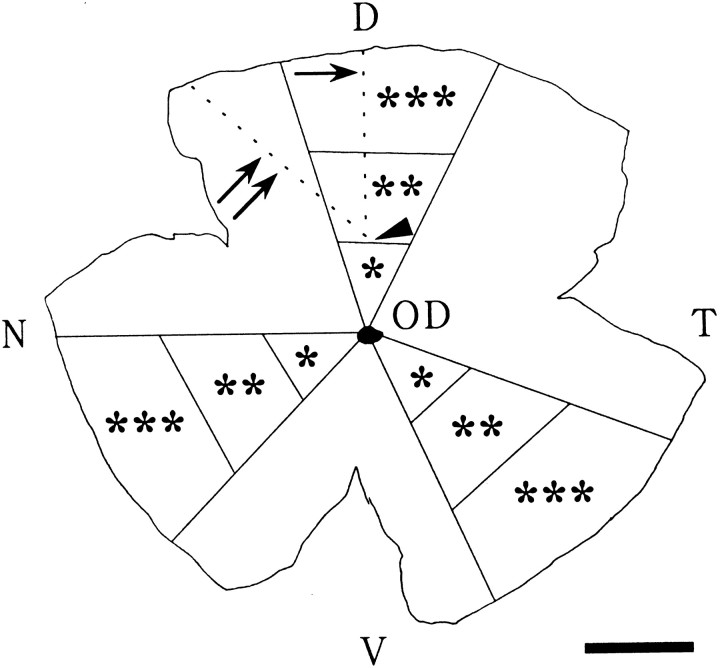
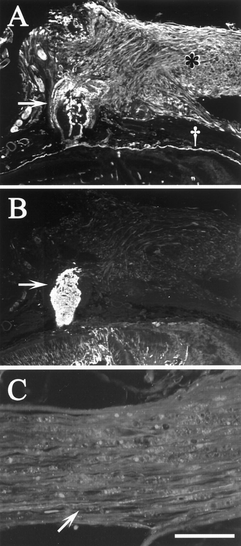
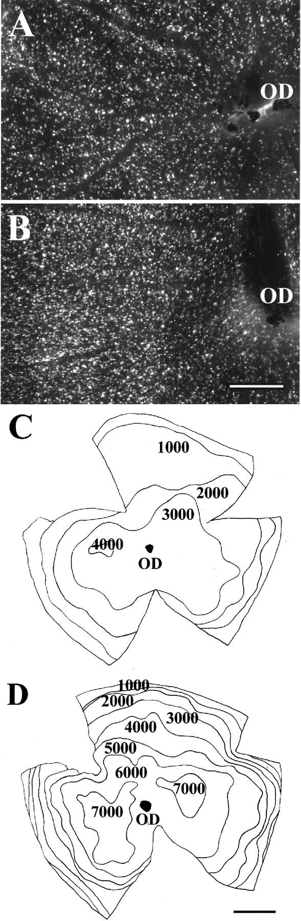
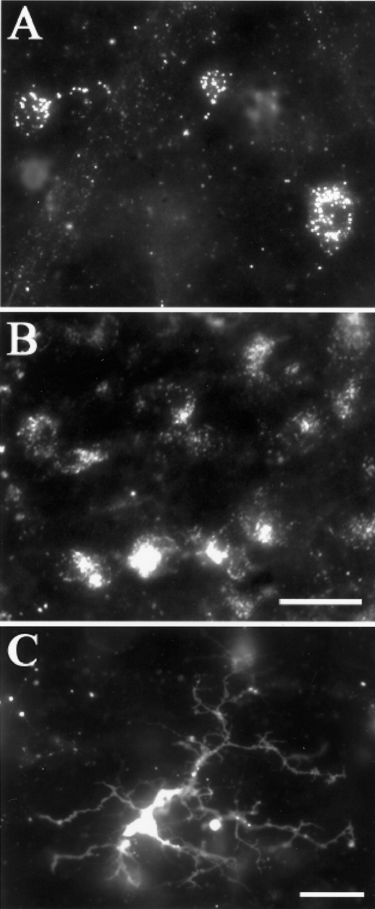
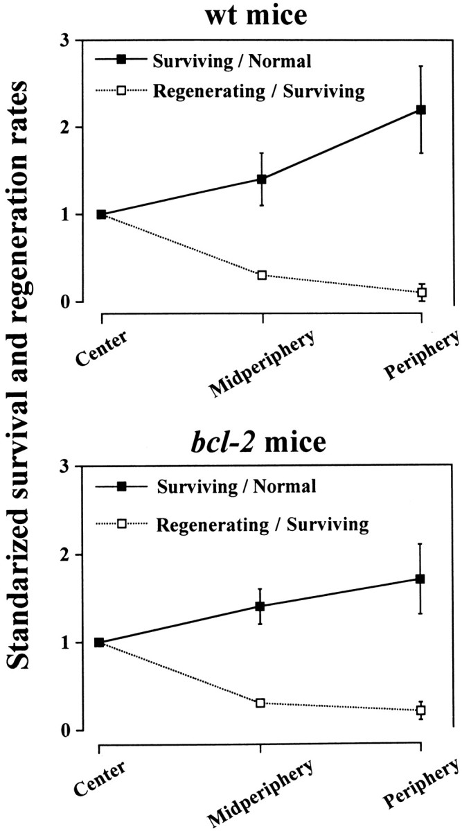
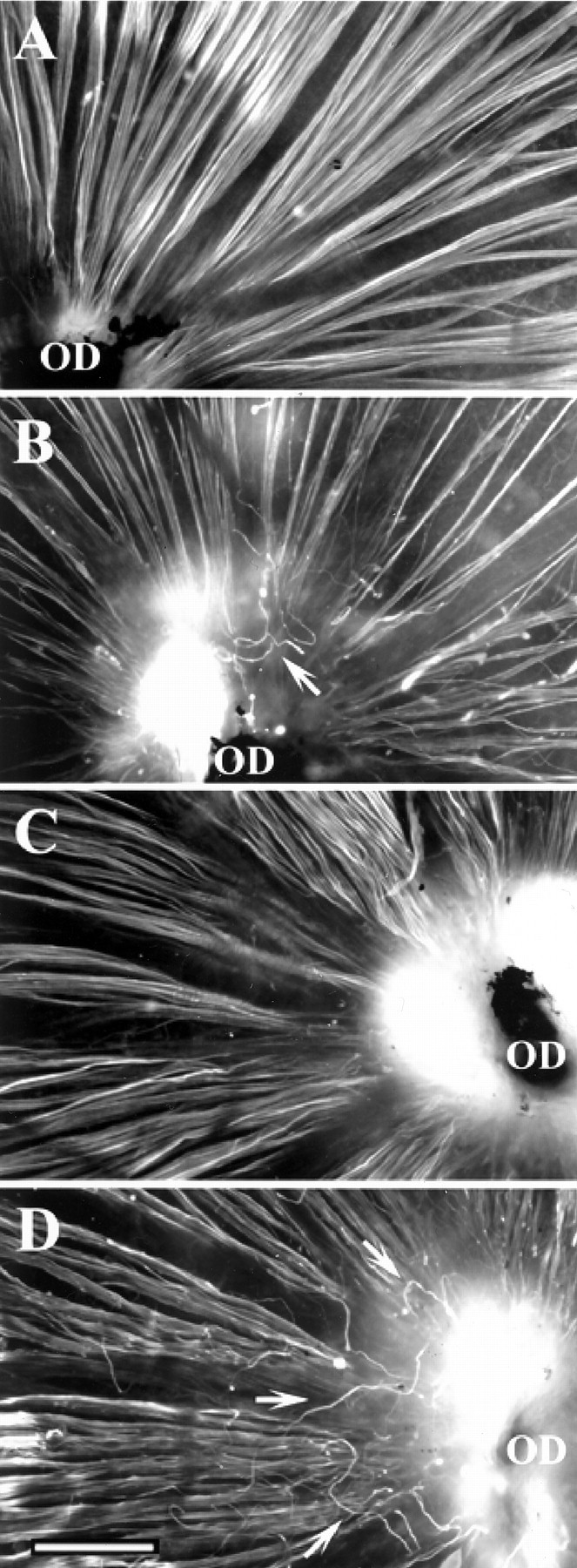
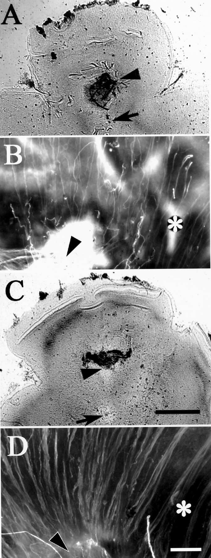
Optic nerve (ON) injury in adult mammals causes retinal ganglion cell (RGC) death and subsequent visual loss. Recovery of vision requires both rescuing axotomized RGCs and inducing their axonal regeneration. Axotomized RGCs are significantly rescued by overexpression of bcl-2, an anti-apoptotic gene. However, whether bcl-2 affects axonal regeneration is controversial. In neonatal bcl-2 transgenic mice (bcl-2 mice), optic tract regeneration after tectal lesion was promoted (Chen et al., 1997), whereas ON regeneration after ON crush was not (Lodovichi et al., 2001). These conflicting results may be attributable to different environments between tectum and ON. We tested here whether bcl-2 overexpression enhances in vivo RGC axonal regeneration in adult mice through a permissive environment in the peripheral nerve (PN) graft. Four weeks after PN transplantation to the proximal ON stump, we assessed the number of surviving and regenerating RGCs by retrograde labeling. Although the survival rate in bcl-2 mice was significantly enhanced compared with that in wild-type (wt) mice, the regeneration rate was not enhanced. In both bcl-2 and wt mice, RT97 immunostaining of the PN-grafted retinas revealed some RGC axons regrowing intraretinally but repulsed at the optic disk. To circumvent this repulsive barrier, we directly transplanted the PN graft to the partially injured retina and compared regeneration rates between these mice. Here again the regeneration rate in bcl-2 mice did not exceed that in wt mice. These findings indicate that bcl-2 overexpression enhances survival but not axonal regeneration of adult RGCs even within a permissive environment.


Figures

Fig. 1.
Fig. 1.
A schematic diagram showing how survival and axonal regeneration of RGCs were evaluated in retinal whole mounts of wt and bcl-2 mice after PN transplantation. In the case of RB transplantation, the survival and regeneration rates of RGCs were estimated by cell counting in three triangular regions bounded by the radial lines drawn from the optic disk (OD) to the retinal margin. For the comparison of survival or axonal regeneration of RGCs among different eccentricities from the OD (see Fig. 5), each triangular region was divided into three congruent parts: center (∗), midperipheral (∗∗), and peripheral (∗∗∗) retina. In the case of IR transplantation, a fan-like region in the dorsonasal retina, bounded by two dotted lines, served for the analysis. Their crossing (arrowhead), where the PN graft was inserted, was set at one-third from the OD to the retinal margin. The angle made by the lines was 55°. The bounded region corresponds to the presumed area of axotomy for IR transplantation. Scale bar, 1 mm.D, Dorsal; V, ventral; N, nasal;T, temporal.
Fig. 2.
Fig. 2.
Longitudinal sections of the ON–PN interface and distal part of the PN graft of one wt mouse. A, The ON–PN interface immunostained with an antibody against laminin. The laminin-positive PN graft (∗) runs horizontally along the eye wall (†) and connects with the proximal ON stump (arrow). In the ON stump, only the meningeal sheath and the blood vessels are laminin-positive. B, The adjacent section immunostained for GFAP. The inner tissue of the ON stump is GFAP-positive (arrow). C, The distal part of the PN graft after anterograde labeling with rhodamine, with the focal point on a long fiber of regenerating RGC axon (arrow). Scale bar: A, B, 160 μm; C, 100 μm.
Fig. 3.
Fig. 3.
A, B, DiI-labeled RGCs in the temporal retina of wt (A) andbcl-2 (B) mice, under epifluorescence illumination. These and all other micrographs are positive prints, in which DiI-fluorescence appearswhite. C, D, Isodensity maps of the retinal whole mounts of wt (C) andbcl-2 (D) mice. The RGC density (cells per millimeters squared) in bcl-2 mice was approximately twice as much as that in wt mice throughout the retina.OD, Optic disk. Scale bar (shown in B):A, B, 500 μm; (shown inD): C, D, 1 mm.
Fig. 4.
Fig. 4.
DiI-positive cells on the retina 4 weeks after RB transplantation, under epifluorescence illumination. A,B, Retrogradely labeled RGCs that contain many DiI granules in the retinas of wt (A) andbcl-2 (B) mice. These are RGCs that have survived the RB transplantation. Many more labeled RGCs appeared in bcl-2 mice than in wt mice.C, A DiI-positive putative microglia showing a small and irregular cell body and some thick processes. Scale bars (shown inB): A, B, 20 μm;C, 20 μm.
Fig. 5.
Fig. 5.
Centri-peripheral gradient of survival and regeneration rates of RGCs after RB transplantation. Thevertical axis plots the rates normalized to the survival and regeneration rates of RGCs in the central retina. The total numbers of regenerated RGCs used in this analysis were 228, 121, and 117 cells in wt mice and 658, 598, and 237 cells in bcl-2 mice. In both wt and bcl-2 mice, the survival rate tends to increase toward the periphery (wt, n = 4;bcl-2, n = 4), whereas the regeneration rate tends to decrease (wt, n = 3;bcl-2, n = 3).
Fig. 6.
Fig. 6.
Epifluorescence micrographs of retinal whole mounts treated with RT97 immunostaining. In the nongrafted retinas of wt (A) and bcl-2(B) mice, all RT97-positive RGC axons showed many fasciculated fibers that run centripetally toward the optic disk (OD). However, 4 weeks after RB transplantation, some regrowing RGC axons form a complex of looping fibers near the OD (seearrows in B for wt and inD for bcl-2 mice). In comparison with the normal fasciculated fibers, these looping fibers do not appear to be more numerous on bcl-2 retina than on wt retina. Scale bar, 200 μm.
Fig. 7.
Fig. 7.
Retinal whole mounts (A,C) and RT97-immunostained retinal axons (B, D) 4 weeks after IR transplantation.A, B, Left retina of a wt mouse.C, D, Right retina of abcl-2 mouse. A, C, In both retinas, the PNs are positioned at similar sites in the dorsonasal retina (see Fig. 1). Positions of the optic disk and the PN graft are indicated with an arrow and an arrowhead, respectively. B, D, Epifluorescence micrographs of RT97-positive RGC axons in each retina: in the dorsal retina distal to the PN graft, many axons reach the graft (arrowhead) in both mice. Apparently no RGC axons that have reached the PN graft loop near the PN graft (compare the photographs in Fig. 6). Because the focus is on the retinal fiber layer, the distal part of the RGC axons that enter the PN graft is not shown clearly. RGC axons in the neighboring area (∗) are uninjured, and they course toward the optic disk. Scale bars: A, C, 1 mm; B,D, 100 μm.

Similar articles

Cited by

References

    1. Bähr M. Live or let die: retinal ganglion cell death and survival during development and in the lesioned adult CNS. Trends Neurosci. 2000;23:483–490. - PubMed
    1. Bates CA, Becker CG, Miotke JA, Meyer RL. Expression of polysialylated NCAM but not L1 or N-cadherin by regenerating adult mouse optic fibers in vitro. Exp Neurol. 1999;155:128–139. - PubMed
    1. Cenni MC, Bonfanti L, Martinou J-C, Ratto GM, Strettoi E, Maffei L. Long-term survival of retinal ganglion cells following optic nerve section in adult bcl-2 transgenic mice. Eur J Neurosci. 1996;8:1735–1745. - PubMed
    1. Chen DF, Schneider GE, Martinou J-C, Tonegewa S. Bcl-2 promotes regeneration of severed axons in mammalian CNS. Nature. 1997;385:434–439. - PubMed
    1. Chierzi S, Strettoi E, Cenni MC, Maffei L. Optic nerve crush: axonal responses in wild-type and bcl-2 transgenic mice. J Neurosci. 1999;19:8367–8376. - PMC - PubMed

Publication types

MeSH terms

Substances

LinkOut - more resources