. 2002 Jun 1;22(11):4550-62.
doi: 10.1523/JNEUROSCI.22-11-04550.2002.

Overexpression of 5-HT1B receptor in dorsal raphe nucleus using Herpes Simplex Virus gene transfer increases anxiety behavior after inescapable stress

Affiliations

Overexpression of 5-HT1B receptor in dorsal raphe nucleus using Herpes Simplex Virus gene transfer increases anxiety behavior after inescapable stress

Michael S Clark et al. J Neurosci. .

Abstract

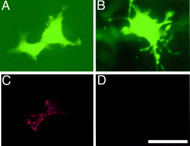
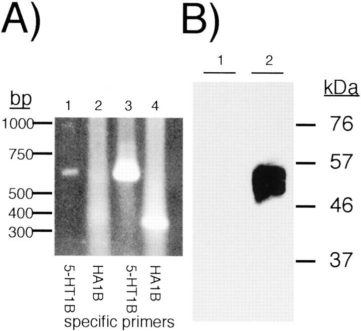
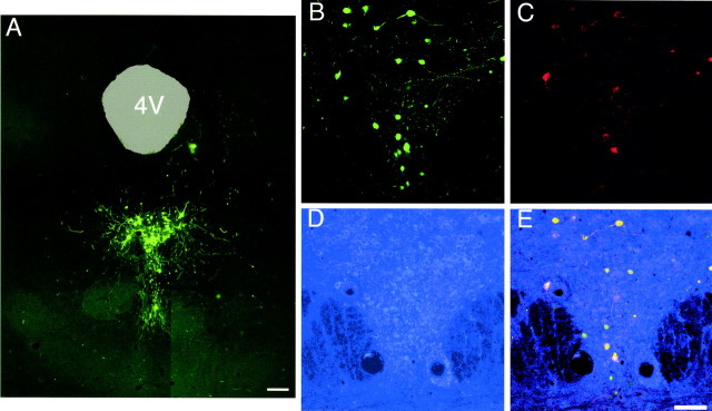
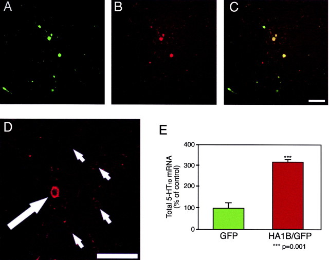
5-HT(1B) autoreceptors have been implicated in animal models of stress and are regulated selectively by serotonin-selective reuptake inhibitors such as fluoxetine. These terminal autoreceptors regulate serotonin release from dorsal raphe nucleus (DRN) projections throughout rat forebrain. However, it has not been previously possible to manipulate 5-HT(1B) autoreceptor activity selectively without also changing 5-HT(1B) activity in other neurons mediating different behavioral responses. Therefore, we have developed a viral-mediated gene transfer strategy to express hemagglutinin-tagged 5-HT(1B) and manipulate these autoreceptors in DRN. Green fluorescent protein (GFP) was coexpressed from a separate transcriptional unit on the same amplicon to assist in monitoring infection and expression. We confirmed the expression and biological activity of both transgenic proteins in vitro. When injected directly into DRN using stereotaxic procedure, HA-5-HT(1B) receptors were expressed in serotonergic neurons and translocated to the forebrain. The effect of DRN expression of HA-5-HT(1B) on stress-induced behaviors was compared with control rats that received GFP-only amplicons. There was no change in immobility in the forced swim test. However, HA-5-HT(1B) expression significantly reduced entrances into the central region of an open-field arena after water-restraint stress without altering overall locomotor activity, but not in the absence of stress exposure. HA-5-HT(1B) expression also reduced entries into the open arms of the elevated plus maze after water restraint. Because these tests are sensitive to increases in anxiety-like behavior, our results suggest that overactivity of 5-HT(1B) autoreceptors in DRN neurons may be an important mediator of pathological responses to stressful events.


Figures

Fig. 1.
Fig. 1.
Amplicon maps used for HA-5-HT1B and GFP expression. The plasmids pHSV-HA1B/GFP and pHSV-GFP were constructed as described in Materials and Methods and were confirmed by sequence analysis. Note that either GFP alone or the HA-5-HT1B and GFP sequences were inserted into the pHSV-PrPUC backbone provided by Dr. Rachael Neve (Neve and Geller, 1995). In the latter case, the HA-5-HT1B and GFP gene sequences were interrupted by an SV40 polyadenylation signal; the two genes have different promoter/enhancers controlling expression (HSV IE 4/5 and CMV IE, respectively), to reduce competition effects.ori S, HSV origin of replication; AmpR, ampicillin resistance gene.
Fig. 2.
Fig. 2.
COS cells transfected with pCIGFP-HA1B show dual expression. Cells were transfected with pCIGFP-HA1B or pCIGFP by electroporation as described. A and C show pCIGFP-HA1B-transfected cells; B andD show pCIGFP-transfected cells.A and B show GFP fluorescence;C and D show hemagglutinin immunoreactivity. The HA-tagged 5-HT1B receptor could be detected only in the pCIGFP-HA1B-transfected cells, and there was no apparent interaction between GFP and HA-5-HT1Bexpression in the same cells. Scale bar, 20 μm.
Fig. 3.
Fig. 3.
HA-5-HT1Breceptors inhibit adenylate cyclase in JEG-3 cells. Adenylate cyclase activity was assayed using a luciferase reporter gene assay as described in Materials and Methods. Data points represent SD of triplicate determinations; two replicate assays were performed. The curve fits and EC50 determinations were calculated using Prism 2.0. HA tagging of the 5-HT1B receptor did not appear to reduce its ability to inhibit adenylate cyclase activity, and coexpression of GFP from the same plasmid did not impair the level of HA-5-HT1B expression or function. RLU, Relative light units.
Fig. 4.
Fig. 4.
In vitro expression of pHSV-HA1B/GFP in CA77 cells. A, Twenty-four hours after infection with pHSV-GFP (lanes 1, 2) or pHSV-HA1B/GFP (lanes 3,4) (3 μl per well in six-well tissue culture plates; 1–2 × 10⁸ infective units per milliliter), CA77 cells were harvested and processed for either immunoblot analysis or poly-A RNA extraction. GFP expression and HA immunoreactivity were robustly detected in ∼50–90% of cells (data not shown). RT-PCR of poly-A RNA from these cells showed the presence of 5-HT1B mRNA using total 5-HT1B primers for amplification (lanes 1, 3) and HA-specific primers (lanes 2, 4). Vehicle and pHSV-GFP-treated CA77 cells express low levels of 5-HT1B RNA (lane 1) and no HA epitope (lane 2). pHSV-HA1B/GFP-treated cells showed dramatically more total 5-HT1B message (lane 3) and a strong HA-5-HT1B-specific PCR product (lane 4). Vehicle-treated CA77 cells showed low levels of 5-HT1B mRNA, similar to pHSV-GFP (data not shown).B, Protein samples (5 μm) from pHSV-GFP (lane 1)- or pHSV-HA1B/GFP (lane 2)-infected CA77 cells were separated by PAGE and immobilized on membranes by Western blot. HA-specific immunoreactive protein was detected only in pHSV-HA1B/GFP-infected cells.
Fig. 5.
Fig. 5.
Coexpression of HA-5-HT1B and GFP in vivo. pHSV-HA1B/GFP viral particles were injected stereotaxically into DRN, and animals were killed 4 d later for evaluation of transgene expression by immunostaining and confocal microscopy. A, A composite image of several 10× fields shows clear GFP localization within the anatomic region of the DRN. The fourth ventricle (4V) has been colored light green for clarity. Scale bar, 100 μm. B, A 20× image of DRN shows GFP fluorescence in cells and beaded fibers.C, HA-5-HT1B immunostaining of the same region shown in B. D, 5-HT1Aimmunostaining of the same region shown in B andC. Because of the lower laser strength available for the secondary dye used (Alexa-633), the intensity of positive staining, typically visible as rings around darker nuclei, is relatively low.E, A composite image of all three signals displays cells positive for GFP, HA-5-HT1B, and 5-HT1A, demonstrating that serotonergic neurons have been infected and that these neurons produce both viral transgene products in vivo. A count of cells positive for both GFP and 5-HT1A in images obtained for this study showed that 80% of GFP-positive neurons were also positive for 5-HT1A. Scale bar, 100 μm.
Fig. 6.
Fig. 6.
Injection of pHSV-HA1B/GFP increases 5-HT1B expression in vivo. Sections through the DRN were taken from animals injected at that site with pHSV-HA1B/GFP viral particles, immunostained for 5-HT1B, and examined by confocal microscopy.A, GFP-containing cells and associated fibers may be clearly seen in the DRN, as shown previously. B, 5-HT1B immunoreactivity is detected within both GFP- and non-GFP-containing cells. C, When GFP fluorescence and 5-HT1B immunoreactivity are overlapped, GFP-positive cells are shown to typically display intense 5-HT1Bimmunoreactivity, as indicated by the yellow-green coloration of these cells. A–C, Magnification: 20×. Scale bar, 100 μm. D, In another focal plane from the same section shown in AC, 5-HT1B immunoreactivity is much more intense in a GFP-positive cell (indicated by a large white arrow) compared with endogenously expressed 5-HT1B protein in non-GFP-positive cells (small arrows). The GFP-positive cell displays much greater 5-HT1B immunoreactivity, although both types of cells display what appears to be membrane-bound localization of 5-HT1B signal. Magnification: 40×. Scale bar, 100 μm. E, DRN punches from animals injected with either pHSV-GFP or pHSV-HA1B/GFP viral particles were analyzed by quantitative RT-PCR for 5-HT1B message content (n = 9 for each group). 5-HT1B mRNA levels, normalized for GAPDH mRNA expression, were 3.16-fold times higher in animals injected with pHSV-HA1B/GFP than in animals injected with pHSV-GFP (***p = 0.001; Student'st test).
Fig. 7.
Fig. 7.
HA-5-HT1Bis translocated to the forebrain. Striatal sections of animals injected in the DRN with either pHSV-HA1B/GFP or pHSV-GFP viral particles were immunostained to detect the presence of HA-5-HT1B immunoreactivity and examined by confocal microscopy. A, In animals injected with pHSV-HA1B/GFP, beaded fibers with anti-HA immunoreactivity may be clearly seen. The fibers demonstrate typical pleiomorphic varicosities, suggesting multiple sites of neurotransmitter release that are characteristic of DRN axons (Kosofsky and Molliver, 1987). B, In animals injected with pHSV-GFP, only background is present. In neither case was GFP detected (data not shown). Images shown are flattened, 60× confocal stacks. Scale bar, 50 μm. C, Western blot of HA-immunostained protein from terminals field of DRN axonal projections to forebrain. Protein samples from frontal cortex (lanes 1, 2) or striatum (lanes 3,4) after DRN injection of pHSV-GFP (−) or pHSV-HA1B/GFP (+) viral particles. The single immunoreactive HA-5-HT1B band migrated at an apparent size of 65 kDa, perhaps reflecting glycosylation and/or other posttranslational modifications of the 49 kDa predicted protein.
Fig. 8.
Fig. 8.
HA-5-HT1Bexpression in DRN neurons did not alter immobility in the forced swim test. Animals received injections of pHSV-HA1B/GFP (n = 11) or pHSV-GFP (n = 8) in DRN and were subjected to the standard FST procedure on days 3 and 4 as described in Materials and Methods. Behaviors were counted as swimming, climbing, or immobile as described in Materials and Methods. Numerical data are as follows and are presented as mean ± SEM (climb: GFP 14 ± 2.0, HA1B/GFP 17 ± 2.5;swim: GFP 14 ± 2.9, HA1B/GFP 12 ± 2.1;tread: GFP 31 ± 4.5, HA1B 31 ± 3.4). There were no significant differences in these behaviors between treatment groups.
Fig. 9.
Fig. 9.
HA-5-HT1B expression in DRN neurons increased avoidance of the center of an open field only after water-restraint stress. Animals received injections of pHSV-HA1B/GFP or pHSV-GFP and either were tested in the OFT 3 d later (A, B) or subjected to water-restraint stress on day 3 and tested in the OFT 24 hr later (C,D). The numbers of entries into the central square during the first 3 min were counted and are shown as mean ± SEM (A, GFP 4.5 ± 0.56, HA1B/GFP 6.5 ± 0.64;C, GFP 8.4 ± 0.86, HA1B/GFP 5.9 ± 0.58). HA-5-HT1B expression increased central square entries in the absence of a specific stress exposure (#p = 0.05) but reduced entries into the central region after stress by 30% (*p = 0.044). The total number of zone crossings, shown as mean ± SEM (B, GFP 102 ± 9, HA1B/GFP 123 ± 10; D, GFP 160 ± 13, HA1B/GFP 162 ± 20), was not different between pHSV-HA1B/GFP and pHSV-GFP.n = 8–14 animals in each treatment condition.
Fig. 10.
Fig. 10.
HA-5-HT1Bexpression in DRN reduced open arm entries in the EPM 24 hr after water-restraint stress. Animals received injections of pHSV-HA1B/GFP (n = 13) or pHSV-GFP (n = 8), were subjected to water-restraint stress 3 d later, and were tested in the EPM 24 hr later. The rat's behavior was recorded and analyzed by computer-assisted video monitoring. A shows the percentage of open arm/total arm entries; HA-5-HT1B-expressing animals had significantly reduced percentage of entries into open arms (GFP 55 ± 4%; HA1B/GFP 44 ± 3%; mean ± SEM shown); *p = 0.047. B, There was no significant difference in total distance traveled between treatment groups (GFP 415 ± 38 cm; HA1B/GFP 450 ± 40 cm; mean ± SEM shown). Other parameters measured include percentage open time (GFP 29.6 ± 8.9; HA1B/GFP 22.3 ± 4.0), percentage closed time (GFP 51.5 ± 8.1, HA1B/GFP 54.2 ± 5.2), open entries (GFP 8.2 ± 1.0, HA1B/GFP 7.2 ± 1.1), and closed entries (GFP 7.4 ± 1.5, HA1B/GFP 9.1 ± 1.2); mean ± SEM shown. Although these parameters did not reach statistical significance (p > 0.05), all trends are consistent with anxiogenic effects in HA-5-HT1B-expressing animals.

References

    1. Adell A, Casanovas JM, Artigas F. Comparative study in the rat of the actions of different types of stress on the release of 5-HT in raphe nuclei and forebrain areas. Neuropharmacology. 1997;36:735–741. - PubMed
    1. Amat J, Matus-Amat P, Watkins LR, Maier SF. Escapable and inescapable stress differentially alter extracellular levels of 5-HT in the basolateral amygdala of the rat. Brain Res. 1998;812:113–120. - PubMed
    1. Anthony JP, Sexton TJ, Neumaier JF. Antidepressant induced regulation of 5-HT1B mRNA in rat dorsal raphe nucleus reverses rapidly following drug discontinuation. J Neurosci Res. 2000;61:82–87. - PubMed
    1. Artigas F, Romero L, de Montigny C, Blier P. Acceleration of the effect of selected antidepressant drugs in major depression by 5-HT1A antagonists. Trends Neurosci. 1996;19:378–383. - PubMed
    1. Barnes NM, Sharp T. A review of central 5-HT receptors and their function. Neuropharmacology. 1999;38:1083–1152. - PubMed

Publication types

MeSH terms