. 2002 Jun 1;22(11):4756-65.
doi: 10.1523/JNEUROSCI.22-11-04756.2002.

Coordinations of locomotor and respiratory rhythms in vitro are critically dependent on hindlimb sensory inputs

Affiliations

Coordinations of locomotor and respiratory rhythms in vitro are critically dependent on hindlimb sensory inputs

Didier Morin et al. J Neurosci. .

Abstract

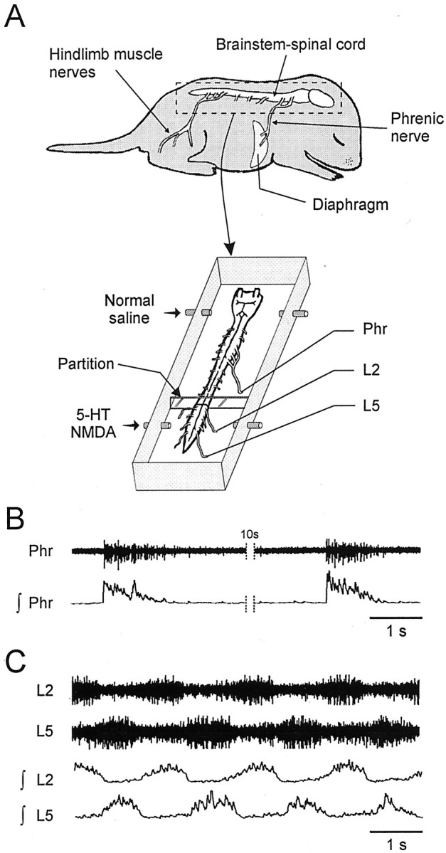
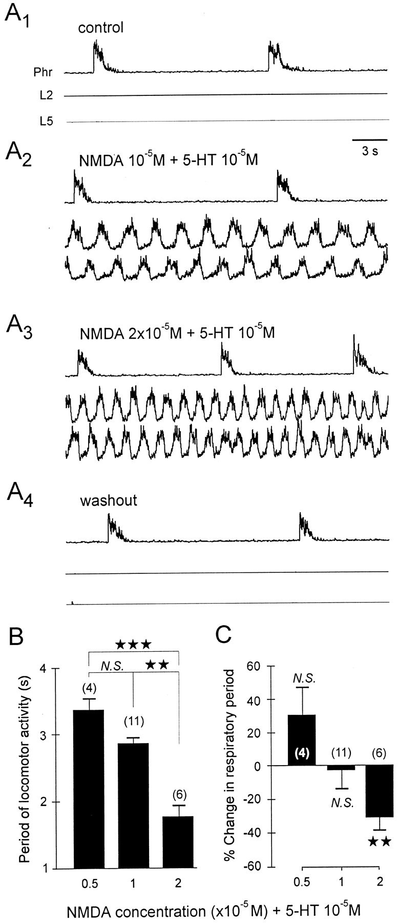
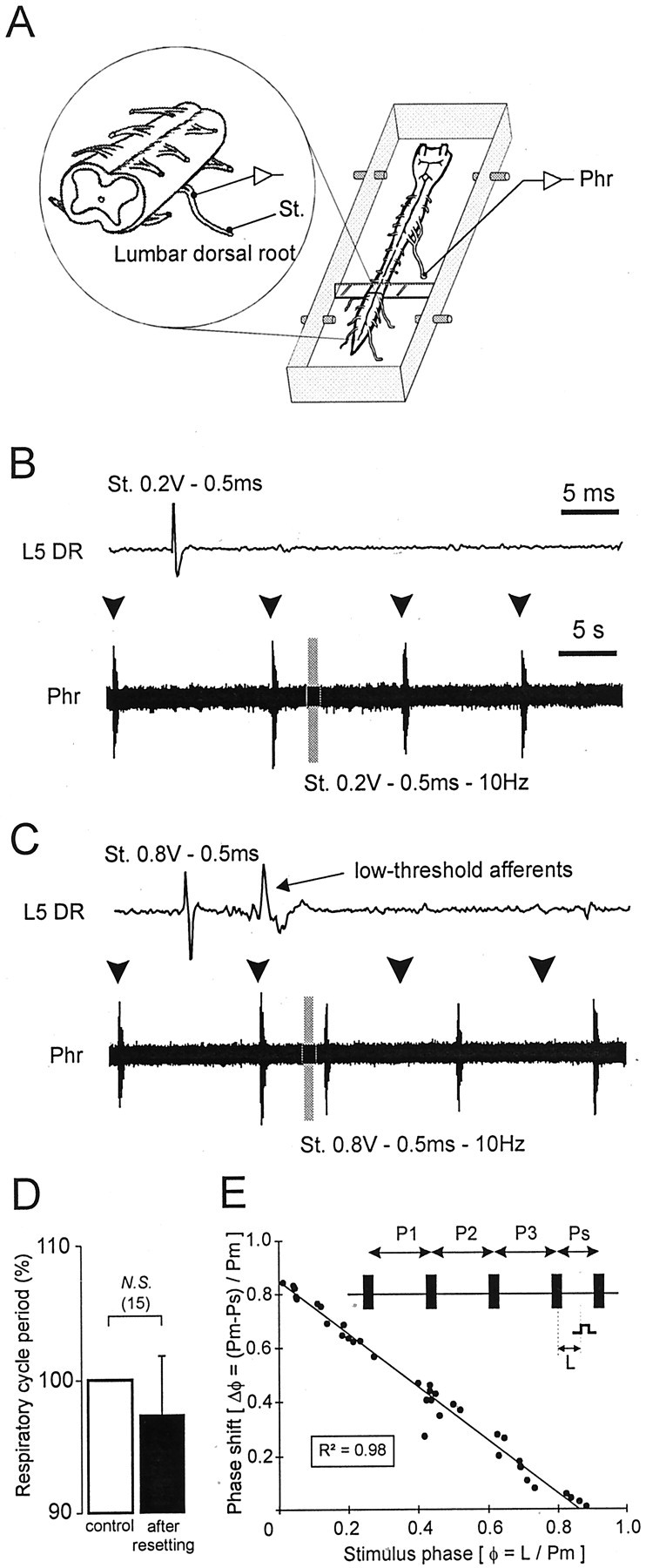
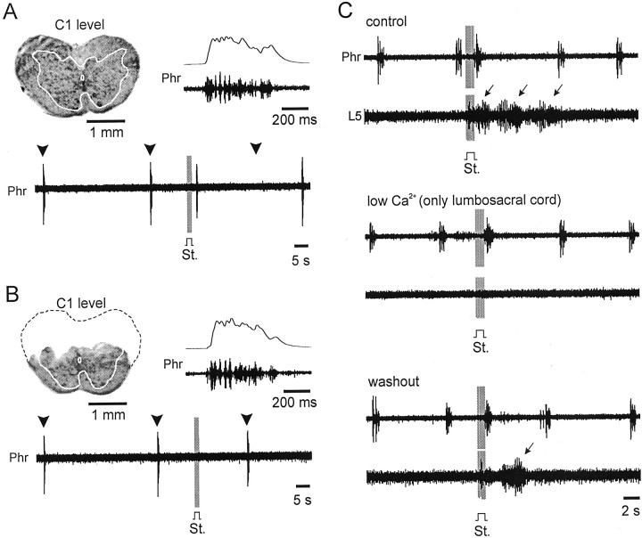
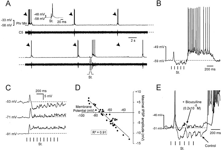
A 1:1 coordination between locomotor and respiratory movements has been described in various mammalian species during fast locomotion, and several mechanisms underlying such interactions have been proposed. Here we use an isolated brainstem-spinal cord preparation of the neonatal rat to determine the origin of this coupling, which could derive either from a direct interaction between the central locomotor- and respiratory-generating networks themselves or from an indirect influence via a peripheral mechanism. We demonstrate that during fictive locomotion induced by pharmacological activation of the lumbar locomotor generators, a concomitant increase in spontaneous respiratory rate occurs without any evident form of phase coupling. In contrast, respiratory motor activity can be fully entrained (1:1 coupling) over a range of periodic electrical stimulation applied to low-threshold sensory pathways originating from hindlimb muscles. Our results provide strong support for the existence of pathways between lumbar proprioceptive afferents, medullary respiratory networks, and phrenic motoneurons that could provide the basis of the locomotor-respiratory coupling in many animals. Thus a peripheral sensory system involved in a well defined rhythmic motor function can be responsible for the tight functional interaction between two otherwise independent motor behaviors.


Figures

Fig. 1.
Fig. 1.
In vitro mammal preparation used to study locomotor–respiratory coupling. A, Schematic drawings of a neonate rat CNS (brainstem–spinal cord) before and after isolation in a recording chamber. Simultaneous recordings were made from a respiratory phrenic nerve (Phr) and locomotor lumbar (L2, L5) ventral roots. Locomotor rhythm-generating networks had to be activated by lumbar cord perfusion of a medium containing 5-HT (10−5m) and NMDA (0.5–2 × 10⁻⁵m).B, Raw (top) and integrated (bottom) phrenic nerve activity showing spontaneous respiratory bursts. C, Episode of 5-HT/NMDA-induced locomotor rhythmicity recorded from homolateral lumbar L2 and L5 ventral roots.
Fig. 2.
Fig. 2.
Modulation of respiratory burst frequency by activation of the lumbosacral locomotor generators. A, Phrenic (Phr) and lumbar (L2,L5) integrated activity under control conditions and during application (restricted to the lumbar cord) of constant 5-HT (10−5m) and increasing NMDA concentrations (from 10−5 to 2 × 10⁻⁵m). B, Histogram showing the relationship between different NMDA concentrations (0.5–2 × 10⁻⁵m plus 10−5m 5-HT) in the lumbosacral bath and the period of the induced locomotor rhythm. C, Histogram showing the resulting change in respiratory rate expressed as percentage of control value in the absence of drugs. Vertical bars indicate mean values; vertical linesindicate the SEM. N.S., Nonsignificant. ★★★p < 0.001; ★★p < 0.01.
Fig. 3.
Fig. 3.
Ability of low-threshold lumbar afferents to reset spontaneous respiratory rhythmicity. A, Schematic representation of the experimental procedure. B, C, Continuous recordings of spontaneous phrenic (Phr) activity during a volley of lumbar (L5) dorsal root (DR) stimulation. Shown above each phrenic trace is a faster time base recording from the corresponding L5 DR during a single shock at the indicated stimulus intensity. The gray barindicates a train stimulation of lumbar afferents. Subthreshold electrical stimulation (0.2 V) of lumbar afferents (B) did not reset the respiratory phrenic rhythmicity, whereas respiratory resetting was obtained when low-threshold lumbar afferents were activated by ≥0.8 V (C). Arrowheads denote the expected time of occurrence of spontaneous phrenic bursts in the absence of resetting. D, Histograms showing lack of significant change in respiratory period (expressed as percentage of the mean control period) after resetting. The control value corresponds to the mean of three successive respiratory periods before the stimulated cycle (white bar), which is compared with the respiratory cycle observed after the stimulated cycle (black bar). N.S., Nonsignificant. E, Phase response plot calculated as follows (also see schematic): the reference period (Pm) was measured from three spontaneous respiratory cycles (P1, P2, and P3); the ratio of the stimulus latency (L) and Pm determined the stimulus phase (φ); the phase shift of the phrenic burst (Δφ) expressed as the difference between Pm and the stimulated period (Ps) and divided again by Pm, was plotted on the ordinate. The solid line indicates linear regression.R2, Coefficient of determination. Standardized data were collected from three different preparations.
Fig. 4.
Fig. 4.
Direct influence of low-threshold lumbar afferents on medullary respiratory networks. A, B, Respiratory resetting (A) results from an action on medullary respiratory centers, because transection of the dorsal spinal cord at C1 (B, see histological control; compare withA) suppressed the ability of the same lumbar afferent stimulation to reset phrenic activity (bottom trace). Note that the pattern of phrenic (Phr) motor bursts was similar in the two experimental conditions (see fast time base raw and integrated records at the top right of eachpanel). Arrowheads denote the expected time of occurrence of phrenic bursts in the absence of resetting. The gray bar in A and Bindicate a train stimulation (St., 0.5 msec, 0.8 V, 10 Hz) of lumbar afferents. The dotted line in B shows the part of the spinal cord removed. C, Effects of lumbar afferent activation (gray bar: St., train stimulation, 0.5 msec, 0.7 V, 10 Hz) on both phrenic nerve (Phr) and lumbar ventral root (L5) activity under control conditions (top), during low Ca2+ perfusion of the lumbosacral cord (middle), and after washout with normal saline (bottom). Note that under normal saline perfusion (top and bottom panels), activation of the lumbar afferents also elicited a short sequence of locomotor bursting (arrows).
Fig. 5.
Fig. 5.
Postsynaptic effects of lumbar afferent activation on phrenic motoneurons. A, Simultaneous whole-cell patch-clamp recording of a phrenic motoneuron (Phr Mn) and raw activity of cervical ventral root (C5) under control conditions (top) and during lumbar dorsal root stimulation (bottom, St.). The traces are contiguous (dashed lines). Arrowheadsdenote the expected time of occurrence of phrenic bursts in the absence of resetting. The inset shows motoneuron identification by antidromic electrical stimulation (St.) of corresponding phrenic ventral root. B, Details of postsynaptic events induced in the phrenic motoneuron by lumbar dorsal root stimulation. Vertical bars: St., Train stimulation (0.5 msec, 0.7 V, 10 Hz). Note the initial occurrence of an EPSP followed by series of IPSPs before a spike burst. Thedashed line indicates the resting membrane potential level. C, Hyperpolarizing current-induced reversal of stimulus-evoked compound IPSP. Dashed lines represent the resting membrane potential. D, Scatter plot illustrating the relationship between maximal IPSP amplitude and motoneuron membrane potential. Note the reversal potential at approximately −70 mV (data from 7 phrenic motoneurons). The solid line indicates linear regression. R2, Coefficient of determination. E, Bicuculline application (0.2 × 10⁻⁵m) blocks lumbar afferent-evoked inhibition of a phrenic motoneuron. Note that the action potentials inE have been truncated.
Fig. 6.
Fig. 6.
Respiratory rhythm entrainment by rhythmic activation of low-threshold lumbar afferents. A, Recordings of phrenic nerve activity (Phr) during electrical stimulation (St.; 0.9 V, 0.5 msec, 10 Hz) of lumbar afferents with different TSPs. A 1:1 coordination occurs with TSPs of 6 sec and 4 sec but fails at a TSP of 3 sec. B, Scatter plot showing the relationships between respiratory cycle period and lumbar afferent TSPs; the solid line indicates a 1:1 coupling. C, Box plots representing phrenic burst latency (lat.) in relation to the train stimulation period (see schematics above). ★★p< 0.01; ★p < 0.05.
Fig. 7.
Fig. 7.
Lumbar motor-sensory loop as a neural substrate for mammalian locomotor–respiratory coupling. A, Spontaneous respiratory activity of phrenic nerve (Phr) and pharmacologically induced locomotor rhythmicity (L2, lumbar ventral root) in the absence of low-threshold lumbar afferent stimulation. The underlying central networks are schematized on theright. B, bottom left, Repetitive activation of low-threshold lumbar afferents (vertical bars: St.) in time with locomotor bursts immediately drives locomotor–respiratory coupling. The two sets oftraces in A and B are from the same experiment. Related synaptic events recorded from a single phrenic motoneuron are shown in B (upper left). B, right, Schematic representation of proposed circuitry involved in the locomotor–respiratory coupling (hindlimb extensor and flexor muscles, which were removed in our preparations, are included to complete thein vivo motor-sensory loop). Connections responsible for different synaptic influences (arrowheads) on phrenic motoneurons are also numbered. Phr Mn, Phrenic motoneuron; Lumbar Mn, lumbar motoneuron;Resp., respiratory; Loco., locomotor.

Similar articles

Cited by

References

    1. Ballion B, Morin D, Viala D. Forelimb locomotor generators and quadrupedal locomotion in the neonatal rat. Eur J Neurosci. 2001;14:1727–1738. - PubMed
    1. Banzett RB, Mead J, Reid MB, Topulos GP. Locomotion in men has no appreciable mechanical effect on breathing. J Appl Physiol. 1992;72:1922–1926. - PubMed
    1. Bechbache RR, Duffin J. The entrainment of breathing frequency by exercise rhythm. J Physiol (Lond) 1977;272:553–561. - PMC - PubMed
    1. Bernasconi P, Kohl J. Analysis of co-ordination between breathing and exercise rhythms in man. J Physiol (Lond) 1993;471:693–706. - PMC - PubMed
    1. Bracci E, Beato M, Nistri A. Afferent inputs modulate the activity of a rhythmic burst generator in the rat disinhibited spinal cord in vitro. J Neurophysiol. 1997;77:3157–3167. - PubMed

Publication types

MeSH terms

LinkOut - more resources