Review
. 2002 Jun;66(2):223-49.
doi: 10.1128/MMBR.66.2.223-249.2002.

Genetics and assembly line enzymology of siderophore biosynthesis in bacteria

Affiliations
Review

Genetics and assembly line enzymology of siderophore biosynthesis in bacteria

Jorge H Crosa et al. Microbiol Mol Biol Rev. 2002 Jun.

Abstract

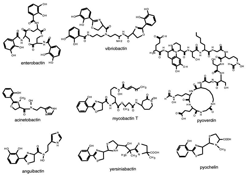
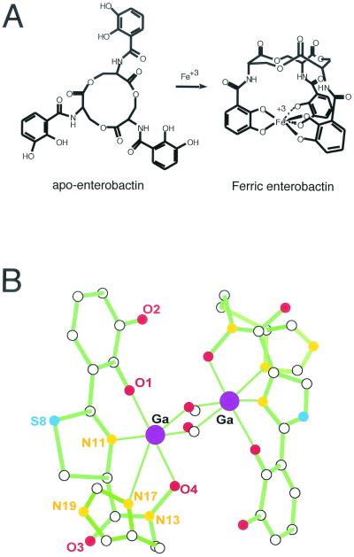
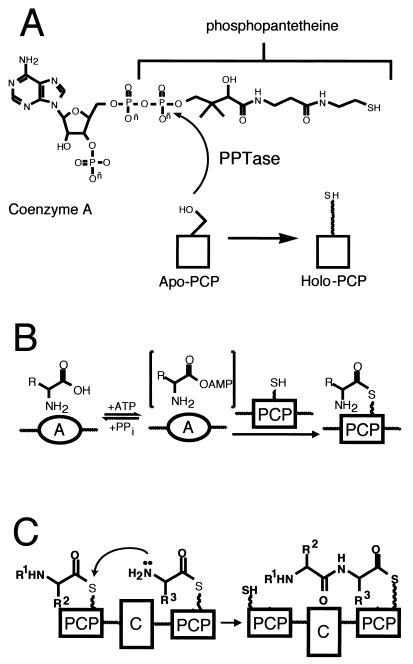
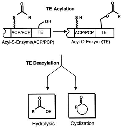
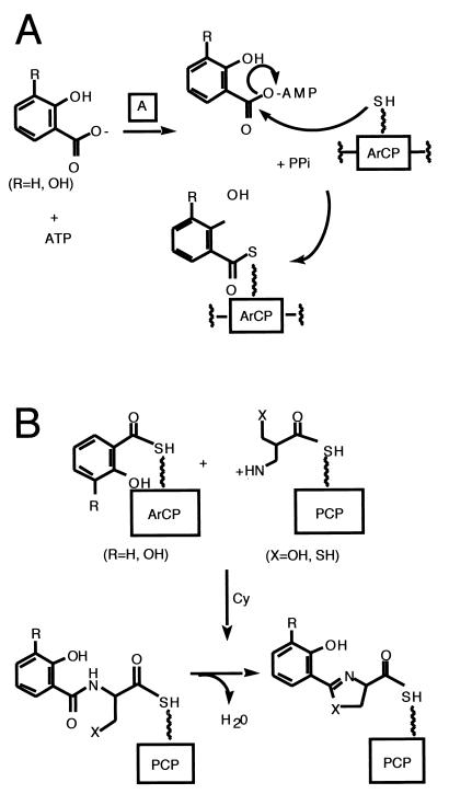
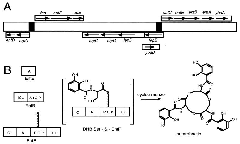
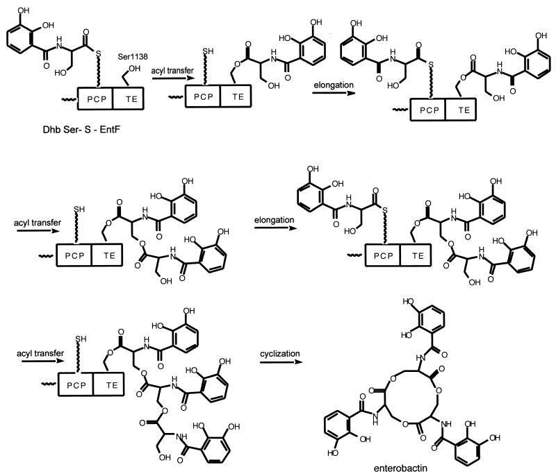
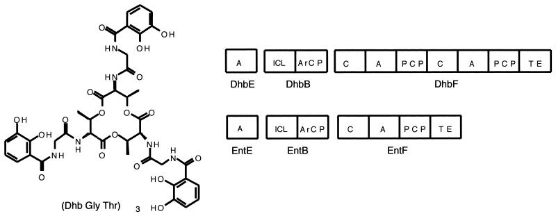
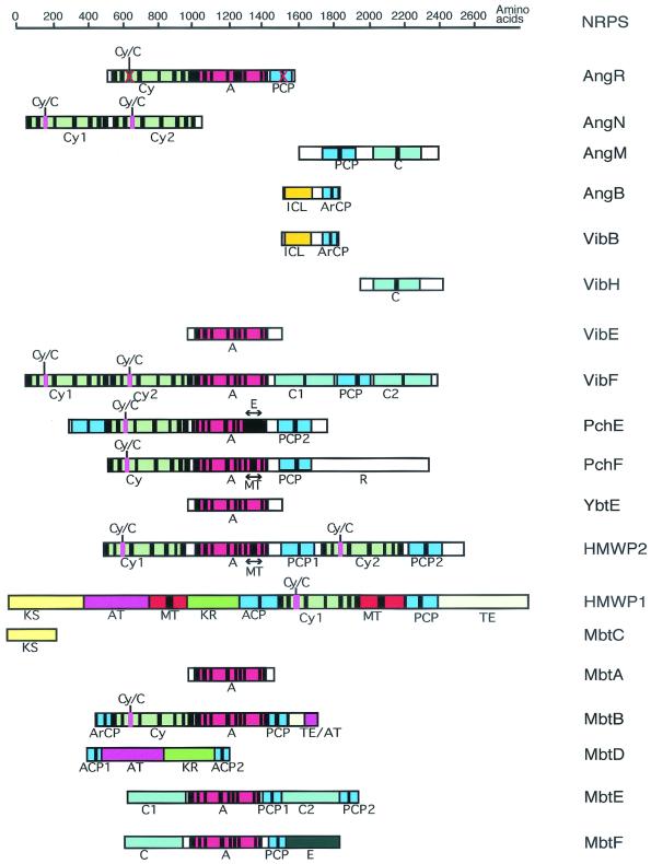
The regulatory logic of siderophore biosynthetic genes in bacteria involves the universal repressor Fur, which acts together with iron as a negative regulator. However in other bacteria, in addition to the Fur-mediated mechanism of regulation, there is a concurrent positive regulation of iron transport and siderophore biosynthetic genes that occurs under conditions of iron deprivation. Despite these regulatory differences the mechanisms of siderophore biosynthesis follow the same fundamental enzymatic logic, which involves a series of elongating acyl-S-enzyme intermediates on multimodular protein assembly lines: nonribosomal peptide synthetases (NRPS). A substantial variety of siderophore structures are produced from similar NRPS assembly lines, and variation can come in the choice of the phenolic acid selected as the N-cap, the tailoring of amino acid residues during chain elongation, the mode of chain termination, and the nature of the capturing nucleophile of the siderophore acyl chain being released. Of course the specific parts that get assembled in a given bacterium may reflect a combination of the inventory of biosynthetic and tailoring gene clusters available. This modular assembly logic can account for all known siderophores. The ability to mix and match domains within modules and to swap modules themselves is likely to be an ongoing process in combinatorial biosynthesis. NRPS evolution will try out new combinations of chain initiation, elongation and tailoring, and termination steps, possibly by genetic exchange with other microorganisms and/or within the same bacterium, to create new variants of iron-chelating siderophores that can fit a particular niche for the producer bacterium.


Figures

FIG. 1.
FIG. 1.
Structures of various bacterial siderophores: enterobactin from E. coli; vibriobactin from V. cholerae; acinetobactin from Acinetobacter calcoaceticus; mycobactin T from M. tuberculosis; pyoverdin and pyochelin from P. aeruginosa; anguibactin from V. anguillarum; and yersiniabactin from Y. pestis.
FIG. 2.
FIG. 2.
Spatial structures of enterobactin and anguibactin. (A) Enterobactin without and with Fe3+. Notice that the groups coordinating the iron are six oxygens from the three diphenolic groups. The complex requires one molecule of enterobactin. (B) X-ray structure of gallium anguibactin. This structure was determined from crystals obtained by complexing with Ga 3+ instead of Fe 3+ (53). Empty spheres: carbon (C); red spheres: oxygen (O); orange spheres: nitrogen (N); dark blue spheres: sulfur (S); purple spheres: gallium (Ga). Notice the coordination of two Ga3+ ions by two molecules of anguibactin using O-4 from the hydroxamate group, N-17 from the imidazole group, N-11 from the thiazoline group, and O-1 from the diphenol group. The complex requires two molecules of anguibactin and two molecules of methoxy since crystallization was carried out in a methanol solution. In biological fluids the complex will consist of two molecules of anguibactin, two Fe 3+ molecules, and two molecules of H2O.
FIG. 3.
FIG. 3.
Initiation of siderophore synthesis. (A) PPTase priming equation on apo PCP; (B) two-step A domain equation; (C) C domain equation.
FIG. 4.
FIG. 4.
Termination of siderophore synthesis.
FIG. 5.
FIG. 5.
Siderophore synthesis. Actions of A and ArCP (A) and Cy (C) domains.
FIG. 6.
FIG. 6.
Genetics and enzymology of enterobactin biosynthesis in E. coli. (A) Scheme of the enterobactin biosynthesis and transport genes. (B) Scheme of the synthetases encoded by the genes shown in panel A and the final product, enterobactin. ICL, isochorismate lyase activity.
FIG. 7.
FIG. 7.
Enterobactin synthetase assembly line in E. coli.
FIG. 8.
FIG. 8.
Bacillobactin from B. subtilis. Comparison of the bacillobactin and enterobactin synthetases.
FIG. 9.
FIG. 9.
Genetics and enzymology of vibriobactin biosynthesis in V. cholerae. (A) Scheme of the vibriobactin biosynthesis and transport genes. (B and C) Vibriobactin assembly line. (B) Oxazoline formation; (C) Incorporation of norspermidine and completion.
FIG. 10.
FIG. 10.
Genetic map of the pJM1 virulence plasmid of V. anguillarum highlighting those genes involved in the regulation, biosynthesis, and uptake of ferric anguibactin.
FIG. 11.
FIG. 11.
Anguibactin biosynthesis in V. anguillarum. (A) AngB/G, provides the isochorismate lyase activity (ICL) and the ArCP for activated 2,3-DHBA. (B) AngM provides condensation and spacer domains.
FIG. 12.
FIG. 12.
Anguibactin biosynthesis in V. anguillarum. (A) AngR may provide the adenylation domain to activate cysteine. (B) AngN may provide the cyclization domain(s) for thiazoline formation.
FIG. 13.
FIG. 13.
Comparison of vibriobactin and anguibactin NRPS. Crossed-out domains indicate that their sequence has mutations in crucial amino acid residues. AngE has not been identified as yet (also crossed out in the scheme).
FIG. 14.
FIG. 14.
Model for the anguibactin synthetase assembly line in V. anguillarum. The asterisk on AngE indicates that this protein has not yet been identified in V. anguillarum.
FIG. 15.
FIG. 15.
Model of regulation of the expression of pyochelin biosynthesis and uptake genes in P. aeruginosa.
FIG. 16.
FIG. 16.
Pyochelin assembly line in P. aeruginosa.
FIG. 17.
FIG. 17.
Genetics and enzymology of yersiniabactin biosynthesis in Y. pestis. (A) Scheme of the yersiniabactin biosynthesis and transport genes. (B and C) Schemes of the synthetases encoded by the genes shown in panel A and initiation steps in yersiniabactin biosynthesis.
FIG. 18.
FIG. 18.
Enzymology of yersiniabactin biosynthesis in Y. pestis. (A) NRPS-PKS switch point; (B) methylation; (C) PKS-NRPS switch point; (D) methylation and termination.
FIG. 19.
FIG. 19.
Mycobacterial siderophores.
FIG. 20.
FIG. 20.
Scheme of the mycobactin biosynthesis and transport genes.
FIG. 21.
FIG. 21.
Mycobactin assembly line in M. tuberculosis. For mycobactin T, R is methyl (Me) and X is 14 to 17. For carboxymycobactins, R is COOH, COOMe and X is 1 to 5.
FIG. 22.
FIG. 22.
Comparison of the domains of siderophore-NRPS from bacterial pathogens and E. coli.

References

    1. Actis, L. A., M. E. Tolmasky, and J. H. Crosa. 1999. Vibriosis, p. 523-557. In P. T. K. Woo and D. W. Bruno (ed.), Fish diseases and disorders, vol. 3. Viral, bacterial and fungal infections. Cab International Publishing, Wallingford, United Kingdom.
    1. Actis, L. A., M. E. Tolmasky, D. H. Farrell, and J. H. Crosa. 1988. Genetic and molecular characterization of essential components of the Vibrio anguillarum plasmid-mediated iron-transport system. J. Biol. Chem. 263:2853-2860. - PubMed
    1. Actis, L. A., W. Fish, J. H. Crosa, K. Kellerman, S. R. Ellenberger, F. M. Jauser, and J. Sanders-Loehr. 1986. Characterization of anguibactin, a novel siderophore from Vibrio anguillarum 775(pJM1). J. Bacteriol. 167:57-65. - PMC - PubMed
    1. Actis, L. A., S. Potter, and J. H. Crosa. 1985. Iron regulated outer membrane protein OM2 of Vibrio anguillarum is encoded by virulence plasmid pJM1. J. Bacteriol. 161:736-742. - PMC - PubMed
    1. Actis, L. A., M. E. Tolmasky, L. M. Crosa, and J. H. Crosa. 1995. Characterization and regulation of the expression of FatB, an iron transport protein encoded by the pJM1 virulence plasmid. Mol. Microbiol. 17:197-204. - PubMed

Publication types

LinkOut - more resources