Review
. 2002:71:635-700.
doi: 10.1146/annurev.biochem.71.110601.135414. Epub 2001 Nov 9.

Lipopolysaccharide endotoxins

Affiliations
Review

Lipopolysaccharide endotoxins

Christian R H Raetz et al. Annu Rev Biochem. 2002.

Abstract

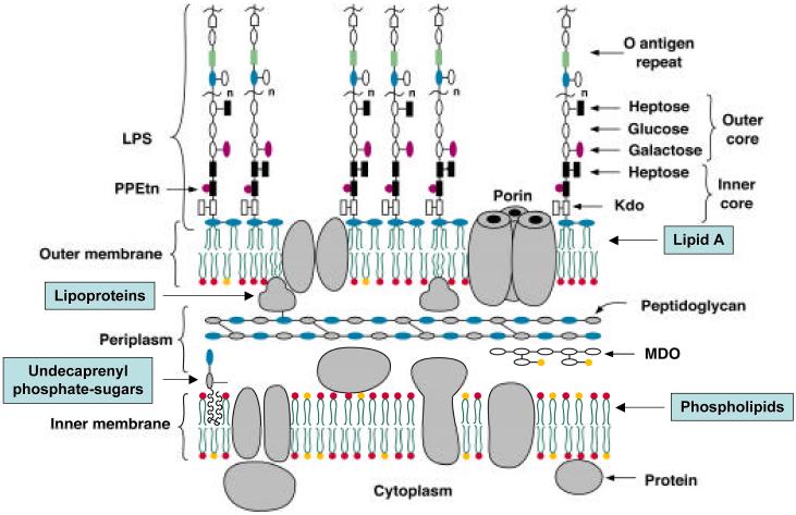
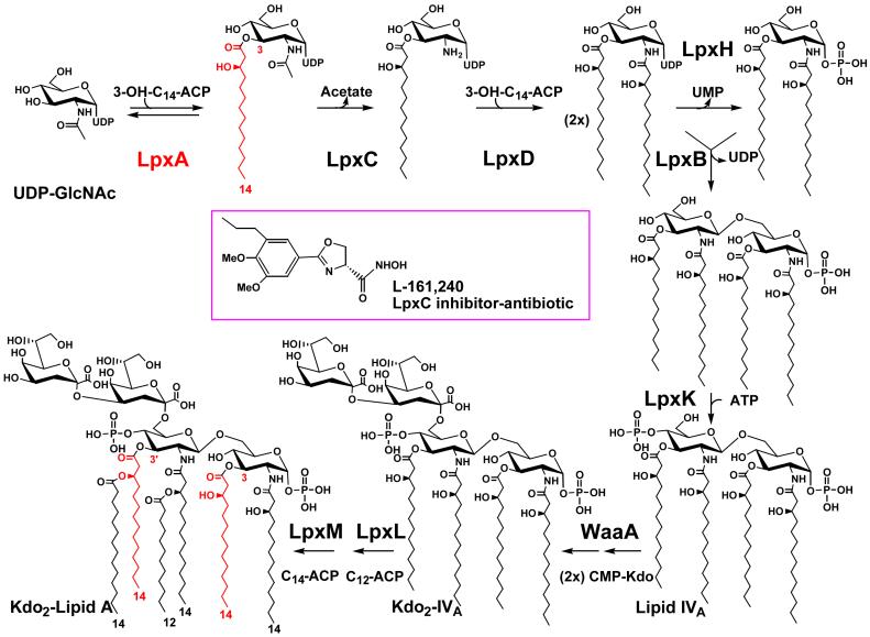
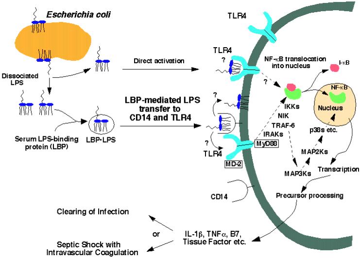
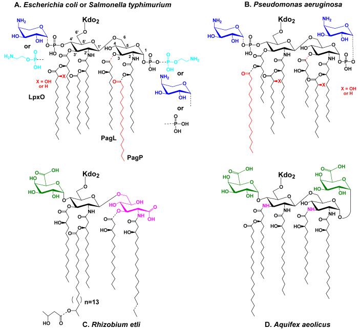
Bacterial lipopolysaccharides (LPS) typically consist of a hydrophobic domain known as lipid A (or endotoxin), a nonrepeating "core" oligosaccharide, and a distal polysaccharide (or O-antigen). Recent genomic data have facilitated study of LPS assembly in diverse Gram-negative bacteria, many of which are human or plant pathogens, and have established the importance of lateral gene transfer in generating structural diversity of O-antigens. Many enzymes of lipid A biosynthesis like LpxC have been validated as targets for development of new antibiotics. Key genes for lipid A biosynthesis have unexpectedly also been found in higher plants, indicating that eukaryotic lipid A-like molecules may exist. Most significant has been the identification of the plasma membrane protein TLR4 as the lipid A signaling receptor of animal cells. TLR4 belongs to a family of innate immunity receptors that possess a large extracellular domain of leucine-rich repeats, a single trans-membrane segment, and a smaller cytoplasmic signaling region that engages the adaptor protein MyD88. The expanding knowledge of TLR4 specificity and its downstream signaling pathways should provide new opportunities for blocking inflammation associated with infection.


Figures

Figure 1
Figure 1. Model of the inner and outer membranes of E. coli K-12
Only the Kdo and lipid A regions of LPS are required for the growth of E. coli and most other Gram-negative bacteria (2). Exceptions to this general rule include certain spirochetes in which all lipid A biosynthesis genes are absent (141), Thermotoga maritima (137), and Neisseria meningitidis type B in which lipid A-deficient lpxA knockouts can be constructed (133), provided the polysialic acid capsule is present.
Figure 2
Figure 2. Structure and biosynthesis of Kdo2-lipid A in E. coli K-12
The symbols indicate the relevant structural genes encoding each of the enzymes (2, 48). A single enzyme catalyzes each reaction. The lipid A system may have evolved only once, as judged by the available genomes. In almost all cases, as illustrated by E. coli (367), the genes encoding the enzymes of lipid A biosynthesis are present in single copy. At the protein level, orthologs of LpxA and LpxC are the most highly conserved among bacteria. The acyl chain incorporated by LpxA is highlighted in red. The LpxC inhibitor L-161,240 displays antibiotic activity against E. coli that is comparable in potency to ampicillin (64, 65).
Figure 3
Figure 3. Detection of lipid A by the TLR4 innate immunity receptor of animal cells
The paradigm is based mainly on studies of the human, mouse and hamster systems (12-15, 27, 31, 368). We speculate that the TLR4 receptor may be oligomerized upon binding of lipid A. The TRL4 mediated inflammatory response is beneficial in combating localized infections, but may be detrimental in overwhelming systemic sepsis. Novel therapies of sepsis directed against the disseminated intravascular coagulation component using recombinant activated protein C have recently shown efficacy in human trials (23). Combination therapy with TLR4 antagonists (369) remains to be explored.
Figure 4
Figure 4. Regulated modifications of lipid A in E. coli and S. typhimurium and unusual lipid A structures in other bacteria
Partial covalent modifications are indicated with the dashed bonds. Panel A. Modifications of E. coli and S. typhimurium lipid A under the control of PmrA are blue, whereas modifications primarily under the control of PhoP are red. For a recent discussion of the structures of the modified lipid A species that can be isolated from various mutants or under different growth conditions see Zhou et al. (112). When present, the L-Ara4N (dark blue) moiety is located mainly at the 4′ position, whereas the phosphoethanolamine (light blue) is mostly at position 1. Certain lipid A species exist in which the same substituents are attached at both sites, or in which their locations are reversed (112, 370). In cells grown with 1-10 mM Mg++ above pH 7.4, the modifications are suppressed, and a pyrophosphate group (the origin of which is unknown) is present at position 1 in about one third of the lipid A molecules (112). Panel B. Lipid A modifications in Pseudomonas under the control of PmrA are shown in blue, whereas modifications primarily under the control of PhoP are red (109). The portion of the lipid A molecule generated by the constitutive pathway is shown in black. Panel C. R. etli lipid A, which lacks phosphate groups, includes a major species in which aminogluconate (magenta) replaces the proximal glucosamine. Aminogluconate is formed in the outer membrane from a lipid A species containing glucosamine (149, 150, 156). The 4′ galacturonic acid moiety is green. Panel D. Like R. etli, Aquifex aeolicus lipid A lacks phosphate moieties (126), but contains two galacturonic acid residues (green). The hydroxyacyl chains at positions 3 and 3′ differ from E. coli in that they are amide-linked (magenta NH atoms).
Figure 5
Figure 5. Pathway for L-Ara4N biosynthesis and mechanism of polymyxin resistance in E. coli and S. typhimurium
In accordance with the proposal of Reeves et al. (9), we have renamed the genes of the polymyxin resistance operon “arn”, given their function in the biosynthesisof the L-Ara4N moiety and its transfer to lipid A (97, 102, 115). The proposed pathway starts with the conversion of UDP-glucose to UDP-glucuronic acid by the well-characterized dehydrogenase, Ugd. Next, ArnA (previously Orf3 or PmrI) (97, 102, 115) catalyzes the oxidative decarboxylation of UDP-glucuronic acid to generate a novel UDP-4-keto-pyranose intermediate, which can be isolated in mg quantities using ArnA (116). In contrast to the proposal of Baker et al. (371), a separate enzyme to catalyze the decarboxylation step is not necessary in our scheme (97, 116). ArnB (previously Orf1 or PmrH) (97, 99, 117) then catalyzes a further transamination to form UDP-L-Ara4N (116). Based upon its homology to dolichyl phosphate-mannose synthase of yeast, we propose that ArnC (PmrF) (97, 99, 117) transfers the L-Ara4N moiety to undecaprenyl phosphate, forming the novel compound undecaprenyl phosphate-α-L-Ara4N (102). After translocation to the outer surface of the inner membrane by unknown mechanisms, ArnT (previously Orf5, PmrK or YfbI) (115) transfers the L-Ara4N unit to lipid A. Other genes of the polymyxin operon (pmrJ, pmrL, and pmrM), as well as the adjacent pmrG gene (99, 117), cannot yet be assigned specific enzymatic or transport functions in our scheme. The ArnA protein has a second catalytic domain (reaction not shown) that can transfer a formyl group from N10-tetrahydrofolate to UDP-L-Ara4N (116), but the significance of this modification is unclear. Addition of the L-Ara4N moiety to lipid A reduces the affinity of lipid A for polymyxin and other cationic anti-microbial peptides.
Figure 6
Figure 6. Structure of the E. coli MsbA dimer at 4.5 Å resolution
This backbone tracing was made from protein data bank file 1 JSQ (120). Transmembrane helices 1-6 are colored purple, blue, yellow, green, red and orange, respectively. The intracellular domain (ICD) is brown, and the nucleotide-binding domain is cyan (120). A schematic model of lipid A is shown in panel A for size comparison. The location of the A270 residue, which is changed to threonine in the temperature sensitive lipid transport mutant WD2 (79), is shown as a red sphere. The putative chamber for binding lipids on the inner surface of the inner membrane is lined with basic residues (120) (not shown).
Figure 7
Figure 7. Model for MsbA mediated lipid export in E. coli
Following the MsbA mediated transport at the inner membrane, additional proteins are likely to be involved in steps 2 and 3, but these have not yet been identified (79).
Figure 8
Figure 8. Structures of the known lipopolysaccharide core oligosaccharides from E. coli and Salmonella
The outer cores are shown, together with one heptose residue of the inner core. The ligation sites for O polysaccharides (O-PS) are indicated where known (reviewed in 201). The inset shows the conserved base structure of the inner core and type-specific non-stoichiometric additions to the inner core are identified by dotted lines. Residues and linkages that are conserved in each of the core oligosaccharides are shown in red. Details of the structures are described elsewhere (168, 236, 372). Unless otherwise noted, all linkages are in the α-anomeric configuration.
Figure 9
Figure 9. Structures of the core oligosaccharides from Klebsiella pneumoniae O1 and P. aeruginosa O5
The β-galacturonic acid residues on the K. pneumoniae core are non-stoichiometric and details of the structures are described elsewhere (178, 179, 232, 373) (and references therein). The boxed regions of the core structures are those that differ between S-LPS and R-LPS, and the details are discussed in the text.
Figure 10
Figure 10. Structure of lipooligosaccharides from Neisseria meningitidis
The structures and immunotypes have been reviewed elsewhere (168, 246). The structure of the single α-chain in each LOS varies and shown here are the sialylated α-chains from immunotypes L1 (374) and L3. Some of the additions to the inner core are missing in some LOS species. Relevant glycosyltransferases involved in the synthesis are identified, and the basis for the structural variations is described in the text. The RfaK enzyme should not be confused with the WaaK (formerly RfaK) glycosyltransferases that adds the terminal GlcNAc residue to the outer core of S. enterica serovar Typhimurium LPS.
Figure 11
Figure 11. Structure and biosynthesis of the E. coli R1 core
Organization of the waa locus is shown. Each gene has been mutated with a non-polar insertion, and the structures of LPSs from the resulting mutants were determined to generate the enzyme assignments shown with the structure (192, 228). Glycosyltransferases that form the inner core backbone are denoted by the yellow boxes and enzymes that modify the structure are in blue. Green boxes identify outer core glycosyltransferases and the ligase enzyme is in pink. The mobility of the mutant LPSs on a silver stained tricine-PAGE gel is also shown.
Figure 12
Figure 12. Biosynthesis pathway for ADP-L-glycero-D-manno-heptose in E. coli
The figure is adapted from recent studies that necessitate revision of the previously proposed pathway (2, 215).
Figure 13
Figure 13. Biosynthesis and assembly of O polysaccharides in a Wzy-dependent pathway
Panel A shows the sequence of reactions involved in the formation of the undecaprenyl-linked repeat units and their polymerization in S. enterica serovar Typhimurium. The individual glycosyltransferase enzymes are identified in green enzymes are identified in the boxes and structural details are presented elsewhere (2, 9). Panel B shows a model for the events in polymerization. Individual undecaprenyl-linked O-repeat units are transferred across the membrane by a process involving Wzx (yellow). These intermediates provide the substrates for the putative polymerase (Wzy- identified in blue) acting in the periplasm. Chain extension occurs at the reducing terminus with the nascent chain being transferred from its undecaprenyl carrier to the non-reducing terminus of the “new” undecaprenyl-linked subunit. The chain-length modality (i.e. the extent of polymerization) is determined by Wzz (also in blue). The nascent polymer is then ligated to lipid A-core and translocated to the outer membrane.
Figure 14
Figure 14. Biosynthesis and assembly of O polysaccharides in an ABC-transporter-dependent pathway
Panel A shows the predicted structures of undecaprenyl-linked intermediates in the biosynthesis of the O9a antigen of E. coli and the D-galactan I polymer found in several serotypes of K. pneumoniae. The glycosyltransferase enzymes involved in each step are indicated below the structures. Biosynthesis is broken down into the formation of a common primer (shown in red), addition of an adaptor region, chain extension of the repeat unit, and addition of a chain terminator (for details see the text). In the O9a structure, the chain is terminated by addition of 3-O-methylmannose but the enzyme responsible has not been identified. In D-galactan I, the structure of the chain terminator has not been established. Chain elongation occurs by processive glycosyl transfer to the non-reducing terminus. Several enzymes in these pathways are bifunctional. In the case of the galactosyltransferase WbbO, both of its activities are required for adaptor synthesis, but only one (WbbO2) participates in chain extension. Panel B provides a model for the trans-membrane assembly system. The glycosyltransferases are shown in green. The ABC-transporter formed by Wzm and Wzt is required for transfer of the undecaprenyl-linked polymer to the periplasmic face of the membrane, where it is ligated to lipid A-core and translocated to the outer membrane. It is presumed that the polymer remains attached to the undecaprenyl carrier throughout the export process. While chain extension and export are separable by mutations in the ABC-transporter, it is conceivable that the two processes are temporally coupled in vivo. Within the nascent polymer, the primer/adaptor is identified by the black-filled hexagon, the residues of the repeating-unit domain by gray-filled circles, and the chain terminator by the open hexagon.
Figure 15
Figure 15. Biosynthesis and assembly of O polysaccharides in a synthase-dependent pathway
The only current example is the plasmid-encoded O:54 antigen of S. enterica serovar Borreze. Panel A shows the predicted structure of the undecaprenyl-linked intermediate. The primer is made by WecA. The WbbE enzyme then adds a single ManNAc residue as an adaptor, but the precise linkage has not been determined. The synthase (WbbF) is then required for chain extension, generating a repeat-unit domain with alternating β(1,3) and β(1,4) linkages. Growth is at the non-reducing terminus. Panel B shows a model for the transmembrane assembly process, with glycosyltransferases identified in green and putative transport functions in yellow. Available data for this system, and information for other bacterial synthases, is consistent with the conclusion that WbbF serves as both a processive glycosyltransferase with dual linkage specificity, as well as an exporter that moves undecaprenyl-linked intermediates to the periplasm. While chain extension and export are shown as separate processes, it is conceivable that they are temporally coupled in vivo. The nascent polymer is then ligated to lipid A-core and translocated to the outer membrane.

References

    1. Raetz CRH. Annual Reviews in Biochemistry. 1990;59:129–70. - PubMed
    1. Raetz CRH. Escherichia coli and Salmonella. In: Niedhardt FC, editor. Cellular and Molecular Biology. American Society for Microbiology; Washington, D.C.: 1996. pp. 1035–63.
    1. Rietschel ET, Kirikae T, Schade FU, Mamat U, Schmidt G, et al. FASEB Journal. 1994;8:217–25. - PubMed
    1. Zähringer U, Lindner B, Rietschel ET. In: Endotoxin in Health and Disease. Brade H, Opal SM, Vogel SN, Morrison DC, editors. Marcel Dekker, Inc.; New York: 1999. pp. 93–114.
    1. Brade H, Opal SM, Vogel SN, Morrison DC, editors. Endotoxin in Health and Disease. Marcel Dekker, Inc.; New York: 1999. p. 950.

Publication types

MeSH terms

LinkOut - more resources