. 2002 Jun 10;157(6):953-62.
doi: 10.1083/jcb.200201060. Epub 2002 Jun 3.

Characterization of Tbc2, a nucleus-encoded factor specifically required for translation of the chloroplast psbC mRNA in Chlamydomonas reinhardtii

Affiliations

Characterization of Tbc2, a nucleus-encoded factor specifically required for translation of the chloroplast psbC mRNA in Chlamydomonas reinhardtii

Andrea H Auchincloss et al. J Cell Biol. .

Abstract

Genetic analysis has revealed that the three nucleus-encoded factors Tbc1, Tbc2, and Tbc3 are involved in the translation of the chloroplast psbC mRNA of the eukaryotic green alga Chlamydomonas reinhardtii. In this study we report the isolation and phenotypic characterization of two new tbc2 mutant alleles and their use for cloning and characterizing the Tbc2 gene by genomic complementation. TBC2 encodes a protein of 1,115 residues containing nine copies of a novel degenerate 38-40 amino acid repeat with a quasiconserved PPPEW motif near its COOH-terminal end. The middle part of the Tbc2 protein displays partial amino acid sequence identity with Crp1, a protein from Zea mays that is implicated in the processing and translation of the chloroplast petA and petD RNAs. The Tbc2 protein is enriched in chloroplast stromal subfractions and is associated with a 400-kD protein complex that appears to play a role in the translation of specifically the psbC mRNA.


Figures
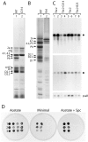
Figure 1.
Figure 1.
Characterization of two new alleles of the TBC2 locus. Pulse labeling of chloroplast-encoded polypeptides with [14C]acetate for 45 min. (A) in wild-type (lane 1) and G314 (lane 2), and for 5 min (B) in wild-type (lane 1) and F64 (lane 2) in the presence of cycloheximide to inhibit protein synthesis in the cytosol. After pulse labeling, thylakoid membranes were isolated and the proteins fractionated on a 7.5–15% SDS-polyacrylamide gel (A) or a 12–18% SDS polyacrylamide gel with 8 M urea (B) (Chua and Bennoun, 1975). The pattern of protein synthesis in B23 was similar to that of G314 (unpublished data). The genes encoding the indicated polypeptides are: 2a, psaB; 2b, psaA; 4.1, atpB; 4.2, atpA; P5, psbB; P6, psbC; cyt.f, petA; D1, psbA; and D2, psbD. (C) RNA blot analysis of the psbC mRNA (*) and the chimeric mRNA psbC(WT)-aadA (+) in RNA preparations from tetrads from cross 1 (FuD50:psbC-aadA, mt+ × G314, mt; lanes 1–4) and from cross 2 (FuD50:psbC-aadA, mt+ × B23, mt; lanes 5–8; Table I). RNA from the TBC2-bearing progeny were loaded in lanes 1, 2, 5, and 6, from the tbc2-G314–bearing progeny in lanes 3 and 4, and from the tbc-B23–bearing progeny in lanes 7 and 8. The level of psaB mRNA was used to standardize for the amount of RNA in each lane. (D) Growth of members of tetrads from a cross of FuD50::psbC(WT)-aadA (mt +) with F64 (mt ), “1”; or G314 (mt ), “2”; or B23 (mt ), “3” on nonselective acetate containing medium (Acetate), on medium that selects for photosynthetic growth (Minimal), and on medium containing acetate and spectinomycin at 750 μg ml−1.
Figure 2.
Figure 2.
Map of the TBC2 locus and its cDNA. (A) Region of the promoter region and 5′ part of the Tbc2 gene. The first three introns are indicated in grey. HinfI sites (HfI, at 273; 1,110; 1,574; and 2,596 bp), and the approximate location of the HindIII sites used to generate probes to clone the cDNA are shown (HdIII). The positions of probes I (1110–1574), II (2011–2346), and III (2273–2767) on the genomic DNA are indicated. Dotted regions indicate unsequenced DNA (this section is not to scale). (B) The map of the longest Tbc2 cDNA (4,098 nucleotides without the polyA tail) is represented. The unique SacI (1771) and NotI (3108) sites as well as the start codon (ATG, 174–176) and stop codon (stop, 3519–3521) are also noted. The location of probes I (195–506), II (630–965), and III (892–1386) relative to the cDNA sequence are shown. The Δ indicates the region deleted in three from eight cDNAs examined (1107–1285). The PCR primers used to insert a HincII site immediately upstream of the stop codon are indicated by arrows. The HA epitope was inserted into this HincII site or at the SacI site. The locations of the first three introns are marked by wedges. The corresponding HindIII, HinfI, and SacI sites on the genomic and cDNA are shown by thin lines.
Figure 3.
Figure 3.
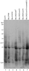
DNA blot analysis of the TBC2 locus in wild-type and the tbc2 mutants. 5 μg of the indicated genomic DNA was digested with HinfI, separated on a 0.7% agarose gel, transferred to Hybond-C extra, and probed with PCR-amplified probe III (Fig. 2). Size markers are indicated in kb on the left of the gel (Eurogentech Smart ladder). The 2-kb fragment in lane 7 is undetectable because of overexposure.
Figure 4.
Figure 4.
Tbc2 protein. A. Schematic view of the Tbc2 protein. The nine 38–40 amino acid internal repeats and the region similar to the Crp1 protein of Zea mays are indicated. The regions of Tbc2 containing the stretches of A, D, L, Q, S, and T residues are shaded. The region corresponding to Δ in Fig. 2 B is marked with a box. The two sites used for HA epitope tagging are indicated. (B) Sequence of the Tbc2 protein. Stretches of A, D, L, P, Q, S, and T residues are highlighted. The region corresponding to Δ in Fig. 2 B is underlined. (C) Alignment of the nine PPPEW repeats. Residues which appear at least four times amongst the nine repeats are highlighted and indicated in the consensus sequence (CONS). (D) Regions of partial sequence identity in Tbc2 and Crp1. The first seven PPPEW repeats are shown in rows and indicated by Arabic numerals, whereas the first seven PPR repeats of Crp1 are shown in separate rows and indicated by Roman numerals. Spacings were introduced to optimize the alignment with the first seven PPR repeats of Crp1. The last six residues of PPPEW repeats 2–6 are shown duplicated in two rows to facilitate the comparison of the PPPEW and PPR repeats. Residues of the repeats that conform to the consensus are shown in bold letters. Identical amino acids are marked with *, similar amino acids are indicated with +. The COOH-terminal consensus (consC) of the PPPEW repeats and the PPR consensus are shown in the lower part. Because of the differences in these two repeats, it is not possible to align them over their whole length simultaneously. These sequence data are available from Genbank/EMBL/DDBJ under accession no AJ427966.
Figure 5.
Figure 5.
Accumulation and subcellular localization of Tbc2. Immunoblot analysis of Tbc2 in wild-type (lanes 1, 3, and 5) and tbc2-F64 rescued with the HA-tagged Tbc2 cDNA (lanes 2, 4, and 6–9); tot, total cell extract (lanes 1, 2); total insoluble fraction (lanes 3 and 4); total soluble fraction (lanes 5, 6); cp total, total chloroplast extract (lane 7), chloroplast insoluble fraction (lane 8), chloroplast soluble fraction (lane 9). After fractionation by SDS-PAGE and blotting, the immunoblots were reacted with antisera raised against HA, D2, SSU Rubisco, and thioredoxin-h (cytosolic protein). For details see Materials and methods.
Figure 6.
Figure 6.
The Tbc2 protein is part of a high molecular weight complex. A total soluble cell extract from a strain carrying the tbc2-F64 allele and expressing the HA-tagged Tbc2 cDNA was concentrated to a final concentration of 15–20 μg/ml and fractionated on a Superose 6 PC3.2/30 column using the SMART system (Amersham Pharmacia Biotech). Fractionation was performed in the presence of MgCl2 and heparin (rows 1 and 4), MgCl2 and RNase (rows 2 and 4), or EDTA and heparin (row 3). 25 fractions were collected and aliquots were electrophoresed on an 8% SDS-polyacrylamide gel and reacted with anti-HA monoclonal antibody (rows 1–3) and antibodies against RB60 (rows 4 and 5).

References

    1. Bairoch, A., and R. Apweiler. 2000. The SWISS-PROT protein sequence database and its supplement TrEMBL. Nucleic Acids Res. 28:45–48. - PMC - PubMed
    1. Barkan, A., and M. Goldschmidt-Clermont. 2000. Participation of nuclear genes in chloroplast gene expression. Biochimie. 82:559–572. - PubMed
    1. Bennoun, P., A. Masson, and M. Delosme. 1980. A method for complementation analysis of nuclear and chloroplast mutants of photosynthesis in Chlamydomonas. Genetics. 95:39–47. - PMC - PubMed
    1. Bennoun, P., M. Spierer-Herz, J. Erickson, J. Girard-Bascou, Y. Pierre, M. Delsome, and J.-D. Rochaix. 1986. Characterization of photosystem II mutants of Chlamydomonas reinhardtii lacking the psbA gene. Plant Mol. Biol. 6:151–160. - PubMed
    1. Blatch, G.L., and M. Lassle. 1999. The tetratricopeptide repeat: a structural motif mediating protein-protein interactions. Bioessays. 21:932–939. - PubMed

Publication types

MeSH terms

Substances