. 2002 Jun 3;195(11):1507-12.
doi: 10.1084/jem.20020207.

Interferon-alpha and interleukin-12 are induced differentially by Toll-like receptor 7 ligands in human blood dendritic cell subsets

Affiliations

Interferon-alpha and interleukin-12 are induced differentially by Toll-like receptor 7 ligands in human blood dendritic cell subsets

Tomoki Ito et al. J Exp Med. .

Abstract

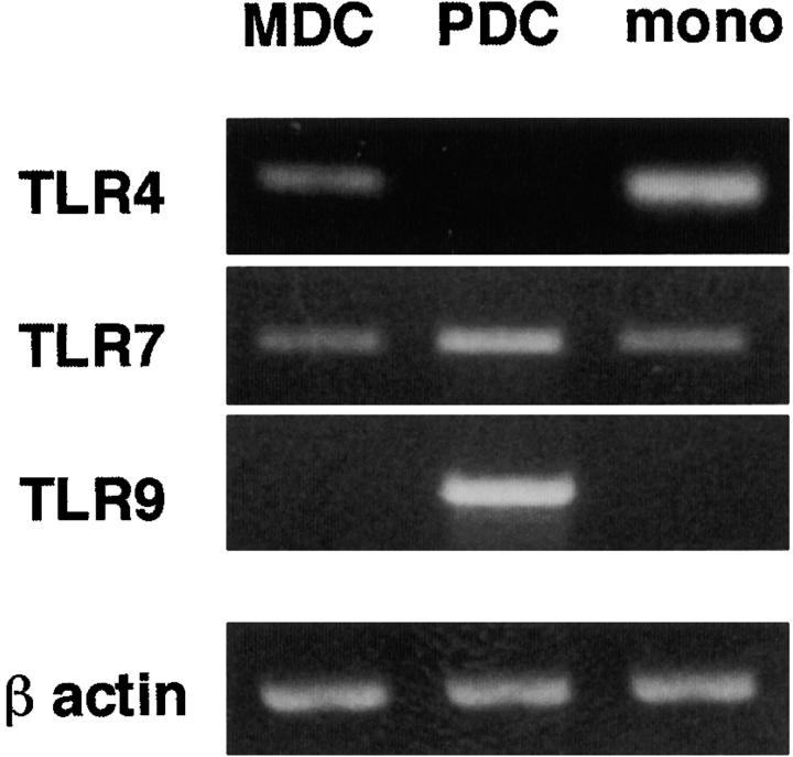
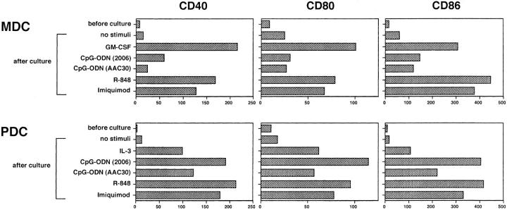
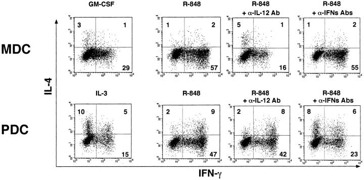
Dendritic cells (DCs) play a crucial role in the immune responses against infections by sensing microbial invasion through toll-like receptors (TLRs). In humans, two distinct DC subsets, CD11c(-) plasmacytoid DCs (PDCs) and CD11c(+) myeloid DCs (MDCs), have been identified and can respond to different TLR ligands, depending on the differential expression of cognate TLRs. In this study, we have examined the effect of TLR-7 ligands on human DC subsets. Both subsets expressed TLR-7 and could respond to TLR-7 ligands, which enhanced the survival of the subsets and upregulated the surface expression of costimulatory molecules such as CD40, CD80, and CD86. However, the cytokine induction pattern was distinct in that PDCs and MDCs produced interferon (IFN)-alpha and interleukin (IL)-12, respectively. In response to TLR-7 ligands, the Th1 cell supporting ability of both DC subsets was enhanced, depending on the cytokines the respective subsets produced. This study demonstrates that TLR-7 exerts its biological effect in a DC subset-specific manner.


Figures

Figure 1.
Figure 1.
mRNA expression of TLRs in MDCs and PDCs. mRNA expression of TLR-4, -7, and -9 was examined in purified blood MDCs, PDCs, and CD14+CD16 monocytes by RT-PCR. For the detection of TLR-7 mRNA, three sets of primers were used, and similar results were obtained when using each set. The results using the first set of TLR-7 primers (as described in Materials and Methods) are shown in the figure. The data shown are representative of two independent experiments.
Figure 2.
Figure 2.
TLR-7 ligands enhance the survival of both MDCs and PDCs. MDCs and PDCs were incubated for 24 h with various stimuli. The viable cells in each DC subset were evaluated as annexin V-negative fractions by flow cytometry. The results are representative of three independent experiments.
Figure 3.
Figure 3.
TLR-7 ligands upregulate the expression of costimulatory molecules on both MDCs and PDCs. After 24-h culture with various stimuli, the expression levels of CD40, CD80, and CD86 on each DC subset were analyzed by flow cytometry. Imiquimod and R-848 were used at concentrations of 10−5 M and 10−7 M, respectively. The results are shown as ΔMFI, which is calculated by subtraction of MFI with the isotype-matched control from that with each mAb. The results shown here are representative of three independent experiments.
Figure 4.
Figure 4.
TLR-7 ligands induce the production of IL-12 and IFN-α from MDCs and PDCs, respectively. (A and B) After 24-h culture with various stimuli, concentration of IL-12 p40+p70 (A) and IFN-α (B) in the culture supernatants of MDCs, PDCs, and PBMCs were measured by ELISA. The data are shown by means ± SEM of five independent experiments. (C) After 5-h culture with R-848 (10−6 M), intracellular staining of IL-12 and IFN-α in MDCs and PDCs, together with the staining of surface expression of CD40, was performed. Percentages of the respective cytokine producing DCs are indicated. This figure represents the results from one of three experiments.
Figure 5.
Figure 5.
MDCs and PDCs stimulated by TLR-7 ligands induce Th1 development depending on each DC-derived cytokine. MDCs and PDCs were preincubated with the cytokines or R-848 (10−6 M) for 24 h, washed, and subsequently cocultured with allogeneic CD4+ naive T cells for 7 d in the presence or absence with neutralizing anti-IL-12 Ab (IL-12α Ab) or a mixture of neutralizing anti–IFN-α Ab, neutralizing anti–IFN-β Ab, and anti-IFN-α/β R mAb (IFN-αs Abs). Intracellular cytokine profiles in the T cells were analyzed by flow cytometry. Percentages of the respective cytokine-producing T cells are indicated. This figure represents the results from one of three experiments.

References

    1. Mellman, I., and R.M. Steinman. 2001. Dendritic cells: specialized and regulated antigen processing machines. Cell. 106:255–258. - PubMed
    1. Liu, Y.-J. 2001. Dendritic cell subsets and lineages, and their functions in innate and adaptive immunity. Cell. 106:259–262. - PubMed
    1. Lanzavecchia, A., and F. Sallusto. 2001. Regulation of T cell immunity by dendritic cells. Cell. 106:263–266. - PubMed
    1. Liu, Y.-J., H. Kanzler, V. Soumelis, and M. Gilliet. 2001. Dendritic cell lineage, plasticity and cross-regulation. Nat. Immunol. 2:585–589. - PubMed
    1. Medzhihtov, R., and C. Janeway, Jr. 1997. Innate immunity: the virtues of a nonclonal system of recognition. Cell. 91:295–298. - PubMed

Publication types

MeSH terms