

Review
doi: 10.1172/JCI15851.
Aquaporin water channels: atomic structure molecular dynamics meet clinical medicine
Affiliations
- PMID: 12045251
- PMCID: PMC151002
- DOI: 10.1172/JCI15851
Item in Clipboard
Review
Aquaporin water channels: atomic structure molecular dynamics meet clinical medicine
J Clin Invest.
2002 Jun.
No abstract available
Figures
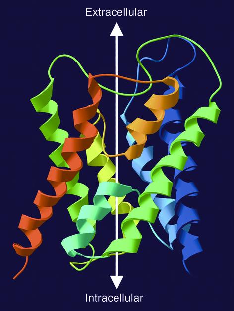
Ribbon diagram of the structure of an AQP1 subunit (sagittal section). The model is based on Protein Data Bank coordinates 1H6I of human red cell AQP1 (8). The amino-terminus is blue and the carboxy-terminus is red. Six membrane-spanning helices surround two hemipores (loops with short helices — cyan and orange) that meet in the center of the bilayer. The white arrow illustrates the aqueous channel through the protein.
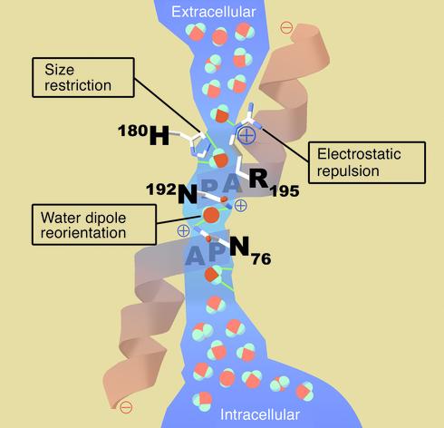
Schematic architecture of the channel within an AQP1 subunit (sagittal section). The shape of the aqueous pore (blue) is derived from calculations based on the structure of bovine AQP1 (9). Four water molecules shown in bold colors represent transient interactions with the pore-lining residues at discrete sites. Bulk water in the extracellular and intracellular vestibules is depicted in pastels. Three features of the channel specify selectivity for water: (a) Size restriction. Eight angstroms above the midpoint of the channel, the pore narrows to a diameter of 2.8 Å (approximately the diameter of a water molecule). (b) Electrostatic repulsion. A conserved residue (Arg-195) at the narrowest constriction of the pore imposes a barrier to cations, including protonated water (H3O+). (c) Water dipole reorientation. Two partial helices meet at the midpoint of the channel, providing positively charged dipoles that reorient a water molecule as it traverses this point. Disrupting hydrogen bonding in the single-file chain water molecules prevents the formation of a proton conductance. A video animation of water molecules passing through the AQP1 protein is available on the internet at http://www.mpibpc.gwdg.de/abteilungen/071/bgroot/presentations/aqp1_dyn/aqp1_mono.html.
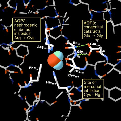
Human disease mutations and pharmacologically active sites projected onto an atomic model of an AQP1 pore at the narrowest constriction point (transverse section). The model is based on Protein Data Bank coordinates 1H6I of human red cell AQP1 (8). The side chains of three residues (Arg-195, His-180, and Phe-56) plus the backbone carbonyl groups of two residues (Gly-188 and Cys-189) line the pore. Mutation in AQP2 of the residue corresponding to Arg-195 results in autosomal recessive nephrogenic diabetes insipidus. The side chain of Cys-189 lies proximal to the pore. Binding of a mercuric ion to the sulfhydryl group of this residue inhibits the water permeability of AQP1. Hydrogen bonding between Glu-142 and the backbone amide groups of Gly-190 and Ile-191 orients the carbonyl groups of these residues toward the pore. Mutation in AQP0 of the residue corresponding to Glu-142 results in dominantly inherited congenital cataracts.
References
-
- Preston GM, Carroll TP, Guggino WB, Agre P. Appearance of water channels in Xenopus oocytes expressing red cell CHIP28 protein. Science. 1992;256:385–387. - PubMed
-
- Borgnia M, Nielsen S, Engel A, Agre P. Cellular and molecular biology of the aquaporin water channels. Annu Rev Biochem. 1999;68:425–458. - PubMed
-
- Jung JS, Preston GM, Smith BL, Guggino WB, Agre P. Molecular structure of the water channel through aquaporin CHIP. The hourglass model. J Biol Chem. 1994;269:14648–14654. - PubMed
-
- Murata K, et al. Structural determinants of water permeation through aquaporin-1. Nature. 2000;407:599–605. - PubMed
Publication types
MeSH terms
Substances
LinkOut - more resources
Full Text Sources
Other Literature Sources
