. 2002 Jun;109(11):1405-15.
doi: 10.1172/JCI15681.

Cyclooxygenase-2 regulates mesenchymal cell differentiation into the osteoblast lineage and is critically involved in bone repair

Affiliations

Cyclooxygenase-2 regulates mesenchymal cell differentiation into the osteoblast lineage and is critically involved in bone repair

Xinping Zhang et al. J Clin Invest. 2002 Jun.

Erratum in

  • J Clin Invest 2002 Oct;110(8):1211

Abstract

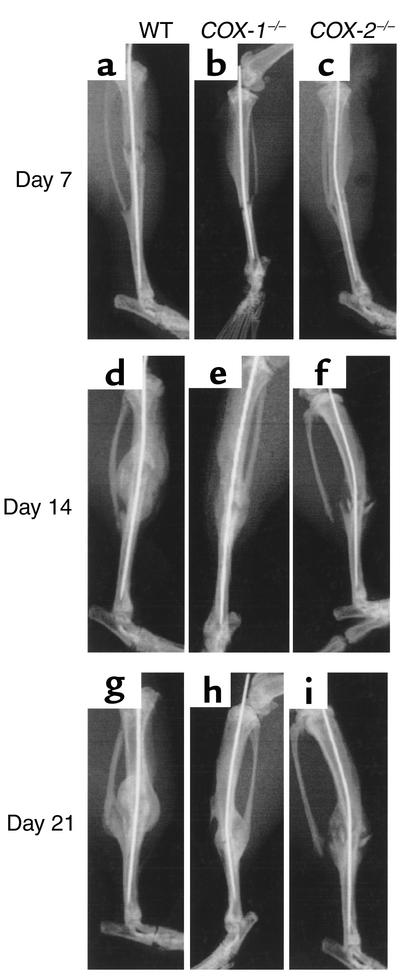
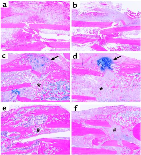
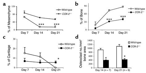
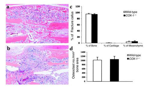
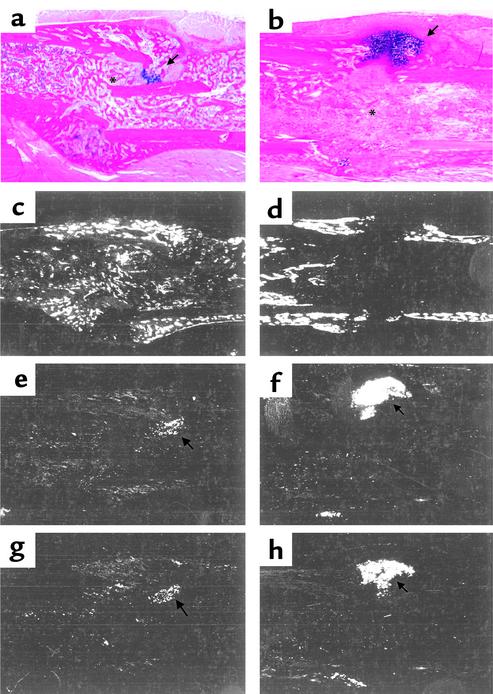
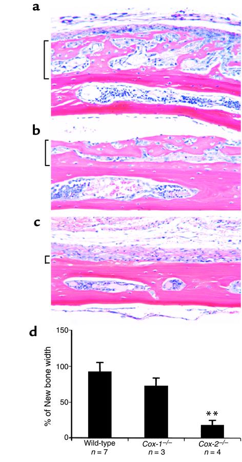
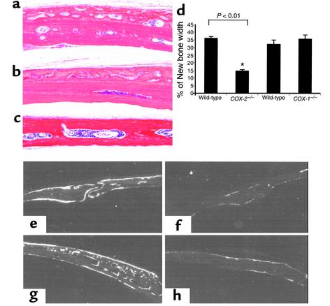
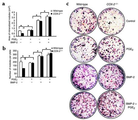
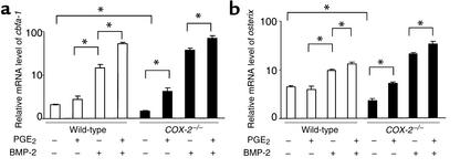
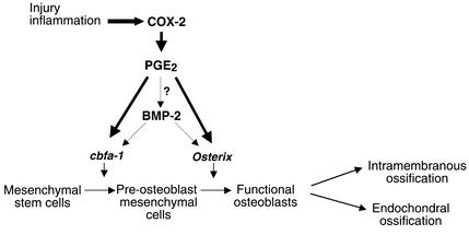
Preclinical and clinical studies suggest a possible role for cyclooxygenases in bone repair and create concerns about the use of nonsteroidal antiinflammatory drugs in patients with skeletal injury. We utilized wild-type, COX-1(-/-), and COX-2(-/-) mice to demonstrate that COX-2 plays an essential role in both endochondral and intramembranous bone formation during skeletal repair. The healing of stabilized tibia fractures was significantly delayed in COX-2(-/-) mice compared with COX-1(-/-) and wild-type controls. The histology was characterized by a persistence of undifferentiated mesenchyme and a marked reduction in osteoblastogenesis that resulted in a high incidence of fibrous nonunion in the COX-2(-/-) mice. Similarly, intramembranous bone formation on the calvaria was reduced 60% in COX-2(-/-) mice following in vivo injection of FGF-1 compared with either COX-1(-/-) or wild-type mice. To elucidate the mechanism involved in reduced bone formation, osteoblastogenesis was studied in bone marrow stromal cell cultures obtained from COX-2(-/-) and wild-type mice. Bone nodule formation was reduced 50% in COX-2(-/-) mice. The defect in osteogenesis was completely rescued by addition of prostaglandin E2 (PGE(2)) to the cultures. In the presence of bone morphogenetic protein (BMP-2), bone nodule formation was enhanced to a similar level above that observed with PGE(2) alone in both control and COX-2(-/-) cultures, indicating that BMPs complement COX-2 deficiency and are downstream of prostaglandins. Furthermore, we found that the defect in COX-2(-/-) cultures correlated with significantly reduced levels of cbfa1 and osterix, two genes necessary for bone formation. Addition of PGE(2) rescued this defect, while BMP-2 enhanced cbfa1 and osterix in both COX-2(-/-) and wild-type cultures. Finally, the effects of these agents were additive, indicating that COX-2 is involved in maximal induction of osteogenesis. These results provide a model whereby COX-2 regulates the induction of cbfa1 and osterix to mediate normal skeletal repair.


Figures

Figure 1
Figure 1
Fracture healing is delayed in COX-2–/– mice. Diaphyseal fractures were created in 2- to 3-month-old wild-type, COX-1–/–, and COX-2–/– mice as described in Methods. Representative radiographs of wild-type (a, d, and g), COX-1–/– (b, e, and h), and COX-2–/– (c, f, and i) mice obtained on days 7 (ac), 14 (df), and 21 (gi) after fracture are shown. Of note is the absence of fracture callus and bony union in the COX-2–/– mice.
Figure 2
Figure 2
Histologic evidence of defective fracture healing in COX-2–/– mice. Histologic sections were obtained from tibial fractures of adult wild-type (a, c, and e) and COX-2–/– mice (b, d, and f) at 7 (a and b), 14, (c and d), and 21 days (e and f) after fracture, and stained with Alcian blue/hematoxylin as described in Methods. Fractures in wild-type mice undergo extensive calcification resulting in little residual cartilage (arrow) and extensive woven bone formation (asterisk) by day 14 (c). This progresses to a remodeling bony union (#) by day 21. In the COX-2–/– mice there is little evidence of endochondral bone formation in the medullary canal, which is filled with undifferentiated mesenchymal tissue (asterisk) on day 14 (d). Concurrently, significant amounts of unmineralized cartilage persists (arrow) (d). By 21 days there are large amounts of fibrotic tissue (#) between the fractured bones, evidence of a fracture nonunion (f).
Figure 3
Figure 3
Quantitative analysis of fracture callus in wild-type and COX-2–/– mice at 7, 14, and 21 days after fracture. Tissue sections from wild-type and COX-2–/– mice harvested on days 7 (n = 5), 14 (n = 7), and 21 (n = 9) after fracture were stained with Alcian blue/hematoxylin or for tartrate resistant acid phosphate (TRAP) as described in Methods. All slides were analyzed using Osteometrics software, (Osteometrics Inc., Atlanta, Georgia, USA) and percentages of mesenchymal tissue (a), bone (b), and cartilage (c) relative to the total fracture area are presented as the mean ± SEM. (d) Osteoclast numbers (TRAP-positive cells) per mm2 bone area were quantified as described in Methods and are presented as the mean ± SEM. Student’s t test was used to determine the statistical differences between each group. *P < 0.05, ***P < 0.001.
Figure 4
Figure 4
Normal fracture healing in COX-1–/– mice. Histologic sections were obtained from tibial fractures of adult wild-type (a) and COX-1–/– (b) mice (n = 5) on day 21 after fracture and stained with Alcian blue/hematoxylin as described in Methods. Quantification of the tissue compositions (c) and osteoclast numbers (d) are presented as described in Figure 3. No significant differences were observed.
Figure 5
Figure 5
Characterization of defective fracture healing in COX-2–/– mice by in situ hybridization. Histologic sections of the fracture callus of wild-type (a, c, e, and g) and COX-2–/– mice (b, d, f, and h), 14 days after fracture, were stained with Alcian blue/hematoxylin (a and b). Serial sections were used for in situ hybridization with probes specific for osteocalcin (c and d), col2 (e and f), and colX (g and h). The gene expression profile confirms the persistence of cartilage (arrows) and the decrease in osteogenesis (asterisks) in COX-2–/– mice.
Figure 6
Figure 6
Inflammation-induced intramembranous bone formation is reduced in COX-2–/– mice. Titanium particles were surgically implanted onto the calvaria of wild-type (a), COX-1–/– (b), and COX-2–/– (c) mice as described in Methods, and representative H&E-stained sections are shown. Ectopic bone formation was measured, and the percentage of new bone width (brackets) is presented as the mean ± SEM (d). Statistical significance compared with wild-type is indicated (**P < 0.01).
Figure 7
Figure 7
Defective intramembranous bone formation in COX-2–/– calvaria in response to FGF-1. FGF-1 was injected subcutaneously (1 μg/d for 3 days) on the calvaria of wild-type, COX-1–/–, and COX-2–/– mice, and osteogenesis was analyzed by histomorphometry 14 days later as described in Methods. The photographs of representative H&E-stained sections from wild-type (a), COX-1–/– (b), and COX-2–/– mice (c) are shown at ×20 magnification. New bone width was measured at ×20 magnification, and the data from five animals in each group are presented as the mean ± SEM (d). Statistical significance compared with wild-type is indicated (*P < 0.01). Histology sections were examined by in situ hybridization using a riboprobe specific for osteocalcin as described in Methods. Dark-field photographs (eh) were taken at ×4 magnification. The signal for osteocalcin mRNA was markedly reduced in COX-2–/– mice (f and h) compared with wild-type (e and g) at both the sagittal suture (e and f) and the lateral region (g and h) of the calvaria.
Figure 8
Figure 8
Defective bone marrow cell osteoblastogenesis can be compensated for by exogenous PGE2 and BMP-2 in vitro. Bone marrow cells from four COX-2–/– mice and four littermate-controlled wild-type mice were pooled and cultured for 21 days in the presence of 50 μM L-ascorbic acid and 10 mM β-glycerophosphate. PGE2 at 10–7 M was added on day 0, and 200 ng/ml BMP-2 was added on day 5. Von Kossa staining was used to examine bone nodule formation on day 21; representative photographs are shown (c). Nodule formation was quantified by counting the number of nodules per plate (b), and by determining the nodule area (a) using NIH image software in triplicate. Statistical significance compared with wild-type is indicated (*P < 0.05).
Figure 9
Figure 9
COX-2 regulates cbfa1 and osterix in bone marrow stromal cell cultures. Bone marrow stromal cells from four wild-type or COX-2–/– mice were pooled and cultured as described previously. Total RNA was extracted on day 13 and real-time PCR was performed as described in the Methods. The relative expression of cbfa1 (a) and osterix (b) mRNA, standardized to actin, was determined as described in Methods and is presented as the mean ± SEM in log scale. Statistical significance compared with wild-type is indicated (*P < 0.05).
Figure 10
Figure 10
Schematic model representing the potential mechanism of COX-2 regulation of mesenchymal cell differentiation in bone repair. In the proposed model, COX-2 is induced in the early phase of the bone reparative process and produces increased amounts of PGE2 in the local milieu. PGE2 may induce BMPs and/or cooperate with BMPs to increase cbfa1 and osterix, two essential transcription factors required for both endochondral and intramembranous bone formation.

References

    1. Kawaguchi H, et al. Regulation of the two prostaglandin G/H synthases by parathyroid hormone, interleukin-1, cortisol, and prostaglandin E2 in cultured neonatal mouse calvariae. Endocrinology. 1994;135:1157–1164. - PubMed
    1. Raisz LG. Physiologic and pathologic roles of prostaglandins and other eicosanoids in bone metabolism. J Nutr. 1995;125:2024S–2027S. - PubMed
    1. Raisz LG. Prostaglandins and bone: physiology and pathophysiology. Osteoarthritis Cartilage. 1999;7:419–421. - PubMed
    1. Okada Y, et al. Prostaglandin G/H synthase-2 is required for maximal formation of osteoclast-like cells in culture. J Clin Invest. 2000;105:823–832. - PMC - PubMed
    1. Zhang X, et al. Evidence for a direct role of COX-2 in implant wear debris induced osteolysis. J Bone Miner Res. 2001;16:660–669. - PubMed

Publication types

MeSH terms