

doi: 10.1086/341283.
Epub 2002 Jun 3.
Identification of microcephalin, a protein implicated in determining the size of the human brain
Affiliations
- PMID: 12046007
- PMCID: PMC419993
- DOI: 10.1086/341283
Item in Clipboard
Identification of microcephalin, a protein implicated in determining the size of the human brain
Am J Hum Genet.
2002 Jul.
Abstract
Primary microcephaly (MIM 251200) is an autosomal recessive neurodevelopmental condition in which there is a global reduction in cerebral cortex volume, to a size comparable with that of early hominids. We previously mapped the MCPH1 locus, for primary microcephaly, to chromosome 8p23, and here we report that a gene within this interval, encoding a BRCA1 C-terminal domain-containing protein, is mutated in MCPH1 families sharing an ancestral 8p23 haplotype. This gene, microcephalin, is expressed in the developing cerebral cortex of the fetal brain. Further study of this and related genes may provide important new insights into neocortical development and evolution.
Figures
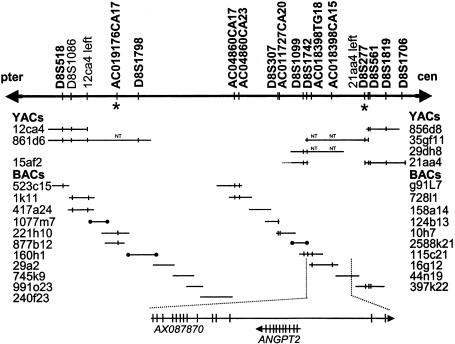
Schematic of the 2.1-mb MCPH1 critical region. Microsatellite markers are in boldface type. An asterisk (*) denotes flanking markers for the ancestral haplotype. Sequenced BAC clones are shown as horizontal lines, and end-sequenced BAC clones as horizontal lines with blackened circles at the ends. The prefix “AC” denotes a novel microsatellite marker. Marker content determined by PCR (or electronically by BLAST) is indicated by vertical lines. NT = STS not tested against clone. The two genes in the critical region are shown, with their directions of transcription indicated by arrows. Vertical lines indicate the positions of individual exons.
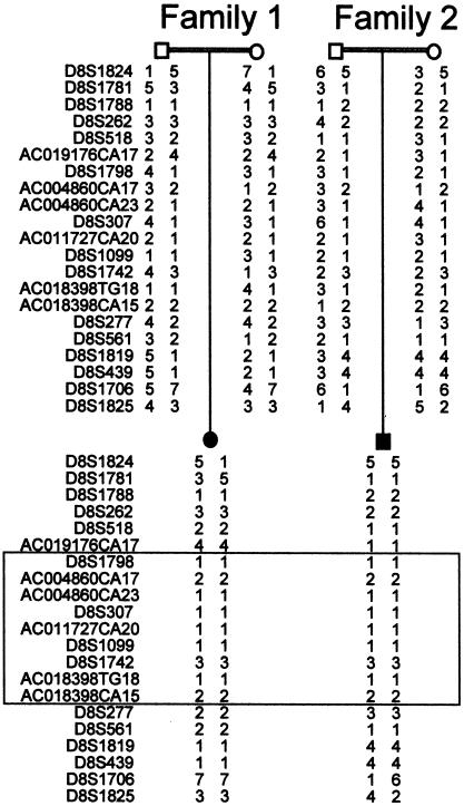
Genotyping results from one affected individual and parents from families 1 and 2, showing a region of identical alleles shared between both families (boxed), indicative of a founder effect.
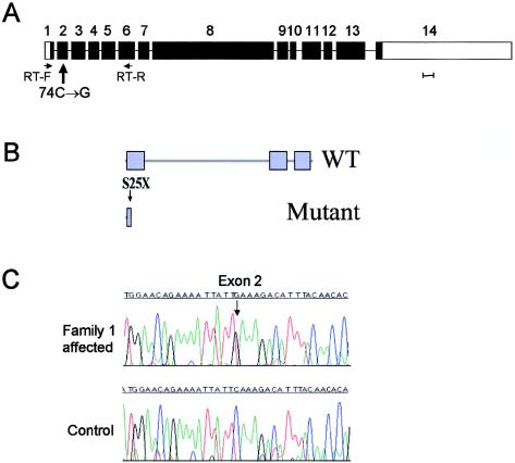
Microcephalin structure and mutation. A, Gene organization. Scale bar represents 100 bp of exon sequence. B, Predicted domain structure of the wild-type (WT) and mutant protein. BRCT domains are indicated by blue boxes. The position of the S25X mutation is indicated by an arrow. C, Nonsense mutation in exon 2 in affected individual from family 1.
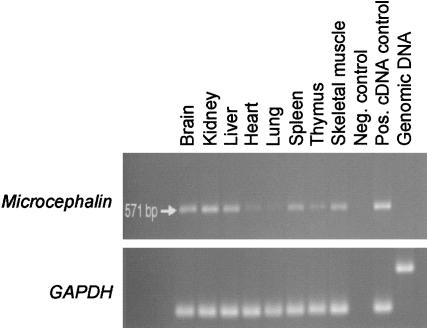
Expression pattern of microcephalin; RT-PCR analysis of human fetal tissues. Upper panel, a 570-bp microcephalin fragment amplified by use of primers from exons 1 and 6, to include the entire first BRCT domain. Lower panel, GAPDH control.
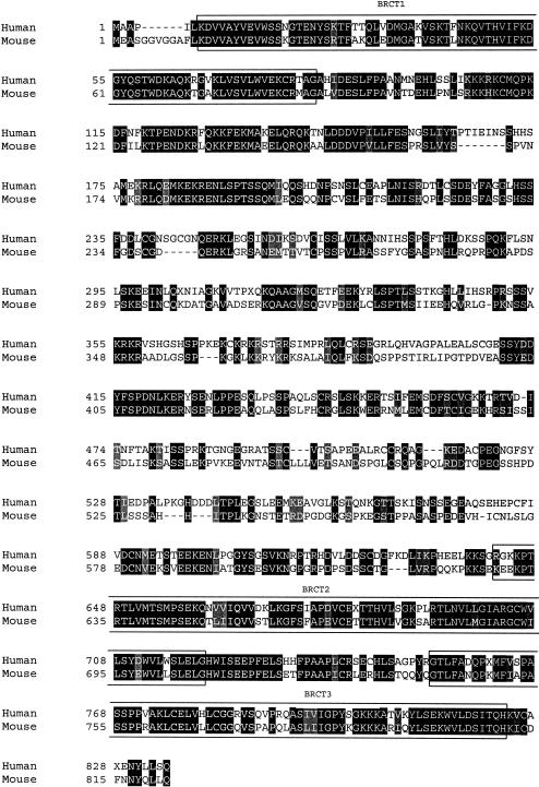
ClustalX alignment of human and mouse microcephalin proteins. Identical amino acids shaded in black, conservative substitutions in gray. BRCT domains (as predicted by SMART) are boxed.
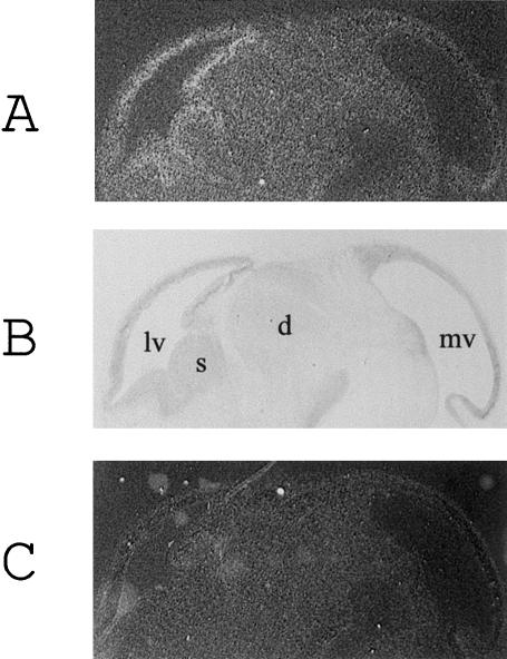
Expression of microcephalin mRNA in the fetal mouse brain. A, B, and C Expression of microcephalin mRNA in 7-μm E13.5 fetal mouse sagittal brain sections. Antisense microcephalin riboprobe (A), toluidine blue counterstained section (B), sense microcephalin riboprobe (C). lv = lateral ventricle; s = corpus striatum; d = diencephalon; mv = mesencephalic vesicle.
References
Electronic-Database Information
-
- Genome Database, http://www.gdb.org/
-
- Human Genome Project Working Draft at University of California Santa Cruz, http://genome.cse.ucsc.edu/
-
- MRC Human Genome Mapping Project Centre, http://www.hgmp.mrc.ac.uk/
-
- NCBI, http://www.ncbi.nlm.nih.gov/ (for microcephalin cDNA, AK022909, AX087870; genomic, AX087869; protein CAC34661; ANGPT2, NM_001147; DNA topoisomerase II binding protein, BAA34202. Mouse microcephalin cDNA, AY070216)
References
-
- Angevine JB Jr, Sidman RL (1961) Autoradiographic study of cell migration during histiogenesis of cerebral cortex in the mouse. Nature 192:766–768 - PubMed
-
- Bundey S (1992) Microcephaly. In: Genetics and neurology: genetics in medicine and surgery. Churchill Livingstone, Edinburgh, pp 20–24
-
- Cox KH, DeLeon DV, Angerer LM, Angerer RC (1984) Detection of mRNAs in sea urchin embryos by in situ hybridization using asymmetric RNA probes. Dev Biol 101:485–502 - PubMed
-
- Dixon J, Hovanes K, Shiang R, Dixon MJ (1997) Sequence analysis, identification of evolutionary conserved motifs and expression analysis of murine tcof1 provide further evidence for a potential function for the gene and its human homologue, TCOF1. Hum Mol Genet 6:727–737 - PubMed
Publication types
MeSH terms
Substances
Associated data
- Actions
- Actions
- Actions
- Actions
- Actions
Grants and funding
LinkOut - more resources
Full Text Sources
Other Literature Sources
Molecular Biology Databases
Miscellaneous
