Changes of NF-kB, p53, Bcl-2 and caspase in apoptosis induced by JTE-522 in human gastric adenocarcinoma cell line AGS cells: role of reactive oxygen species
- PMID: 12046064
- PMCID: PMC4656415
- DOI: 10.3748/wjg.v8.i3.431
Changes of NF-kB, p53, Bcl-2 and caspase in apoptosis induced by JTE-522 in human gastric adenocarcinoma cell line AGS cells: role of reactive oxygen species
Abstract
Aim: To identify whether JTE-522 can induce apoptosis in AGS cells and ROS also involved in the process, and to investigate the changes in NF-kB, p53, bcl-2 and caspase in the apoptosis process.
Methods: Cell culture, MTT, Electromicroscopy, agarose gel electrophoresis, lucigenin, Western blot and electrophoretic mobility shift assay (EMSA) analysis were employed to investigate the effect of JTE-522 on cell proliferation and apoptosis in AGS cells and related molecular mechanisms.
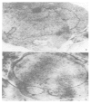
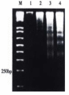
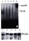
Results: JTE-522 inhibited the growth of AGS cells and induced the apoptosis. Lucigenin assay showed the generation of ROS in cells under incubation with JTE-522. The increased ROS generation might contribute to the induction of AGS cells to apoptosis. EMSA and Western blot revealed that NF-kB activity was almost completely inhibited by preventing the degradation of IkBalpha. Additionally, by using Western blot we confirmed that the level of bcl-2 was decreased, whereas p53 showed a great increase following JTE-522 treatment. Their changes were in a dose-dependent manner.
Conclusion: These findings suggest that reactive oxygen species, NF-kB, p53, bcl-2 and caspase-3 may play an important role in the induction of apoptosis in AGS cells after treatment with JTE-522.
Figures


Similar articles
-
JTE-522-induced apoptosis in human gastric adenocarcinoma [correction of adenocarcinoma] cell line AGS cells by caspase activation accompanying cytochrome C release, membrane translocation of Bax and loss of mitochondrial membrane potential.World J Gastroenterol. 2002 Apr;8(2):217-23. doi: 10.3748/wjg.v8.i2.217. World J Gastroenterol. 2002. PMID: 11925595 Free PMC article.
-
Functional p53 is required for triptolide-induced apoptosis and AP-1 and nuclear factor-kappaB activation in gastric cancer cells.Oncogene. 2001 Nov 29;20(55):8009-18. doi: 10.1038/sj.onc.1204981. Oncogene. 2001. PMID: 11753684
-
JTE-522, a selective COX-2 inhibitor, inhibits cell proliferation and induces apoptosis in RL95-2 cells.Acta Pharmacol Sin. 2002 Jul;23(7):631-7. Acta Pharmacol Sin. 2002. PMID: 12100758
-
Acacetin induces apoptosis in human gastric carcinoma cells accompanied by activation of caspase cascades and production of reactive oxygen species.J Agric Food Chem. 2005 Feb 9;53(3):620-30. doi: 10.1021/jf048430m. J Agric Food Chem. 2005. PMID: 15686411
-
Mechanical stress-induced apoptosis in the cardiovascular system.Prog Biophys Mol Biol. 2002 Feb-Apr;78(2-3):105-37. doi: 10.1016/s0079-6107(02)00008-1. Prog Biophys Mol Biol. 2002. PMID: 12429110 Review.
Cited by
-
Docetaxel inhibits SMMC-7721 human hepatocellular carcinoma cells growth and induces apoptosis.World J Gastroenterol. 2003 Apr;9(4):696-700. doi: 10.3748/wjg.v9.i4.696. World J Gastroenterol. 2003. PMID: 12679913 Free PMC article.
-
Effect of Helix aspersa extract on TNFα, NF-κB and some tumor suppressor genes in breast cancer cell line Hs578T.Pharmacogn Mag. 2017 Apr-Jun;13(50):281-285. doi: 10.4103/0973-1296.204618. Epub 2017 Apr 18. Pharmacogn Mag. 2017. PMID: 28539722 Free PMC article.
-
Inhibitory effects of tea polyphenols by targeting cyclooxygenase-2 through regulation of nuclear factor kappa B, Akt and p53 in rat mammary tumors.Invest New Drugs. 2011 Apr;29(2):225-31. doi: 10.1007/s10637-009-9349-y. Epub 2009 Nov 20. Invest New Drugs. 2011. PMID: 19936622
-
Morphology and infectivity of virus that persistently caused infection in an AGS cell line.Med Mol Morphol. 2011 Dec;44(4):213-20. doi: 10.1007/s00795-010-0530-3. Epub 2011 Dec 17. Med Mol Morphol. 2011. PMID: 22179184
-
Intravenous chemotherapy for resected gastric cancer: meta-analysis of randomized controlled trials.World J Gastroenterol. 2002 Dec;8(6):1023-8. doi: 10.3748/wjg.v8.i6.1023. World J Gastroenterol. 2002. PMID: 12439918 Free PMC article.
References
-
- Peng XM, Peng MM, Chen Q, Yao JL. Apoptosis, Bcl-2 and p53 protein expression in tissues from hepatocellular carcinoma. Huaren Xiaohua Zazhi. 1998;6:834–836.
-
- Hua JS. Effect of Hp: cell proliferation and apoptosis on stomach cancer. Shijie Huaren Xiaohua Zazhi. 1999;7:647–648.
-
- Xue XC, Fang GE, Hua JD. Gastric cancer and apoptosis. Shijie Huaren Xiaohua Zazhi. 1999;7:359–361.
-
- Qin LF, Wang RN. Prognostic significance of FCM DNA analysis in carcinoma of stomach. Shanghai Dier Yike Daxue Xuebao. 1992;12:198–202.
Publication types
MeSH terms
Substances
LinkOut - more resources
Full Text Sources
Medical
Molecular Biology Databases
Research Materials
Miscellaneous

