. 2002 Jun;8(3):469-75.
doi: 10.3748/wjg.v8.i3.469.

The promoting molecular mechanism of alpha-fetoprotein on the growth of human hepatoma Bel7402 cell line

Affiliations

The promoting molecular mechanism of alpha-fetoprotein on the growth of human hepatoma Bel7402 cell line

Meng-Sen Li et al. World J Gastroenterol. 2002 Jun.

Abstract

Aim: The goal of this study was to characterize the AFP receptor, its possible signal transduction pathway and its proliferative functions in human hepatoma cell line Bel 7402.

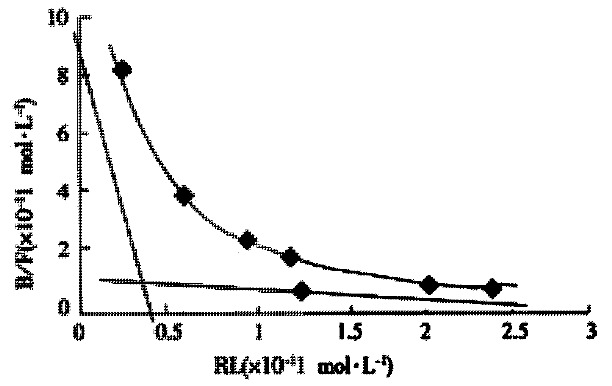
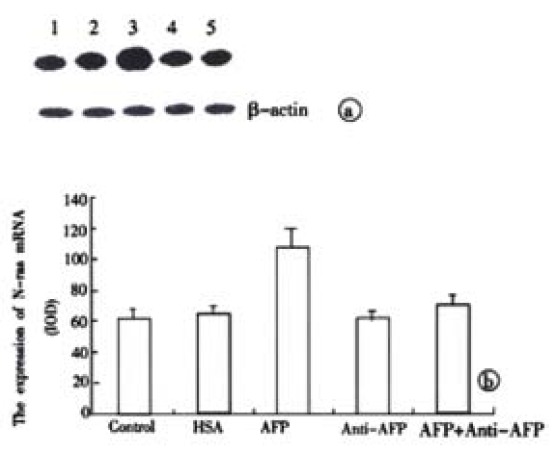
Methods: Cell proliferation enhanced by AFP was detected by MTT assay, 3H-thymidine incorporation and S-stage percentage of cell cycle analysis. With radioactive labeled 125I-AFP for receptor binding assay; cAMP accumulation, protein kinase A activity were detected by radioactive immunosorbent assay and the change of intracellular free calcium (Ca2+i) was monitored by scanning fluorescence intensity under TCS-NT confocal microscope. The expression of oncogenes N- ras, p 53, and p21( ras ) in the cultured cells in vitro were detected by Northern blotting and Western blotting respectively.

Results: It was demonstrated that AFP enhanced the proliferation of human hepatoma Bel 7402 cell in a dose dependent fashion as shown in MTT assay, (3)H-thymidine incorporation and S-phase percentage up to 2-fold. Two subtypes of AFP receptors were identified in the cells with Kds of 1.3 x 10(-9)mol.L(-1) and 9.9 x10(-8)mol. (-1)L respectively. Pretreatment of cells with AFP resulted in a significant increase (625%) in cAMP accumulation. The activity of protein kinase A activity were increased up to 37.5, 122.6, 73.7 and 61.2% at treatment time point 2, 6, 12 and 24 hours. The level of intracellular calcium were elevated after the treatment of alpha-fetoprotein and achieved to 204% at 4 min. The results also showed that AFP(20mg.L(-1)) could upregulate the expression of N- ras oncogenes and p 53 and p21( ras ) in Bel 7402 cells. In the later case,the alteration were 81.1%(12h) and 97.3%(12h) respectively compared with control.

Conclusion: These results demonstrate that AFP is a potential growth factor to promote the proliferation of human hepatoma Bel 7402 cells. Its growth-regulatory effects are mediated by its specific plasma membrane receptors coupled with its transmembrane signaling transduction through the pathway of cAMP-PKA and intracellular calcium to regulate the expression of oncogenes.


Figures

Figure 1
Figure 1
The effects of different concentration of AFP on the proliferation of cells. Bel 7402 cells were incubated with different concentrations of AFP for 48 h and the cell proliferation was measured by MTT assay. The data represented the mean values of six independent experiments performed each in triplicate. aP < 0.05 and bP < 0.01 vs control (0 mol·L-1).
Figure 2
Figure 2
The blockage of anti-AFP to the effect of AFP on the proliferation of cells. A. The data of MTT assay. B. The data of 3H-TdR incorporation. The cells were respectively treated with either AFP (20 mg·L-1), anti-AFP (40 mg·L-1), AFP (20 mg·L-1) + anti-AFP (40 mg·L-1) or HSA (20 mg·L-1) for 48 h. (MTT assay) or 18 h (3H-TdR incorporation). The data represented as the mean value of four independent experiments performed each in triplicate. aP < 0.05 and bP < 0.01 vs control (0 mol·L-1).
Figure 3
Figure 3
Scatchard analysis of 125I-AFP binding to Bel 7402 cells. The properties of AFP receptor in Bel 7402 cells was detected with receptor binding assay and analyzed by a program of Radioligand Binding Assay of Receptor. The data were selected from three independent experiments.
Figure 4
Figure 4
The effects of AFP on the cAMP concentration in cytosol of human hepatoma Bel 7402 cells. 4 × 10⁴ cells were respectively treated with AFP (20 mg·L-1), anti-AFP (40 mg·L-1), AFP (20 mg·L-1) + anti-AFP (40 mg·L-1) or HSA (20 mg·L-1). The data represented the mean values of four independent experiments performed each in triplicate. bP < 0.01 vs control (0 mol·L-1).
Figure 5
Figure 5
The effects of AFP on the activity of PKA in Bel 7402 cells. 4 × 10⁵ cells per ml were respectively treated with either AFP (20 mg·L-1), anti-AFP (40 mg·L-1), AFP (20 mg·L-1) + anti-AFP (40 mg·L-1) or HAS (20 mg·L-1) for differ-ent time and the activities of intracellular PKA were then detected. The data represented the mean values of four independent experiments performed each in triplicate. aP < 0.05 and bP < 0.01 vs control (0 mol·L-1).
Figure 6
Figure 6
A: The change of Ca2+ concentration in human hepatoma Bel7402 cells was measured by confocal microscopic scanning. Cells were incubated with 5 μmol·L-1 fluo-3/AM at 37 °C for 30 min and then stimulated with Hank’s. (1) AFP (20 mg·L-1); (2) HSA (20 mg·L-1); (3) Anti-AFP; (40 mg·L-1); (4) or AFP (20 mg·L-1) + Anti-AFP (40 mg·L-1); (5) The arrow indicate the stimulated time point. B: The graph shows the scanning results. The data represented as the mean value of 10 cells ± s. aP < 0.05 and bP < 0.01 vs control (0 mol·L-1)
Figure 7
Figure 7
The effects of AFP on the expression of p53 (A) and p21 (B) proteins in Bel 7402 cells. 1 × 10⁵ cells were respectively treated with AFP (20 mg·L-1), anti-AFP (40 mg·L-1), AFP (20 mg·L-1) + anti-AFP (40 mg·L-1) or HSA (20 mg·L-1) for 24 hours and the expression of p53 and p21protein were detected by Western blot assay. Lane 1: control group; lane 2: HSA treated group; lane 3: AFP treated group; lane 4: anti-AFP treated group; lane 5: AFP plus anti-AFP treated group. The data was selected from 3 indepen-dent experiments. A:p53; B:p21. The columns represent the means of triplicate determinations ± s.
Figure 8
Figure 8
The effects of AFP on the expression of N-ras mRNA in Bel 7402 cells. 1 × 10⁵ cells were respectively treated with AFP (20 mg·L-1), anti-AFP (40 mg·L-1), AFP (20 mg·L-1) + anti-AFP (40 mg·L-1) or HSA (20 mg·L-1) for 12 hours and expression of N-ras mRNA was detected by Northern blot assay. Lane 1: control group; Lane 2: HSA treated group; Lane 3: AFP treated group; Lane 4: anti-AFP treated group; Lane 5: AFP plus anti-AFP treated group. The data was selected from 3 independent experiments. A: Auto-radiograph of Northern blot. B: Quantitated by densitometric scanning of N-ras mRNA expression blot in Bel7402 cells (relative IOD units). The columns represent the means of triplicate determinations ± s.

Similar articles

Cited by

References

    1. Mizejewski GJ, Warner AS. Alpha-fetoprotein can regulate growth in the uterus of the immature and adult ovariectomized mouse. J Reprod Fertil. 1989;85:177–185. - PubMed
    1. Keel BA, Eddy KB, Cho S, May JV. Synergistic action of purified alpha-fetoprotein and growth factors on the proliferation of porcine granulosa cells in monolayer culture. Endocrinology. 1991;129:217–225. - PubMed
    1. Wang W, Alpert E. Downregulation of phorbol 12-myristate 13-acetate-induced tumor necrosis factor-alpha and interleukin-1 beta production and gene expression in human monocytic cells by human alpha-fetoprotein. Hepatology. 1995;22:921–928. - PubMed
    1. Mizejewski GJ. alpha-fetoprotein as a biologic response modifier: relevance to domain and subdomain structure. Proc Soc Exp Biol Med. 1997;215:333–362. - PubMed
    1. Dudich E, Semenkova L, Gorbatova E, Dudich I, Khromykh L, Tatulov E, Grechko G, Sukhikh G. Growth-regulative activity of human alpha-fetoprotein for different types of tumor and normal cells. Tumour Biol. 1998;19:30–40. - PubMed

Publication types

MeSH terms