

TGF beta1 expression and angiogenesis in colorectal cancer tissue
- PMID: 12046078
- PMCID: PMC4656429
- DOI: 10.3748/wjg.v8.i3.496
TGF beta1 expression and angiogenesis in colorectal cancer tissue
Abstract
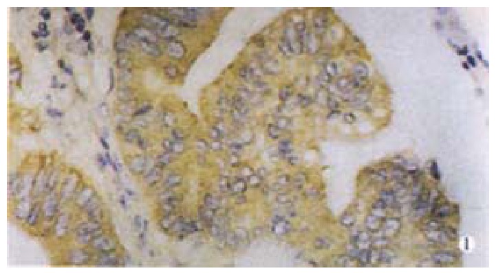
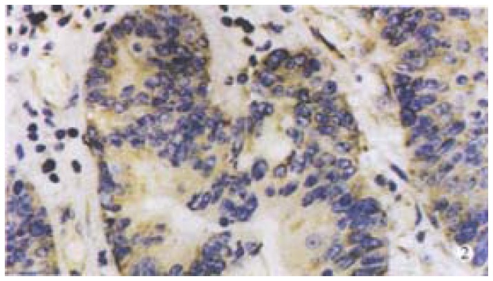
Aim: Transforming growth factor TGF beta1 is involved in a variety of important cellular functions,including cell growth and differentiation, angiogenesis, immune function and extracellular matrix formation. However, the role of TGF beta(1) as an angiogenic factor in colorectal cancer is still unclear. We investigate the relationship between transforming growth factor beta(1) and angiogenesis by analyzing the expression of transforming growth factor TGF beta(1) in colorectal cancer, as well as its association with VEGF and MVD.
Methods: The expression of TGF beta(1),VEGF, as well as MVD were detected in 98 colorectal cancer by immunohistochemical staining. The relationship between the TGF beta(1) expression and VEGF expression,MVD was evaluated. To evaluate the effect of TGF beta(1) on the angiogenesis of colorectal cancers.
Results: Among 98 cases of colorectal cancer,37 were positive for TGF beta(1) 37.8% 36 for VEGF 36.7% respectively. The microvessel counts ranged from 19 to 139.8, with a mean of 48.7(standard deviation,21.8). The expression of TGF beta(1) was correlated significantly with the depth of invasion, stage of disease, lymph node metastasis, VEGF expression and MVD. Patients in T3-T4, stage III-IV and with lymph node metastasis had much higher expression of TGF beta(1) than patients in T1-T2, stage I-II and without lymph node metastasis (P<0.05). The positive expression rate of VEGF(58.3%) in the TGF-beta(1) positive group is higher than that in the TGF-beta(1) negative group(41.7%, P<0.05). Also, the microvessel count (54+/-18) in TGF-beta(1) positive group is significantly higher than that in TGF-beta(1) negative group(46+/-15, P<0.05). The microvessel count in tumors with both TGF-beta(1) and VEGF positive were the highest (58+/-20 36-140, P<0.05). Whereas that in tumors with both TGF-beta(1) and VEGF negative were the lowest (38+/-16, 19-60, P<0.05).
Conclusion: TGF beta(1) might be associated with tumor progression by modulating the angiogenesis in colorectal cancer and TGF beta(1) may be used as a possible biomarker.
Figures
References
-
- Grunstein J, Roberts WG, Mathieu-Costello O, Hanahan D, Johnson RS. Tumor-derived expression of vascular endothelial growth factor is a critical factor in tumor expansion and vascular function. Cancer Res. 1999;59:1592–1598. - PubMed
-
- Karpanen T, Egeblad M, Karkkainen MJ, Kubo H, Ylä-Herttuala S, Jäättelä M, Alitalo K. Vascular endothelial growth factor C promotes tumor lymphangiogenesis and intralymphatic tumor growth. Cancer Res. 2001;61:1786–1790. - PubMed
-
- Siemeister G, Schirner M, Weindel K, Reusch P, Menrad A, Marmé D, Martiny-Baron G. Two independent mechanisms essential for tumor angiogenesis: inhibition of human melanoma xenograft growth by interfering with either the vascular endothelial growth factor receptor pathway or the Tie-2 pathway. Cancer Res. 1999;59:3185–3191. - PubMed
-
- Masood R, Cai J, Zheng T, Smith DL, Hinton DR, Gill PS. Vascular endothelial growth factor (VEGF) is an autocrine growth factor for VEGF receptor-positive human tumors. Blood. 2001;98:1904–1913. - PubMed
-
- Leenders W, van Altena M, Lubsen N, Ruiter D, De Waal R. In vivo activities of mutants of vascular endothelial growth factor (VEGF) with differential in vitro activities. Int J Cancer. 2001;91:327–333. - PubMed
Publication types
MeSH terms
Substances
LinkOut - more resources
Full Text Sources
Other Literature Sources
Medical
Molecular Biology Databases
Miscellaneous
