

Salvia miltiorrhiza monomer IH764-3 induces hepatic stellate cell apoptosis via caspase-3 activation
- PMID: 12046082
- PMCID: PMC4656433
- DOI: 10.3748/wjg.v8.i3.515
Salvia miltiorrhiza monomer IH764-3 induces hepatic stellate cell apoptosis via caspase-3 activation
Abstract
Aim: To investigate the effects of IH764-3 on HSC apoptosis and the expression of caspase-3 protein in HSC apoptotic process.
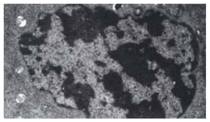
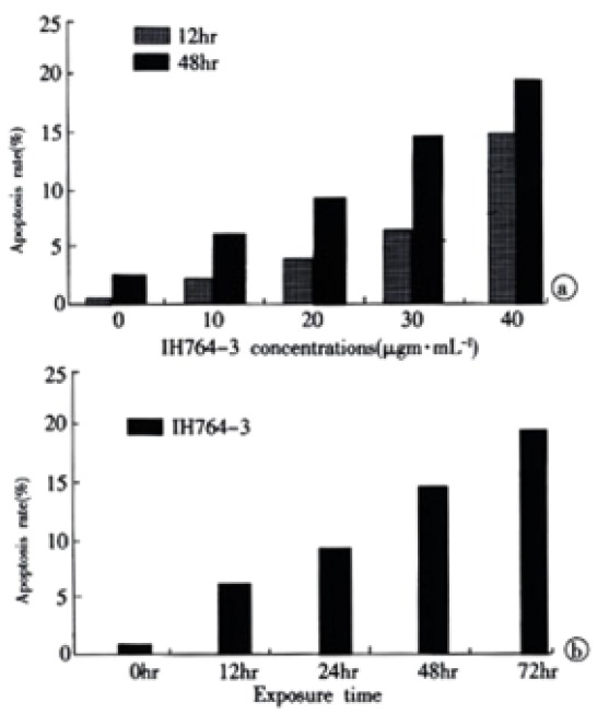
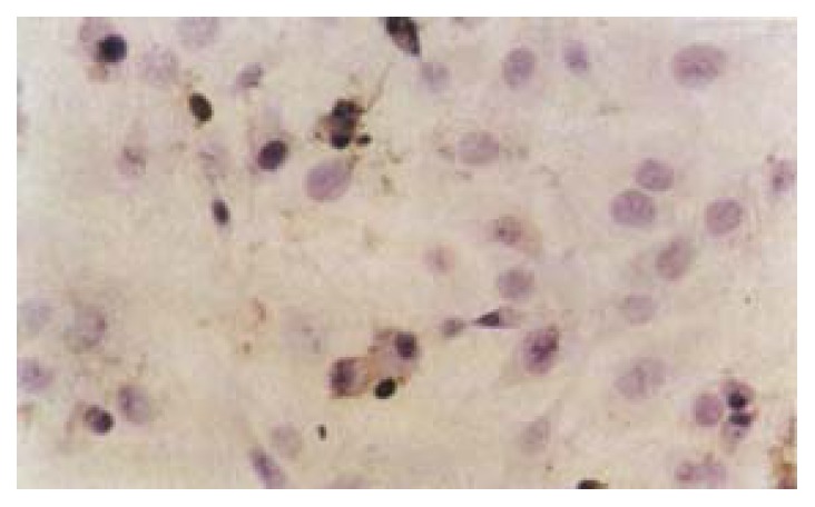
Methods: HSCs were cultured in medium with different IH764-3 doses(10 microg.mL(-1) 20 microg.mL(-1) 30 microg.mL(-1) 40 microg.mL(-1)) and without IH764-3 and HSC proliferation was quantitatively measured by (3)H-thymidine incorporation. The morphological changes of HSCs were observed with transmission electron microscope after exposure to the dose of 40 microg.mL(-1) of IH764-3 for 48 hr. The apoptosis rates were detected by annexin V/PI and TdT-mediated dUTP nick end labeling (TUNEL). The expression of caspase-3 protein was determined by flow cytometry.
Results: (1) HSC proliferation rates induced with different IH764-3 doses (10 microg.mL(-1) 20 microg.mL(-1) 30 microg.mL(-1) 40 microg.mL(-1)) were significantly reduced compared with that of the control group (P<0.01). (2)With the doses above,IH764-3 dose-dependently produced HSC apoptosis rates of 6.7%(9.4%) 9.3%(21.6%) 15.1%(27.2%) and 19.0%(28.4%) respectively by annexin V and PI-labeled flow cytometry assay or TUNEL while it was only 2.3%(6.7%) in the control. (3) The expression of caspase-3 protein in IH764-3 groups was significantly higher than that of the control (P<0.05).
Conclusion: Within the dose range used in present study IH764-3 can inhibit HSC proliferation as well as enhance HSC apoptosis. Furthermore IH764-3 can significantly increase the caspase-3 protein expression.
Figures
Similar articles
-
The Salvia miltiorrhiza monomer IH764-3 induces apoptosis of hepatic stellate cells in vivo in a bile duct ligation-induced model of liver fibrosis.Mol Med Rep. 2012 Dec;6(6):1231-8. doi: 10.3892/mmr.2012.1076. Epub 2012 Sep 11. Mol Med Rep. 2012. PMID: 22971838
-
[The role of extracellular signal-regulated kinase in induction of apoptosis with salvia miltiorrhiza monomer IH764-3 in hepatic stellate cells].Zhongguo Ying Yong Sheng Li Xue Za Zhi. 2011 Nov;27(4):402-6. Zhongguo Ying Yong Sheng Li Xue Za Zhi. 2011. PMID: 22295510 Chinese.
-
[The effect and mechanism of Salvia miltiorrhiza monomer IH764-3 on proliferation and collagen synthesis of hepatic stellate cells stimulated by H2O2].Zhongguo Ying Yong Sheng Li Xue Za Zhi. 2003 Feb;19(1):78-81. Zhongguo Ying Yong Sheng Li Xue Za Zhi. 2003. PMID: 21207864 Chinese.
-
Effects of Yigan Decoction on proliferation and apoptosis of hepatic stellate cells.World J Gastroenterol. 2002 Jun;8(3):511-4. doi: 10.3748/wjg.v8.i3.511. World J Gastroenterol. 2002. PMID: 12046081 Free PMC article.
-
[Effect of Salvia miltiorrhiza monomer IH764-3 on MMP-13 and TIMP-1 by downregulating the expression of focal adhesion kinase in hepatic stellate cell stimulated by H2O2].Zhongguo Ying Yong Sheng Li Xue Za Zhi. 2007 Nov;23(4):482-6. Zhongguo Ying Yong Sheng Li Xue Za Zhi. 2007. PMID: 21180139 Chinese.
Cited by
-
Serum from rabbit orally administered cobra venom inhibits growth of implanted hepatocellular carcinoma cells in mice.World J Gastroenterol. 2003 Nov;9(11):2441-4. doi: 10.3748/wjg.v9.i11.2441. World J Gastroenterol. 2003. PMID: 14606072 Free PMC article.
-
Ginkgo biloba extract reverses CCl4-induced liver fibrosis in rats.World J Gastroenterol. 2004 Apr 1;10(7):1037-42. doi: 10.3748/wjg.v10.i7.1037. World J Gastroenterol. 2004. PMID: 15052689 Free PMC article.
-
Effects of Chinese Jianpi herbs on cell apoptosis and related gene expression in human gastric cancer grafted onto nude mice.World J Gastroenterol. 2002 Oct;8(5):792-6. doi: 10.3748/wjg.v8.i5.792. World J Gastroenterol. 2002. PMID: 12378617 Free PMC article.
-
Protective mechanisms of medicinal plants targeting hepatic stellate cell activation and extracellular matrix deposition in liver fibrosis.Chin Med. 2014 Dec 24;9(1):27. doi: 10.1186/s13020-014-0027-4. eCollection 2014. Chin Med. 2014. PMID: 25606051 Free PMC article.
-
Antiproliferative and proapoptotic effects of somatostatin on activated hepatic stellate cells.World J Gastroenterol. 2004 Apr 1;10(7):1015-8. doi: 10.3748/wjg.v10.i7.1015. World J Gastroenterol. 2004. PMID: 15052685 Free PMC article.
References
-
- Jiang HQ, Zhang XL. Progress in the study of pathogenesis in hepatic fibrosis. Shijie Huaren Xiaohua Zazhi. 2000;8:687–689.
-
- Friedman SL. Molecular mechanisms of hepatic fibrosis and principles of therapy. J Gastroenterol. 1997;32:424–430. - PubMed
Publication types
MeSH terms
Substances
LinkOut - more resources
Full Text Sources
Other Literature Sources
Research Materials
Miscellaneous
