

Distribution of constitutive nitric oxide synthase in the jejunum of adult rat
- PMID: 12046087
- PMCID: PMC4656438
- DOI: 10.3748/wjg.v8.i3.537
Distribution of constitutive nitric oxide synthase in the jejunum of adult rat
Abstract
Aim: To study the distribution of the constitutive nitric oxide synthase (NOS) in the jejunum of adult rat.
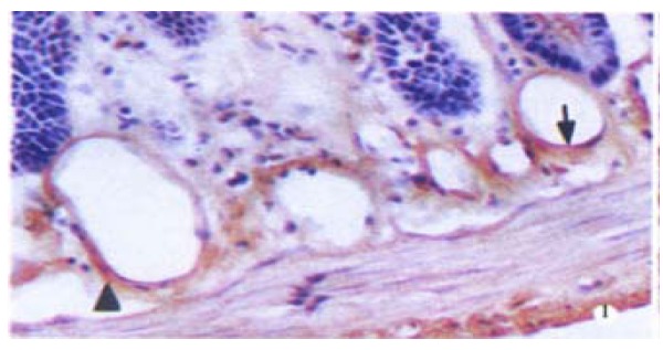
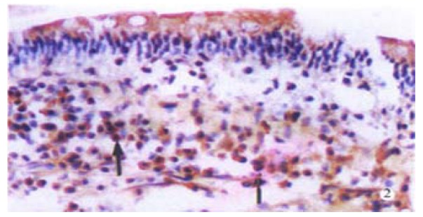
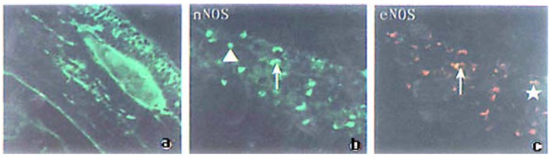
Methods: The distribution of endothelial NOS (eNOS) was detected by immunohistochemistry. Immunofluorescence histochemical dual staining technique were used for studying the distribution of neuronal NOS (nNOS) and eNOS. The dual stained slides were observed under a confocal laser scanning microscope.
Results: Positive neuronal NOS (nNOS) and endothelial NOS (eNOS) cells were found to be distributed in lamina propria of villi, and the epithelial cell was not stained. eNOS was mainly located in submucosal vascular endothelia, while nNOS was mainly situated in myenteric plexus. Some cells in the villi had both nNOS and eNOS. More than 80% of the cells were positive for both nNOS and eNOS, the rest cells were positive either for nNOS or for eNOS.
Conclusion: The two constitutive nitric oxide synthases are distributed differently in the jejunum of rat. nNOS distributed in myenteric plexus is a neurotransmitter in the non-adrenergic non-cholinergic (NANC) inhibitory nerves. eNOS distributed in endothelial and smooth muscle cells of blood vessels plays vasodilator role. eNOS and nNOS are coexpressed in some cells of lamina propria of villi. NO generated by those NOS is very important in the physiological and pathological process of small intestine.
Figures
References
-
- Eskandari MK, Kalff JC, Billiar TR, Lee KK, Bauer AJ. LPS-induced muscularis macrophage nitric oxide suppresses rat jejunal circular muscle activity. Am J Physiol. 1999;277:G478–G486. - PubMed
-
- Konomi H, Meedeniya AC, Simula ME, Toouli J, Saccone GT. Characterization of circular muscle motor neurons of the duodenum and distal colon in the Australian brush-tailed possum. J Comp Neurol. 2002;443:15–26. - PubMed
-
- Peng X, Wang SL. Nitric oxide and gastrointestinal movement. Shijie Huaren Xiaohua Zazhi. 1998;6:445–446.
-
- Nichols K, Staines W, Krantis A. Nitric oxide synthase distribution in the rat intestine: a histochemical analysis. Gastroenterology. 1993;105:1651–1661. - PubMed
Publication types
MeSH terms
Substances
LinkOut - more resources
Full Text Sources
