. 2002 Jun 1;16(11):1339-44.
doi: 10.1101/gad.987602.

Human CRSP interacts with RNA polymerase II CTD and adopts a specific CTD-bound conformation

Affiliations

Human CRSP interacts with RNA polymerase II CTD and adopts a specific CTD-bound conformation

Anders M Näär et al. Genes Dev. .

Abstract

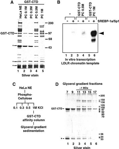
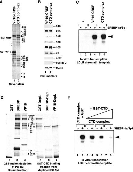
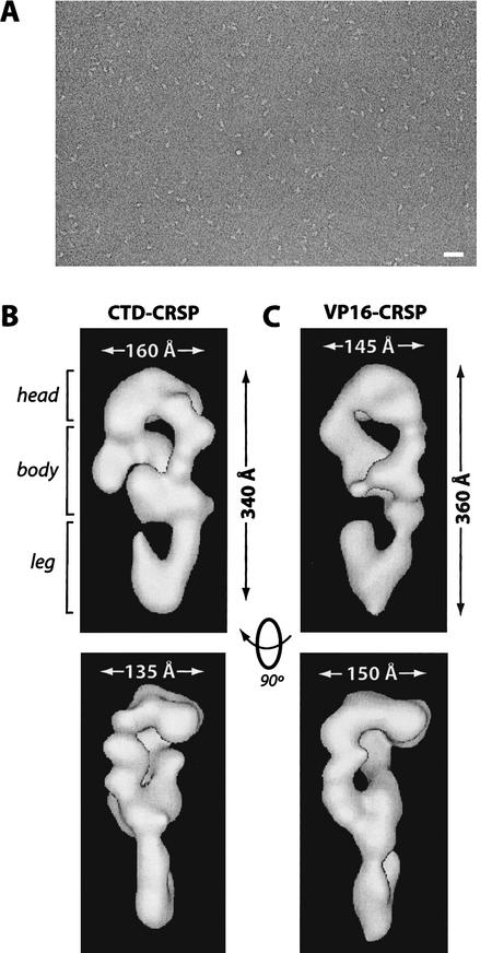
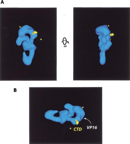
Activation of gene transcription in mammalian cells requires several classes of coactivators that participate in different steps of the activation cascade. Using conventional and affinity chromatography, we have isolated a human coactivator complex that interacts directly with the C-terminal domain (CTD) of RNA polymerase II (Pol II). The CTD-binding complex is structurally and functionally indistinguishable from our previously isolated CRSP coactivator complex. The closely related, but transcriptionally inactive, ARC-L complex failed to interact with the CTD, indicating a significant biochemical difference between CRSP and ARC-L that may, in part, explain their functional divergence. Electron microscopy and three-dimensional single-particle reconstruction reveals a conformation for CTD-CRSP that is structurally distinct from unliganded CRSP or CRSP bound to SREBP-1a, but highly similar to CRSP bound to the VP16 activator. Together, our findings suggest that the human CRSP coactivator functions, at least in part, by mediating activator-dependent recruitment of RNA Pol II via the CTD.


Figures

Figure 1
Figure 1
(A) Silver-stained SDS-polyacrylamide gel of GST–CTD-purified material from HeLa cell nuclear extract (HeLa NE, lane 1) and Phosphocellulose (PC) fractions eluting at 0.1 M (lane 2), 0.3 M (lane 3), 0.5 M (lane 4), and 1 M KCl (lane 5). The GST–CTD coeluting with associated proteins is indicated at left. (*) Nonspecific polypeptides. The migration of molecular weight standards (in kilodaltons) is indicated at right. (B) In vitro transcription analysis of coactivator activity associated with GST–CTD-bound polypeptides isolated from the PC 0.5M (lanes 3,4) and PC 1M (lanes 5,6) fractions. Transcriptional activity from the Low Density Lipoprotein Receptor (LDLR)-derived chromatin-assembled DNA template was assessed in the absence (lanes 1,3,5) or presence (lanes 2,4,6) of activators (SREBP-1a/Sp1). The expected location of the primer extension product is indicated by the arrowhead at right. (C) Purification scheme of the CTD-binding coactivator. HeLa NE was separated by PC chromatography. The 1 M fraction was incubated with GST–CTD resin and eluted with glutathione after extensive washing. Eluted material was separated on a 2-mL 15%–40% glycerol gradient. (D) SDS-PAGE and silver-stain analysis of glycerol gradient-purified CTD–CRSP. Estimated subunit sizes are shown at right (in kilodaltons) and the migration of GST–CTD and a nonspecific protein (*) are indicated at left.
Figure 2
Figure 2
Comparative analysis of VP16-purified CRSP and the PC 1M-derived CTD-binding complex. (A) Silver stain of SDS-PAGE-separated VP16–CRSP (lane 1) and the CTD-binding complex (lane 2). The bands corresponding to GST–CTD and GST–VP16 are indicated at left, along with nonspecific proteins (*). The molecular weights of the subunits are indicated at right (in kilodaltons). (B) Immunoblot analysis of VP16–CRSP (lane 1) and the PC 1M-derived CTD-binding complex (lane 2). The ARC-L/CRSP subunits analyzed are indicated at right. (C) Direct comparison of coactivator activity associated with equal quantities of VP16-purified CRSP and CTD-purified complex. SREBP-1a/Sp1-dependent activation of the LDLR-derived chromatin template (lanes 1,3,5, no activator; lanes 2,4,6, SREBP-1a and Sp1) was analyzed in the absence of added protein (lanes 1,2), in the presence of 0.5 nM CTD-binding complex (lanes 5,6), or in the presence of 0.5 nM VP16-purified CRSP (lanes 3,4). The primer extension product is indicated by the arrowhead at right. (D) Analysis of presence of CTD-binding complex in activator- or control-depleted PC 1M. Silver-stain analysis shows depletion of CTD-complex from PC 1M by CRSP-targeting activation domains. The PC 1M fraction was depleted (see Materials and Methods) using resins containing GST (lane 1), GST–SREBP1a (lane 2), or GST–VP16 (lane 3). Lanes 1–3 show bound material after first depletion. After depletion, the PC 1M fractions were incubated with GST–CTD resin and bound material was then analyzed by SDS-PAGE and silver staining (lanes 4–6). (E) Addition of exogenous CTD inhibits gene activation dependent on the CTD-purified complex. SREBP-1a/Sp1-dependent activation was analyzed as in C after the addition of 5 pmole (lanes 3,4), 15 pmole (lanes 5,6), or 50 pmole (lanes 7,8) of GST–CTD, or 50 pmole of GST alone (lanes 1,2). The primer extension product is indicated at right by an arrowhead.
Figure 3
Figure 3
CTD–CRSP and VP16–CRSP are structurally similar. (A) Negatively stained electron micrograph of CTD–CRSP sample. Bar, 800 Å. (B,C) Three-dimensional reconstruction of CTD–CRSP and VP16–CRSP at 32 Å resolution. Complexes are rendered to 1.25 MD, their approximate predicted molecular weight. Dimensions shown. Rotation of the volumes 90° gives the second side view of the coactivator.
Figure 4
Figure 4
(A) Localization of the CTD binding site (yellow) on the CRSP coactivator. This site was identified via EM analysis and difference mapping of structures generated from CTD–CRSP samples incubated with anti-GST antibodies, which target the GST–CTD fusion protein bound to the CRSP complex. (B) VP16 and CTD bind similar regions on the CRSP complex. As in A, the CTD-binding site is shown in yellow. The VP16-binding site is indicated by the white arrowhead.

References

    1. Albright SR, Tjian R. TAFs revisited: More data reveal new twists and confirm old ideas. Gene. 2000;242:1–13. - PubMed
    1. Andel F, Ladurner AG, Inouye C, Tjian R, Nogales E. Three-dimensional structure of the human TFIID-IIA-IIB complex. Science. 1999;286:2153–2156. - PubMed
    1. Boube M, Faucher C, Joulia L, Cribbs DL, Bourbon H. Drosophila homologs of transcriptional mediator complex subunits are required for adult cell and segment identity specification. Genes & Dev. 2000;14:2906–2917. - PMC - PubMed
    1. Boyer TG, Martin MED, Lees E, Ricciardi RP, Berk AJ. Mammalian Srb/Mediator complex is targeted by adenovirus E1A protein. Nature. 1999;399:276–279. - PubMed
    1. Chiba N, Suldan Z, Freedman LP, Parvin JD. Binding of liganded vitamin D receptor to the vitamin D receptor interacting protein coactivator complex induces interaction with RNA polymerase II holoenzyme. J Biol Chem. 2000;275:10719–10722. - PubMed

Publication types

LinkOut - more resources