. 2002 Jun 1;16(11):1371-82.
doi: 10.1101/gad.971402.

Phosphorylation of the mitotic regulator Pds1/securin by Cdc28 is required for efficient nuclear localization of Esp1/separase

Affiliations

Phosphorylation of the mitotic regulator Pds1/securin by Cdc28 is required for efficient nuclear localization of Esp1/separase

Ritu Agarwal et al. Genes Dev. .

Abstract

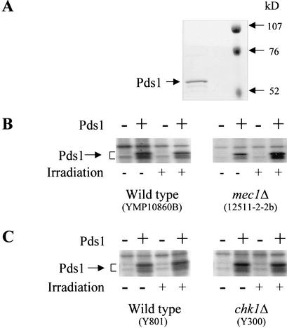
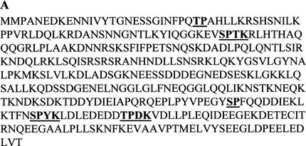
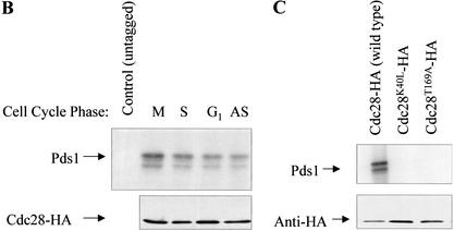
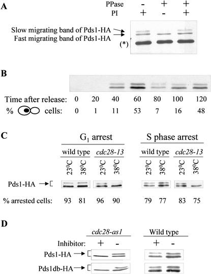
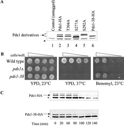
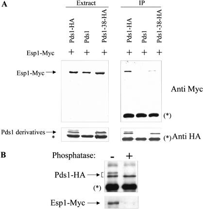
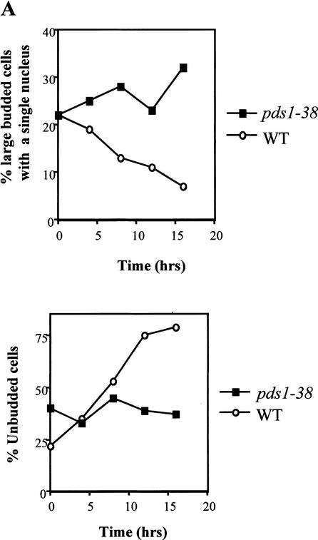
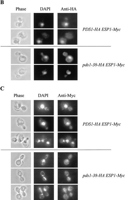
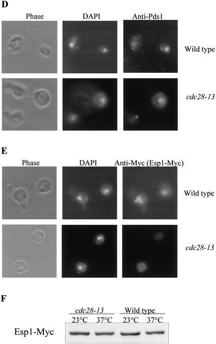
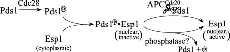
Sister chromatid separation at the metaphase-to-anaphase transition is induced by the proteolytic cleavage of one of the cohesin complex subunits. This process is mediated by a conserved protease called separase. Separase is associated with its inhibitor, securin, until the time of anaphase initiation, when securin is degraded in an anaphase-promoting complex/cyclosome (APC/C)-dependent manner. In budding yeast securin/Pds1 not only inhibits separase/Esp1, but also promotes its nuclear localization. The molecular mechanism and regulation of this nuclear targeting are presently unknown. Here we show that Pds1 is a substrate of the cyclin-dependent kinase Cdc28. Phosphorylation of Pds1 by Cdc28 is important for efficient binding of Pds1 to Esp1 and for promoting the nuclear localization of Esp1. Our results uncover a previously unknown mechanism for regulating the Pds1-Esp1 interaction and shed light on a novel role for Cdc28 in promoting the metaphase-to-anaphase transition in budding yeast.


Figures

Figure 1
Figure 1
Pds1 is phosphorylated in vitro in a Mec1- and Chk1-independent manner. (A) Purification of Pds1. Pds1 was expressed in Escherichia coli and purified as described under Materials and Methods. The purified protein was resolved by 10% SDS-PAGE and visualized by Coommassie blue staining. (B,C) Modification of Pds1 in vitro. In vitro kinase reactions were carried out as described under Materials and Methods in the absence or presence of purified Pds1, as indicated. The protein extract was made from the following strains that were either irradiated or left untreated: (B) wild type and mec1Δ (YMP10860b and 12511-2-2b, respectively); (C) wild type and chk1Δ (Y801 and Y300, respectively). Radiolabeled proteins were resolved by 10% SDS-PAGE and detected by autoradiography.
Figure 2
Figure 2
Pds1 is a Cdc28 substrate in vitro. (A) The assignment of the putative Cdc28 phosphorylation sites was done according to Songyang et al. (1994), Zhang et al. (1994), and Srinivasan et al. (1995). (B) Pds1 is phosphorylated in vitro by Cdc28 isolated from cells arrested in different cell cycle phases. HA-tagged Cdc28 was immunoprecipitated from protein extracts made from cells that were either grown to mid-log phase (asynchronous, AS), or arrested in G1 (with α-factor, 200 nM final concentration), in S phase (with hydroxyurea, 0.2 M final concentration), or in mitosis, M, (with nocodazole, 15 μg/mL final concentration). Asynchronous cells expressing untagged Cdc28 were used as a control. Immunoprecipitated Cdc28 was used in the in vitro phosphorylation reaction as described under Materials and Methods. The reaction products were resolved by 10% SDS-PAGE followed by autoradiography to detect 32P-labeled Pds1 (upper panel) or Western blot analysis with anti-HA antibody to show the relative amounts of the Cdc28 protein in each reaction (lower panel). (C) Pds1 is phosphorylated by wild-type but not by mutant Cdc28. Cells carrying plasmids expressing Cdc28-HA, Cdc28T169A-HA, or Cdc28K40L-HA were grown to mid-log phase. Protein extraction, immunoprecipitation, and in vitro kinase reactions were carried out and analyzed as described for panel B.
Figure 2
Figure 2
Pds1 is a Cdc28 substrate in vitro. (A) The assignment of the putative Cdc28 phosphorylation sites was done according to Songyang et al. (1994), Zhang et al. (1994), and Srinivasan et al. (1995). (B) Pds1 is phosphorylated in vitro by Cdc28 isolated from cells arrested in different cell cycle phases. HA-tagged Cdc28 was immunoprecipitated from protein extracts made from cells that were either grown to mid-log phase (asynchronous, AS), or arrested in G1 (with α-factor, 200 nM final concentration), in S phase (with hydroxyurea, 0.2 M final concentration), or in mitosis, M, (with nocodazole, 15 μg/mL final concentration). Asynchronous cells expressing untagged Cdc28 were used as a control. Immunoprecipitated Cdc28 was used in the in vitro phosphorylation reaction as described under Materials and Methods. The reaction products were resolved by 10% SDS-PAGE followed by autoradiography to detect 32P-labeled Pds1 (upper panel) or Western blot analysis with anti-HA antibody to show the relative amounts of the Cdc28 protein in each reaction (lower panel). (C) Pds1 is phosphorylated by wild-type but not by mutant Cdc28. Cells carrying plasmids expressing Cdc28-HA, Cdc28T169A-HA, or Cdc28K40L-HA were grown to mid-log phase. Protein extraction, immunoprecipitation, and in vitro kinase reactions were carried out and analyzed as described for panel B.
Figure 3
Figure 3
Pds1 is phosphorylated in vivo in a Cdc28-dependent manner. (A) Pds1-HA was immunoprecipitated from mid-log-phase wild-type OCF1522 cells using anti-HA antibodies. The immunoprecipitates were subjected to treatment with alkaline phosphatase (PPase) in the presence and absence of phosphatase inhibitors (PI). Pds1 mobility was analyzed by 10% SDS-PAGE followed by Western blot analysis using anti-HA antibodies. The asterisk in brackets indicates the heavy chain of the antibody used for immunoprecipitation. (B) Wild-type cells (OCF1522) expressing Pds1-HA from its native promoter were grown at 30°C, arrested in G1 with α-factor mating pheromone and then released into media lacking pheromone. Samples were taken at the indicated time points, processed for Western blot analysis, and scored for cell morphology by DIC and DAPI staining. The percent of large budded cells with a single nucleus, indicative of a post-S phase/pre-anaphase state, is shown. (C) cdc28-13 and wild-type strains (RA2817-8a and RA2817-1d, respectively), both carrying a plasmid encoding for PDS1-HA expressed from a galactose-inducible promoter (pOC42), were grown to mid-log phase at 23°C, arrested in G1 with α factor (5 μM) or in S phase with hydroxyurea (200 mM), and then shifted to 38°C for 3.5 h. Following the temperature shift, the expression of Pds1-HA was induced for 1 h (still at 38°C), after which protein extracts were prepared and analyzed by Western blot analyses. The percent of cells in G1 or S phase is indicated. (D) Centromeric plasmids encoding for Pds1-HA or Pds1db-HA (Pds1 lacking a destruction box; Cohen-Fix et al. 1996) expressed from a galactose-inducible promoter (pOC42 and pOC57-HA, respectively) were introduced into either a wild-type strain or a cdc28-as1 strain (Bishop et al. 2000; see Materials and Methods). Following an arrest in G1 with α factor, cells were either mock-treated or treated with 10 μM of PP1 (analog 9) for 4.5 h. The expression of Pds1-HA or Pds1-db-HA was induced for 1 h, after which protein extracts were prepared and analyzed by Western blot analysis.
Figure 4
Figure 4
The effect of alanine substitutions at putative Cdc28 phosphorylation sites on the electrophoretic mobility of Pds1. (A) Protein extracts from wild type (lane 2) or strains carrying mutations in PDS1 as indicated (lanes 36) were prepared from logarithmically growing cells, resolved by 10% SDS-PAGE, and analyzed by Western blot analysis using anti-HA antibodies. The Pds1-38-HA protein (lane 6) contains all three mutations (S277A, S292A, and T304A). (B) The pds1-38 strain is temperature-sensitive but not benomyl-sensitive. Wild-type, pds1Δ, and pds1-38 strains were grown to log phase and spotted by serial dilutions onto YPD plates incubated at 23°C or 37°C and onto YPD plates containing benomyl (12.5 μg/mL). (C) Pds1-38 stability at 37°C. Cells expressing Pds1-HA or Pds1-38-HA were synchronized in S phase using hydroxyurea, temperature-shifted for 1 h after a full arrest was achieved, and then released into prewarmed YPD (37°C) containing α factor to arrest the cells at the subsequent G1. Protein extracts were prepared every 20 min and analyzed by Western blot analysis. The asterisk represents an anti-HA cross-reacting band in the extracts.
Figure 5
Figure 5
Pds1 phosphorylation is required for its efficient binding to Esp1. (A) Pds1-38 has reduced affinity for Esp1. Protein extracts were prepared from strains expressing Esp1-Myc and untagged Pds1 (OCF1548-6D), Pds1-HA (OCF1548-5C), or Pds1-38-HA (RA2806-2b), all grown to mid-log phase at 23°C. The extracts were subjected to immunoprecipitation using anti-HA antibodies. Total extracts (left panel) and immunoprecipitates (IP, right panel) were separated by SDS-PAGE followed by Western blot analyses using anti-HA antibodies (bottom panel) or anti-myc antibodies (top panel). Densitometric scans showed that the ratio of the immunoprecipitated Pds1-HA to Pds1-38-HA was ∼1.2, whereas the ratio of the Esp1-Myc that coimmunoprecipitated with Pds1-HA to the Esp1-Myc that coimmunoprecipitated with Pds1-38-HA was ∼6.8. The asterisk indicates an anti-HA cross-reacting band in the extracts, whereas an asterisk in brackets represents the cross-reacting band of the heavy chain of the antibody used for immunoprecipitation. (B) Nonphosphorylated Pds1 binds weakly to Esp1 in vitro. Pds1-HA was immunoprecipitated from extracts made from mid-log phase wild-type cells expressing Pds1-HA (OCF1522), using anti-HA antibodies. The immunoprecipitates were split in two and were either treated with alkaline phosphatase or mock-treated, after which they were mixed with an extract prepared from the pds1Δ ESP1-Myc strain (RA2804-25c). The immunoprecipitates were then washed extensively, and the bound proteins were eluted using SDS sample buffer. The proteins were resolved by SDS-PAGE followed by Western blot analyses of Pds1-HA (top panel) and Esp1-Myc (bottom panel).
Figure 6
Figure 6
Cdc28-dependent phosphorylation of Pds1 is required for Esp1 accumulation in the nucleus. (A) The distribution of cell types in a wild-type versus a pds1-38 strain grown at 37°C. Wild-type (OCF1522) and pds1-38 mutant (RA2806-2b) strains were grown to log phase at 23°C and then shifted to 37°C. Samples were collected every 4 h and fixed in 70% ethanol; cell morphology was analyzed by microscopy. The percentage of large budded cells with a single nucleus (upper panel) and unbudded cells (lower panel) in wild-type cells (circles) and pds1-38 mutant cells (squares) is shown. (B,C) Esp1 does not accumulate in the nucleus in the pds1-38 strain. An HA-tagged Pds1 strain (OCF1548-5C, upper panels) and an HA-tagged Pds1-38 strain (RA2806-2b, lower panels), both of which also carried Myc-tagged Esp1 expressed from its own promoter, were grown to early-log phase and then shifted to 37°C for 4 h. Cells were fixed with 4% formaldehyde and processed for indirect immunofluorescence as described under Materials and Methods using anti-HA antibodies for detecting Pds1-HA or Pds1-38-HA (B) and anti-Myc antibodies for detecting Esp1-Myc (C). In each case several typical examples are shown. (D,E) The nuclear localization of Esp1 is Cdc28-dependent. Wild-type (RA2817-8a, upper panel) or cdc28-13 (RA2817-1d, lower panel) cells carrying Myc-tagged Esp1 expressed from its own promoter and Pds1-HA expressed from a galactose-inducible promoter were grown to early log phase at 23°C in media containing raffinose, after which they were arrested in G1 with α factor (5 μM). After a complete arrest the cultures were shifted to 37°C for 3.5 h. Pds1 expression was then induced from a galactose promoter for 2 h. Cells were fixed and processed for indirect immunofluorescence as described under Materials and Methods using anti-Pds1 antibodies to detect Pds1 (D) and anti-Myc antibodies to detect Esp1 (E). (F) The levels of Esp1-Myc in wild-type and cdc28-13 cells at 23°C and 37°C.
Figure 6
Figure 6
Cdc28-dependent phosphorylation of Pds1 is required for Esp1 accumulation in the nucleus. (A) The distribution of cell types in a wild-type versus a pds1-38 strain grown at 37°C. Wild-type (OCF1522) and pds1-38 mutant (RA2806-2b) strains were grown to log phase at 23°C and then shifted to 37°C. Samples were collected every 4 h and fixed in 70% ethanol; cell morphology was analyzed by microscopy. The percentage of large budded cells with a single nucleus (upper panel) and unbudded cells (lower panel) in wild-type cells (circles) and pds1-38 mutant cells (squares) is shown. (B,C) Esp1 does not accumulate in the nucleus in the pds1-38 strain. An HA-tagged Pds1 strain (OCF1548-5C, upper panels) and an HA-tagged Pds1-38 strain (RA2806-2b, lower panels), both of which also carried Myc-tagged Esp1 expressed from its own promoter, were grown to early-log phase and then shifted to 37°C for 4 h. Cells were fixed with 4% formaldehyde and processed for indirect immunofluorescence as described under Materials and Methods using anti-HA antibodies for detecting Pds1-HA or Pds1-38-HA (B) and anti-Myc antibodies for detecting Esp1-Myc (C). In each case several typical examples are shown. (D,E) The nuclear localization of Esp1 is Cdc28-dependent. Wild-type (RA2817-8a, upper panel) or cdc28-13 (RA2817-1d, lower panel) cells carrying Myc-tagged Esp1 expressed from its own promoter and Pds1-HA expressed from a galactose-inducible promoter were grown to early log phase at 23°C in media containing raffinose, after which they were arrested in G1 with α factor (5 μM). After a complete arrest the cultures were shifted to 37°C for 3.5 h. Pds1 expression was then induced from a galactose promoter for 2 h. Cells were fixed and processed for indirect immunofluorescence as described under Materials and Methods using anti-Pds1 antibodies to detect Pds1 (D) and anti-Myc antibodies to detect Esp1 (E). (F) The levels of Esp1-Myc in wild-type and cdc28-13 cells at 23°C and 37°C.
Figure 7
Figure 7
A model for the regulation of the Pds1–Esp1 interaction by Cdc28. See text for details.

References

    1. Alexandru G, Zachariae W, Schleiffer A, Nasmyth K. Sister chromatid separation and chromosome re-duplication are regulated by different mechanisms in response to spindle damage. EMBO J. 1999;18:2707–2721. - PMC - PubMed
    1. Amon A. Regulation of B-type cyclin proteolysis by Cdc28-associated kinases in budding yeast. EMBO J. 1997;16:2693–2702. - PMC - PubMed
    1. Bishop AC, Ubersax JA, Petsch DT, Matheos DP, Gray NS, Blethrow J, Shimizu E, Tsien JZ, Schultz PG, Rose MD, et al. A chemical switch for inhibitor-sensitive alleles of any protein kinase. Nature. 2000;407:395–401. - PubMed
    1. Ciosk R, Zachariae W, Michaelis C, Shevchenko A, Mann M, Nasmyth K. An ESP1/PDS1 complex regulates loss of sister chromatid cohesion at the metaphase to anaphase transition in yeast. Cell. 1998;93:1067–1076. - PubMed
    1. Cohen-Fix O. Making and breaking sister chromatid cohesion. Cell. 2001;106:137–140. - PubMed

Publication types

MeSH terms