. 2002 Jun 1;16(11):1383-96.
doi: 10.1101/gad.982902.

Saccharomyces Rrm3p, a 5' to 3' DNA helicase that promotes replication fork progression through telomeric and subtelomeric DNA

Affiliations

Saccharomyces Rrm3p, a 5' to 3' DNA helicase that promotes replication fork progression through telomeric and subtelomeric DNA

Andreas S Ivessa et al. Genes Dev. .

Abstract

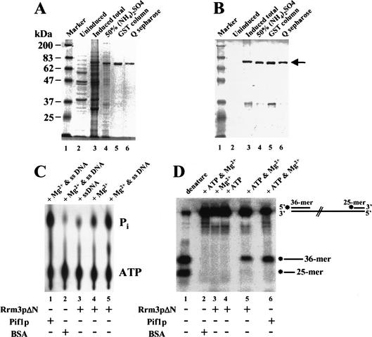
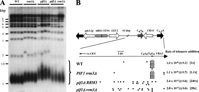
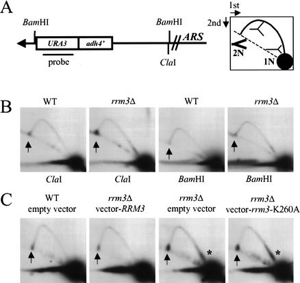
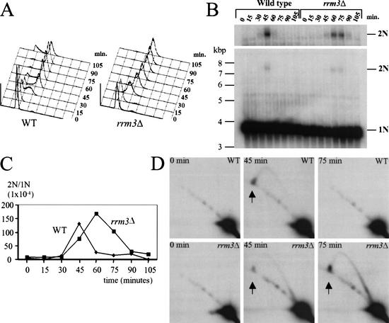
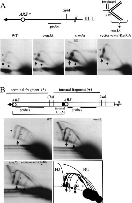
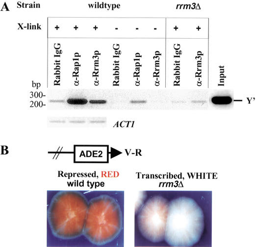
In wild-type Saccharomyces cerevisiae, replication forks slowed during their passage through telomeric C(1-3)A/TG(1-3) tracts. This slowing was greatly exacerbated in the absence of RRM3, shown here to encode a 5' to 3' DNA helicase. Rrm3p-dependent fork progression was seen at a modified Chromosome VII-L telomere, at the natural X-bearing Chromosome III-L telomere, and at Y'-bearing telomeres. Loss of Rrm3p also resulted in replication fork pausing at specific sites in subtelomeric DNA, such as at inactive replication origins, and at internal tracts of C(1-3)A/TG(1-3) DNA. The ATPase/helicase activity of Rrm3p was required for its role in telomeric and subtelomeric DNA replication. Because Rrm3p was telomere-associated in vivo, it likely has a direct role in telomere replication.


Figures

Figure 1
Figure 1
Recombinant Rrm3 protein has ATPase and 5′ to 3′ DNA helicase activity. A truncated form of Rrm3p (Rrm3pΔN) was fused to GST and expressed in Saccharomyces cerevisiae under control of the galactose-inducible GAL1 promoter. Yeast proteins were resolved by 8% SDS-PAGE and detected by (A) Coomassie blue staining or (B) immunoblotting. In A and B, lane 1 is size markers in kilodaltons. (Because covalent coupling of marker proteins to the blue dye used for visualization affects their mobility in SDS-PAGE, the 83-kD marker protein has a slower mobility than the 87-kD Rrm3pΔN.) Lanes 2 and 3 contain a crude extract of proteins from glucose grown (lane 2) or galactose grown (lane 3) cells. Lanes 46 contain proteins from galactose-grown cells after sequential purification by 50% ammonium sulfate precipitation (lane 4), fractionation on Glutathione sepharose 4B (lane 5), and fractionation on Q-sepharose (lane 6). (C) The ATPase reaction products were developed in a polyethylimine (PEI) cellulose plate and visualized on a Molecular Dynamics PhosphorImager. Lane 5 contains [γ-P32]ATP, M13 single-stranded DNA, Mg2+, and Rrm3pΔN. The other lanes were the same except lane 1 had Pif1p in place of Rrm3pΔN; lane 2 had BSA in place of Rrm3pΔN; lane 3 had no Mg2+; and lane 4 had no single-stranded DNA. (D) For the helicase assay, the DNA substrate was linear M13 single-stranded DNA to which kinase labeled 36-mer and 25-mer oligonucleotides had been annealed. Lane 1 contains the heat-denatured DNA substrate; lane 5 contains Rrm3pΔN, the DNA substrate, ATP and Mg2+. The other lanes are the same as lane 5 except lane 2 contains BSA in place of Rrm3pΔN; lane 3 has no ATP; lane 4 has no Mg2+; and lane 6 contains Pif1p in place of Rrm3pΔN.
Figure 2
Figure 2
Rrm3p affects telomere length and de novo telomere addition but not in the same ways as Pif1p. (A) DNA was prepared from four individual colonies from each strain: PIF1 RRM3, PIF1 rrm3Δ, pif1Δ RRM3, and pif1Δ rrm3Δ cells. The DNA was digested with XhoI, separated by electrophoresis in a 0.7% agarose gel, prepared for Southern analysis, and probed with a C1–3A/TG1–3 telomeric probe. The large curly brace indicates the terminal XhoI fragments from Y‘-bearing telomeres. Molecular weight markers are in kilobase pairs. (B) The rate of de novo telomere formation on a YAC was determined using 10 plate fluctuation tests (Lea and Coulson 1949), conducted 2–4 times per strain, as described in Schulz and Zakian (1994). Because Leu+ FOAR cells can also be generated by point mutations in URA3, the sites of telomere addition in multiple independent Leu+ FOAR clones were mapped to determine the fraction of these events that were due to de novo telomere addition. Each filled circle marks the physical end of a YAC in one such colony. Leu+ FOAR colonies that contained a YAC of unaltered length were presumed to arise from point mutations in URA3. Numbers in parentheses indicate the range of values seen in independent experiments. Numbers in brackets are the fold difference relative to the wild-type strain.
Figure 3
Figure 3
Replication of telomere VII-L is impaired in the absence of Rrm3p. (A) Structure of the left telomere of Chromosome VII after insertion of URA3 in a strain in which URA3 was deleted from its normal location. The URA3 probe indicated in A was used in B and C as well as in Figure 4. Digestion with ClaI generates a 3.8-kb fragment containing the VII-L telomere. A has a cartoon of the expected pattern of replication intermediates for simple forks moving leftward toward the telomere after their separation in 2D gels. The arc of linear molecules is denoted by the dotted line, 1N marks the position of an unreplicated restriction fragment, and 2N is the same molecule immediately before its replication is complete. The 2N intermediate is drawn in thick lines to emphasize that it was more abundant than other forked replication intermediates. (B) DNA from wild-type (WT) or rrm3Δ cultures was digested with either ClaI (left two panels) or BamHI (right two panels). The arrows in this and subsequent gels denote the position of the 2N spot. (C) DNA was prepared from cultures of wild-type or rrm3Δ strains carrying the plasmid YCplac111, YCplac111 containing RRM3, or YCplac111 containing the rrm3 K260A allele. DNA was digested with ClaI and analyzed by 2D gels. Asterisks mark the region downstream of ADH4 where replication forks slow in the absence of Rrm3p.
Figure 4
Figure 4
The 2N spot is cell cycle regulated, appearing at the time of telomere replication. Wild-type or rrm3Δ cells were G1 arrested with α factor and then removed from the α factor and allowed to progress through the cell cycle. Samples were prepared at the time of removal from α factor (0 min) and then at 15-min intervals. (A) Cells from each time point were analyzed by fluorescent activated cell sorting (FACs). (B) DNA samples from each time point were digested with ClaI, separated by conventional agarose gel electrophoresis, and hybridized with the URA3 probe (Fig. 3A). In a conventional gel, 2N-spot DNA migrates as a 7.6-kb linear fragment. The top panel shows a longer exposure of the 2N spot region of the gel. (C) The data in panel B were quantitated; the 2N to 1N ratio is shown for each time point. (D) Each ClaI-digested DNA sample was also analyzed by 2D gel analysis; representative time points from both strains are shown.
Figure 5
Figure 5
Rrm3p affects replication of natural, X-, and Y‘-bearing telomeres. (A) Structure of the left telomere of Chromosome III. An inactive ARS in the subtelomeric X element, 1 kb from the chromosome end, is indicated by an asterisk. Genomic DNA was digested with SphI, which generates a 4-kb terminal fragment from Chromosome III-L, and analyzed by 2D gels using the probe shown in A. Pausing at the inactive ARS is denoted by an asterisk. A diamond marks a molecule on the arc of simple linears that has a mass of ∼6 kb. A 6-kb, almost linear fragment is generated by breakage in front of the fork stalled at the ARS (see cartoon). The third panel is a longer exposure of the second panel that makes it easier to see replication bubbles (BU). The fourth panel is the pattern in an rrm3Δ strain that carries the rrm3-K260A allele on plasmid YCplac111 (as in Fig. 3C). (B) Structure of Y‘ elements. Up to four tandem Y‘ elements are found at a given telomere: the bracket indicates the position of the most terminal Y‘ element on a hypothetical telomere containing two Y‘ repeats. Genomic DNA from asynchronous cells was digested with ClaI and analyzed by 2D gels using the combination of probes shown in the cartoon. The pattern of replication intermediates for three strains is shown: RRM3 (WT), rrm3Δ, and rrm3Δ carrying the rrm3-K260A allele on plasmid YCplac111. The arc labeled with an asterisk is the 5.3-kb ClaI fragment from terminal Y‘ long elements; the arc labeled with a diamond is the 6.2-kb ClaI fragment from internal Y‘ long elements (two size variants of this fragment are visible in wild-type DNA). The rightmost arrow in the rrm3Δ gel is at the position for the 2N spot for Y‘ short telomeres. In the schematic of the 2D gel for rrm3Δ Y‘ DNA, arrows point to the 2N spot for Y‘ long (leftmost arrow) and Y‘ short (rightmost arrow) telomeres. Pauses in rrm3Δ DNA are labeled 1–6 (see text). (HJ) Holliday junctions between internal Y‘ long elements; (BU) bubble-containing replication intermediates for internal Y‘ long elements.
Figure 6
Figure 6
Internal tracts of C1–3A/TG1–3 cause replication fork pausing, and this pausing is increased in an rrm3Δ strain. (A) The cartoon shows replication of a yeast telomere. The telomeric C1–3A/TG1–3 tract is in bold. There are two models that can explain the appearance of the 2N spot. In Model I, the 2N spot is formed by replication forks pausing before or within the C1–3A/TG1–3 tract. In this model, Rrm3p promotes semiconservative replication of telomeres. After DNA replication, the C strand of telomeric DNA is degraded to form long single-strand TG1–3 tails (Wellinger et al. 1993a,b, 1996). If the TG1–3 tails on sister chromatids interacted by stable G–G base pairing, they would generate DNA that behaves like 2N-spot DNA. In Model II, Rrm3p would promote dissociation of G-base-paired telomeres. Only the first model predicts that Rrm3p should promote replication through internal C1–3A/TG1–3 tracts. (B) Three 276-bp tracts of C1–3A/TG1–3 DNA, each separated from the adjacent tract by 19 bp of polylinker DNA, were inserted within the LYS2 locus (Stavenhagen and Zakian 1994). DNA from wild-type and rrm3Δ cells containing the C1–3A/TG1–3 tract was digested with BglII and SalI, and examined by 2D gels. The probe detects a 7-kb fragment. (C) DNA from wild-type or rrm3Δ cells with or without (no tract) a 500-bp C1–3A/TG1–3 tract inserted within HIS4 was digested with AatII and analyzed by 2D gels, using the indicated probe. The probe detects a 3.6-kb AatII fragment in the no-tract control strains and a 4.1-kb AatII fragment in strains with the C1–3A/TG1–3 tract. The asterisks mark an ∼8-kb AatII fragment that cross-reacts with the hybridization probe. This cross-reacting band falls fortuitously near the position of 2N-spot DNA in the strains with the C1–3A/TG1–3 tract. The linear fragment of ∼5.5 kb that is marked by a diamond is seen only in the presence of the 0.5-kb C1–3A/TG1–3 tract. Because its abundance is proportional to the amount of pausing at the C1–3A/TG1–3 tract, it is probably caused by breakage of stalled replication intermediates immediately ahead of the replication fork as in Figure 5A. In panels B and C, brackets indicate the position of the C1–3A/TG1–3 tracts.
Figure 7
Figure 7
Rrm3p is associated with telomeric DNA in vivo and has a modest effect on TPE. (A) Chromatin was prepared from otherwise isogenic wild-type or rrm3Δ cells that had been cross-linked (+ cross-link) or not (− cross-link) with formaldehyde in vivo. Immunoprecipitation was carried out using either protein-A-purified preimmune IgG (rabbit IgG), a polyclonal Rap1p antiserum (α-Rap1p; Conrad et al. 1990), or affinity-purified anti-Rrm3p polyclonal antibodies (Ivessa et al. 2000). The DNA in the precipitate was PCR-amplified for 28 cycles using Y‘ primers that detect a 233-bp portion of the subtelomeric Y‘ element that begins 30 bp upstream of the terminal C1–3A/TG1–3 tracts or for 31 cycles using ACT1 primers. The PCR products were separated in a 2.3% agarose gel and visualized by staining with ethidium bromide. PCR amplification of the input DNA with telomeric primers is also shown (Input). Although Rrm3p association with telomeric DNA was eliminated in the absence of in vivo cross-linking, some Rap1p association with telomeric DNA was detected in the no cross-linking control. (B) TPE was measured in a strain with URA3 next to the left telomere of Chromosome VII (Gottschling et al. 1990) and ADE2 next to the right telomere of Chromosome V (Wiley and Zakian 1995). Wild-type or rrm3Δ cells were plated on media containing low amounts of adenine, and the color of the resulting colonies was examined.

References

    1. Bennett RJ, Sharp JA, Wang JC. Purification and characterization of the Sgs1 DNA helicase activity of Saccharomyces cerevisiae. J Biol Chem. 1998;273:9644–9650. - PubMed
    1. Bessler JB, Torres JZ, Zakian VA. The Pif1p subfamily of helicases: Region specific DNA helicases. Trends Cell Biol. 2001;11:60–65. - PubMed
    1. Bourns BD, Alexander MK, Smith AM, Zakian VA. Sir proteins, Rif proteins and Cdc13p bind Saccharomyces telomeres in vivo. Mol Cell Biol. 1998;18:5600–5608. - PMC - PubMed
    1. Brewer BJ, Fangman WL. The localization of replication origins on ARS plasmids in S. cerevisiae. Cell. 1987;51:463–471. - PubMed
    1. Button LL, Astell CR. The Saccharomyces cerevisiae chromosome III left telomere has a type X, but not a type Y‘, ARS region. Mol Cell Biol. 1986;6:1352–1356. - PMC - PubMed

Publication types