. 2002 Jun 1;16(11):1433-40.
doi: 10.1101/gad.981202.

Desert Hedgehog/Patched 1 signaling specifies fetal Leydig cell fate in testis organogenesis

Affiliations

Desert Hedgehog/Patched 1 signaling specifies fetal Leydig cell fate in testis organogenesis

Humphrey Hung-Chang Yao et al. Genes Dev. .

Abstract

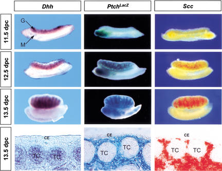
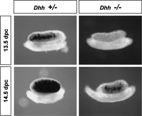
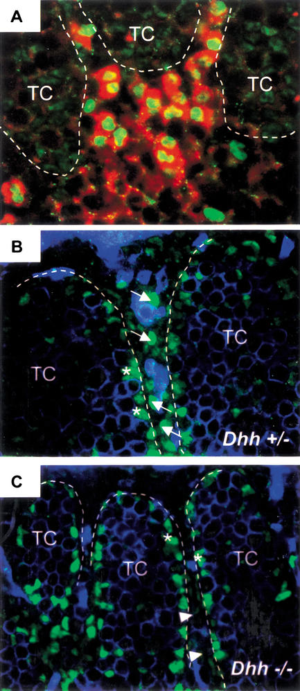
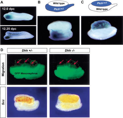
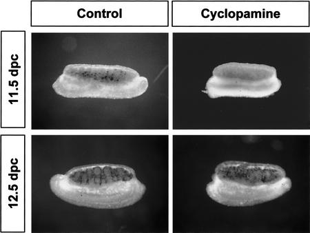
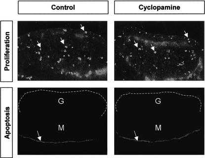
Establishment of the steroid-producing Leydig cell lineage is an event downstream of Sry that is critical for masculinization of mammalian embryos. Neither the origin of fetal Leydig cell precursors nor the signaling pathway that specifies the Leydig cell lineage is known. Based on the sex-specific expression patterns of Desert Hedgehog (Dhh) and its receptor Patched 1 (Ptch1) in XY gonads, we investigated the potential role of DHH/PTCH1 signaling in the origin and specification of fetal Leydig cells. Analysis of Dhh(-/-) XY gonads revealed that differentiation of fetal Leydig cells was severely defective. Defects in Leydig cell differentiation in Dhh(-/-) XY gonads did not result from failure of cell migration from the mesonephros, thought to be a possible source of Leydig cell precursors. Nor did DHH/PTCH1 signaling appear to be involved in the proliferation or survival of fetal Leydig precursors in the interstitium of the XY gonad. Instead, our results suggest that DHH/PTCH1 signaling triggers Leydig cell differentiation by up-regulating Steroidogenic Factor 1 and P450 Side Chain Cleavage enzyme expression in Ptch1-expressing precursor cells located outside testis cords.


Figures

Figure 1
Figure 1
Expression patterns of Dhh (dark purple), Ptch1 (blue), and the Leydig cell marker Scc (red) in XY gonads from 11.5 dpc to 13.5 dpc. Expression of Dhh and Scc were detected by whole-mount in situ hybridization. Ptch1 expression was detected by analyzing β-galactosidase activity in the Ptchtm1Mps XY gonads. Sections of 13.5 dpc whole-mount samples are shown at the bottom to confirm the cell-specific expression patterns of Dhh, Ptch1, and Scc in the gonads. CE, coelomic epithelium; G, gonad; M, mesonephros; TC, testis cords.
Figure 2
Figure 2
Expression of Scc in Dhh+/− and Dhh−/− XY gonads. The mRNA for the Leydig cell marker Scc (black stain) is present at 13.5 and 14.5 dpc in Dhh+/+ (data not shown) and Dhh+/− but is reduced or absent in Dhh−/− XY gonads.
Figure 3
Figure 3
Expression of SF1 and Scc in 13.5 dpc Dhh+/− and Dhh−/− XY gonads. (A) Colocalization of SF1 (green nuclear staining) and Scc (red cytoplasmic staining) in Leydig cells in normal 13.5 dpc XY gonads by immunocytochemistry for SF1 and in situ hybridization for Scc. (B) Strong nuclear SF1 staining (arrows) in Leydig cells in Dhh+/− gonads. (C) Absence of strong nuclear SF1 staining in Leydig cells in Dhh−/− gonads. Weak nuclear SF1 staining was still present (arrowheads). SF1 staining was also detected in Sertoli cells (asterisks) in testis cords (TC, outlined by dotted lines). Germ cells and endothelial cells were stained with an anti-PECAM antibody (blue staining in B and C).
Figure 4
Figure 4
DHH/PTCH1 signaling is not responsible for induction of mesonephric cell migration into XY gonads. (A) Analysis of PtchLacZ expression in 12.0 and 12.25 dpc XY gonads. (B) Recombinant organ culture using a wild-type gonad apposed to a PtchLacZ mesonephros. (C) Reciprocal organ culture with a PtchLacZ gonad apposed to a wild-type mesonephros. Recombinant organ cultures in both B and C were assembled at 11.25 dpc, cultured for 48 h, and assayed for PtchLacZ expression. (D) Mesonephric cell migration and Scc expression in Dhh+/− and Dhh−/− gonads: Recombinant gonad cultures were assembled with an 11.5 dpc Dhh+/− or Dhh−/− XY gonad apposed to an 11.5 dpc mesonephros expressing GFP. Cell migration (green cell, red arrows) was detected 48 h after culture. Samples were fixed, and then expression of Scc was detected by in situ hybridization (red staining in gonads).
Figure 5
Figure 5
Stage-specific effects of the hedgehog inhibitor cyclopamine on expression of Scc mRNA in Leydig cells. XY gonads (11.5 or 12.5 dpc) were cultured in the presence or absence of cyclopamine (25 μM) for 24 h followed by whole-mount in situ hybridization for Scc (black staining in gonads).
Figure 6
Figure 6
Effects of the hedgehog inhibitor cyclopamine on proliferation and apoptosis in 11.5 dpc gonads. Gonads (11.5 dpc) were cultured for 24 h in the presence or absence of cyclopamine (25 μM) followed by immunocytochemistry for phosphorylated Histone H3 (arrows) or LysoTracker staining for apoptosis (arrows indicate position of the Müllerian duct). G, gonad (outlined by a dotted line); M, mesonephros.

References

    1. Albrecht KH, Eicher EM. Evidence that Sry is expressed in pre-Sertoli cells and Sertoli and granulosa cells have a common precursor. Dev Biol. 2001;240:92–107. - PubMed
    1. Ariyaratne SHB, Mendis-Handagama CS, Hales BD, Mason IJ. Studies on the onset of Leydig precursor cell differentiation in the prepubertal rat testis. Biol Reprod. 2000;63:165–171. - PubMed
    1. Bitgood MJ, McMahon AP. Hedgehog and Bmp genes are coexpressed at many diverse sites of cell-cell interaction in the mouse embryo. Dev Biol. 1995;172:126–138. - PubMed
    1. Bitgood MJ, Shen L, McMahon AP. Sertoli cell signaling by Desert Hedgehog regulates the male germline. Curr Biol. 1996;6:298–304. - PubMed
    1. Buehr M, Gu S, McLaren A. Mesonephric contribution to testis differentiation in the fetal mouse. Development. 1993;117:273–281. - PubMed

Publication types

MeSH terms