. 2002 Aug 30;277(35):32099-108.
doi: 10.1074/jbc.M203639200. Epub 2002 Jun 5.

Involvement of both G(q/11) and G(s) proteins in gonadotropin-releasing hormone receptor-mediated signaling in L beta T2 cells

Affiliations

Involvement of both G(q/11) and G(s) proteins in gonadotropin-releasing hormone receptor-mediated signaling in L beta T2 cells

Fujun Liu et al. J Biol Chem. .

Abstract

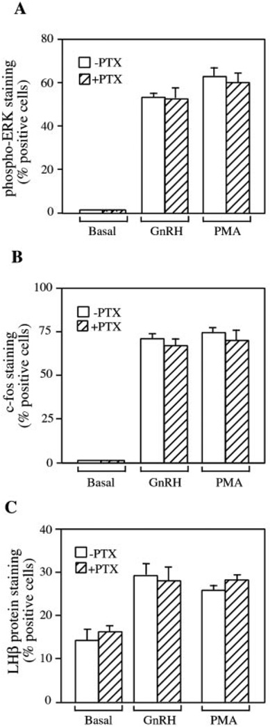
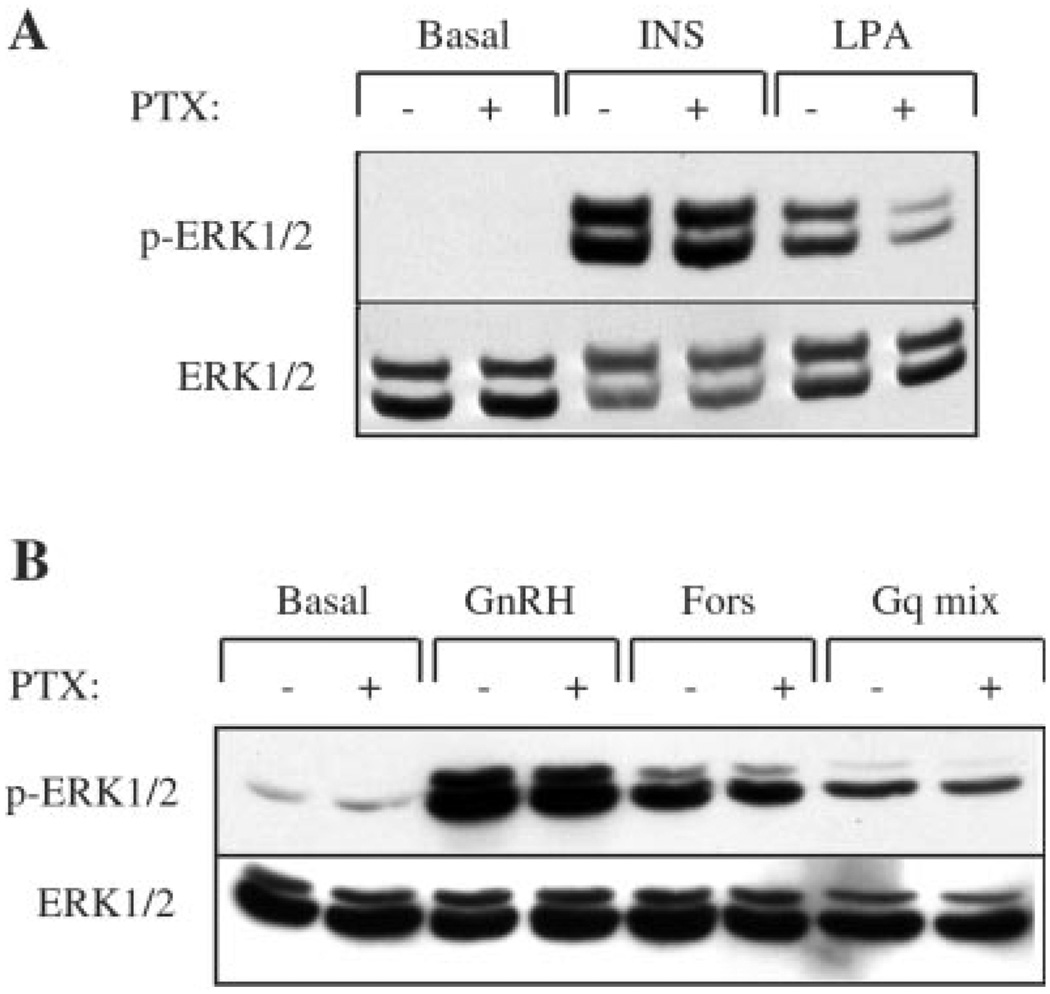
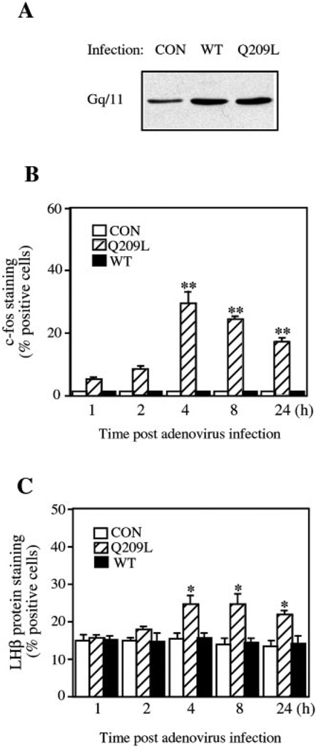
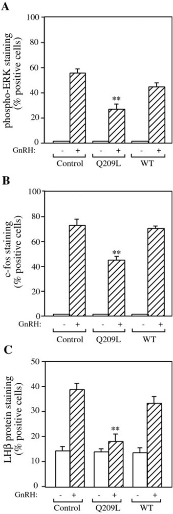
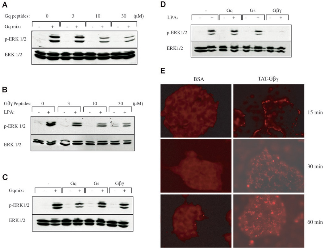
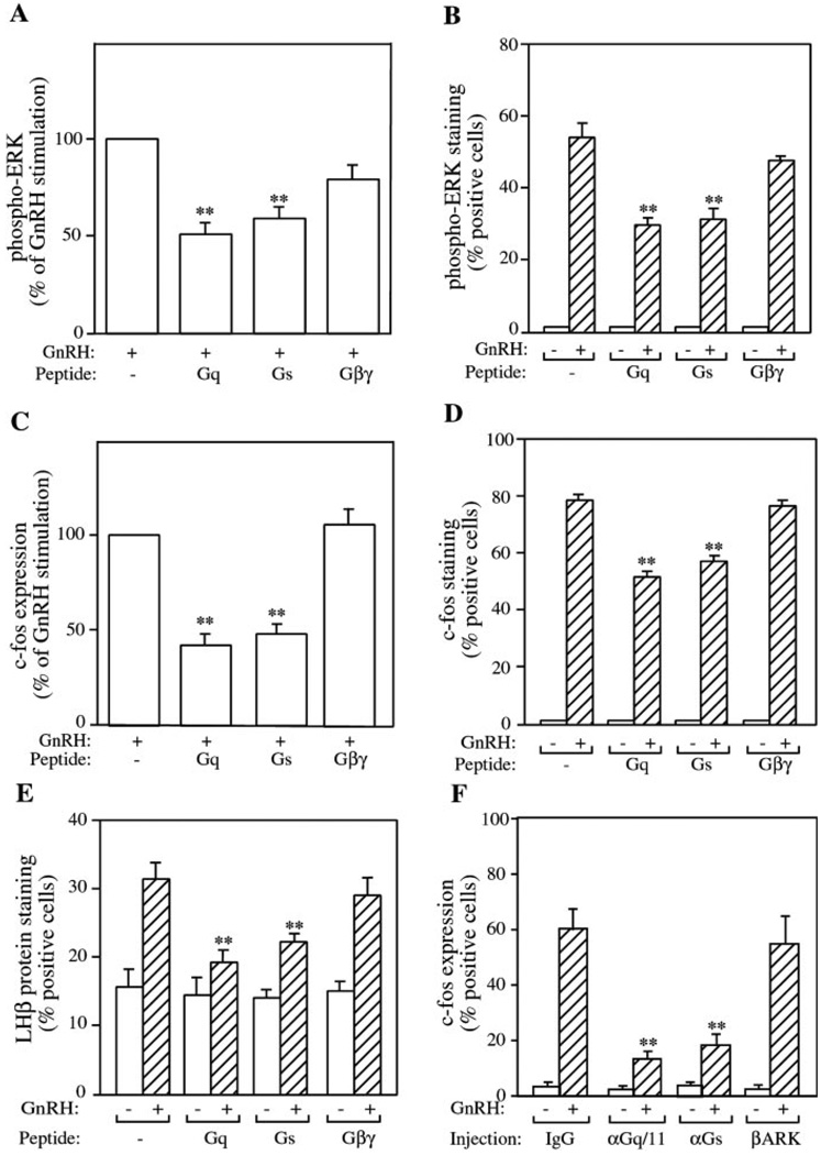
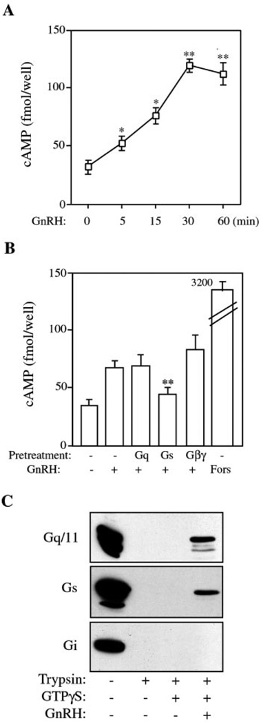
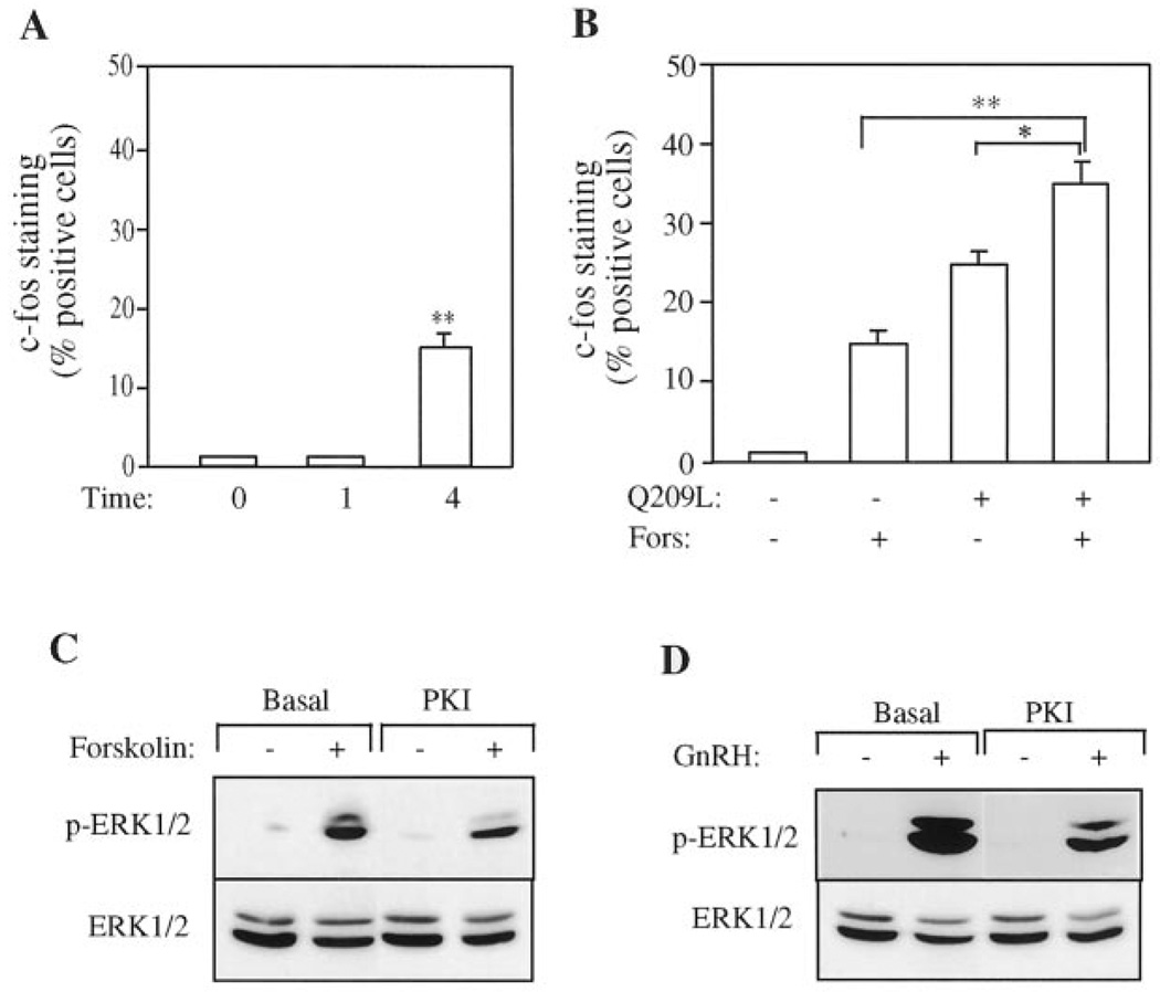
The hypothalamic hormone gonadotropin-releasing hormone (GnRH) stimulates the synthesis and release of the pituitary gonadotropins. GnRH acts through a plasma membrane receptor that is a member of the G protein-coupled receptor (GPCR) family. These receptors interact with heterotrimeric G proteins to initiate downstream signaling. In this study, we have investigated which G proteins are involved in GnRH receptor-mediated signaling in L beta T2 pituitary gonadotrope cells. We have shown previously that GnRH activates ERK and induces the c-fos and LH beta genes in these cells. Signaling via the G(i) subfamily of G proteins was excluded, as neither ERK activation nor c-Fos and LH beta induction was impaired by treatment with pertussis toxin or a cell-permeable peptide that sequesters G beta gamma-subunits. GnRH signaling was partially mimicked by adenoviral expression of a constitutively active mutant of G alpha(q) (Q209L) and was blocked by a cell-permeable peptide that uncouples G alpha(q) from GPCRs. Furthermore, chronic activation of G alpha(q) signaling induced a state of GnRH resistance. A cell-permeable peptide that uncouples G alpha(s) from receptors was also able to inhibit ERK, c-Fos, and LH beta, indicating that both G(q/11) and G(s) proteins are involved in signaling. Consistent with this, GnRH caused GTP loading on G(s) and G(q/11) and increased intracellular cAMP. Artificial elevation of cAMP with forskolin activated ERK and caused a partial induction of c-Fos. Finally, treatment of G alpha(q) (Q209L)-infected cells with forskolin enhanced the induction of c-Fos showing that the two pathways are independent and additive. Taken together, these results indicate that the GnRH receptor activates both G(q) and G(s) signaling to regulate gene expression in L beta T2 cells.


Figures

FIG. 1
FIG. 1. Effect of PTX on GnRH-induced ERK activation and c-Fos and LHβ protein expression in LβT2 cells
A, effect of PTX on GnRH-induced ERK activation. LβT2 cells were plated on acid-washed coverslips and incubated in serum-free DMEM overnight with or without 100 ng/ml PTX. Cells were then stimulated with 100 nm GnRH or 100 nm PMA for 5 min at 37 °C, fixed, and processed for immunofluorescence. Active ERK was visualized with an antibody against dually phosphorylated ERK (Thr202/Tyr204) and TRITC-labeled secondary antibody. Nuclei were counterstained with Hoechst 33258 DNA dye. Cells with nuclear fluorescence were scored as positive for ERK activation. B, effect of PTX on GnRH-induced c-Fos expression. LβT2 cells on coverslips were starved overnight in serum-free medium with or without 100 ng/ml PTX. Cells were then stimulated with 100 nm GnRH or 100 nm PMA for 60 min. Nuclear c-Fos expression was visualized using a rabbit anti-c-Fos antibody, followed by a TRITC-conjugated secondary antibody. Nuclei were counterstained with Hoechst 33258 DNA dye. Cells with nuclear c-Fos immunofluorescence were counted as positive for c-Fos expression. C, effect of PTX on GnRH-induced LHβ protein expression. LβT2 cells on coverslips were starved in serum-free DMEM. Cells were stimulated with 100 nm GnRH or 100 nm PMA overnight. LHβ protein expression was visualized using a rabbit anti-LHβ antibody, followed by a TRITC-conjugated secondary antibody. Nuclei were counterstained with Hoechst 33258 DNA dye. Cells with perinuclear LHβ staining were counted as positive for LHβ protein expression. All results are the mean ± S.E. of three experiments and are presented as the percentage of cells positive for immunofluorescence.
FIG. 2
FIG. 2. Effect of PTX on ERK activation in response to other agonists
A, effect of PTX on LPA and insulin-induced ERK activation. hIRcB cells were incubated overnight in serum-free medium in the presence or absence of PTX (100 ng/ml), and cells were then stimulated with 100 ng/ml insulin (INS) or 10 µm LPA for 5 min at 37 °C. Whole-cell lysates were separated by SDS-PAGE and immunoblotted with the antibody against phospho-ERK (Thr202/Tyr204). The blots were then stripped and re-blotted for ERK protein to verify equal loading. B, effect of PTX on Gq-induced ERK activation. LβT2 cells were starved with serum-free DMEM overnight before treatment with 100 nm GnRH, 10 µm forskolin (Fors), or a mixture of Gq agonists (Gqmix: 50 nm bombesin, 50 nm bradykinin, and 10 nm endothelin-1) for 5 min at 37 °C. Whole-cell lysates were separated by SDS-PAGE and immunoblotted as above. Blots were stripped and re-blotted for ERK protein to determine equal total protein loading. Blots are representative of two experiments with similar results.
FIG. 3
FIG. 3. Expression and effect of Gαq on c-Fos and LHβ protein expression in LβT2 cells
A, LβT2 cells were infected with recombinant adenoviruses expressing wild-type Gαq (WT), Q209L-Gαq (Q209L), or lacZ control (CON) at an m.o.i. of 10. After infection for 16 h, whole-cell lysates were analyzed by Western blotting with an anti-Gαq/11 C-terminal antibody. B and C, effect of Gαq expression on c-Fos and LHβ protein expression in LβT2 cells. LβT2 cells on acid-washed coverslips were infected with the adenoviruses expressing wild-type (WT), Q209L-Gαq (Q209L), or lacZ control (CON) at an m.o.i. of 10. After infection for 16 h, the medium was changed, and cells were allowed to express the viral protein for 1, 2, 4, 8, or 24 h. The cells were then fixed and processed for immunofluorescence as Fig. 1. Data are mean ± S.E. of three experiments and are presented as percentage of cells positive for c-Fos or LHβ expression. Asterisks indicate statistical significance relative to 1 h group (*, p < 0.05; **, p < 0.01).
FIG. 4
FIG. 4. Effect of chronic Gαq expression on GnRH-induced ERK activation, c-Fos and LHβ protein expression in LβT2 cells
LβT2 cells on coverslips were infected with recombinant adenoviruses expressing WT-Gαq (WT), Q209L-Gαq (Q209L), or lacZ control (CON) at an m.o.i. of 10 for 16 h. Cells were allowed to express the viral protein for a further 60 h, then stimulated with 100 nm GnRH, fixed, and stained. A, cells were stimulated for 5 min and stained with the antibody to phospho-ERK. B, cells were stimulated for 60 min and stained for c-Fos. C, cells were stimulated overnight and stained with the LHβ antibody. Results are the mean ± S.E. of three experiments and are presented as percentage of cells positive for immunofluorescence. Asterisks indicate statistical significance versus GnRH-stimulated control cells (**, p < 0.01).
FIG. 5
FIG. 5. Effect of cell-permeable inhibitory peptides on Gq and LPA-induced ERK activation
A, effect of TAT-GqCT peptide on Gq-induced ERK activation. hIRcB cells were starved with serum-free medium overnight and then pretreated with TAT-GqCT inhibitory peptide (Gq) at various concentrations for 45 min. Cells were then stimulated with a mixture of Gq agonists (Gqmix: 50 nm bombesin, 50 nm bradykinin, and 10 nm endothelin-1) for 5 min. Whole-cell lysates were separated by SDS-PAGE and immunoblotted with the antibody to phospho-ERK. Blots were stripped and re-blotted for ERK protein to verify equal loading. B, effect of TAT-Gβγ peptide on LPA-induced ERK activation. Serum-starved hIRcB cells were pretreated with TAT-Gβγ peptide (Gβγ) for 45 min and then stimulated with 10 µm LPA for 5 min. Whole-cell lysates were analyzed by immunoblotting as above. Blots are representative of three experiments with similar results. C, effect of TAT peptides on Gq-induced ERK activation. hIRcB cells were starved with serum-free medium overnight and then pretreated with 30 µm TAT-GqCT (Gq), TAT-GsCT (Gs), or TAT-Gβγ (Gβγ) inhibitory peptide for 45 min. Cells were then stimulated with a mixture of Gq agonists (Gqmix: 50 nm bombesin, 50 nm bradykinin, and 10 nm endothelin-1) for 5 min. Whole-cell lysates were analyzed by immunoblotting as above. D, effect of TAT peptides on LPA-induced ERK activation. Serum-starved hIRcB cells were pretreated with 30 µm TAT-GqCT (Gq), TAT-GsCT (Gs), or TAT-Gβγ (Gβγ) inhibitory peptide for 45 min and then stimulated with 10 µm LPA for 5 min. Whole-cell lysates were analyzed by immunoblotting as above. E, rhodamine-labeled TAT-Gβγ loading into LβT2 cells. LβT2 cells were plated on acid-washed coverslips and serum-starved overnight. BSA and TAT-Gβγ labeled with rhodamine (30 µm) were added to the medium for 15, 30, or 60 min. The cells were fixed, and the uptake of labeled peptide was determined by fluorescence microscopy. Representative fields of cells are shown.
FIG. 6
FIG. 6. Effect of inhibitory peptides on GnRH receptor signaling in LβT2 cells
LβT2 cells plated on coverslips or 24-well plates were starved with serum-free DMEM overnight and then pretreated with various peptides for 45 min before stimulation with 100 nm GnRH at 37 °C. A and B, cells were stimulated for 5 min. ERK activation was monitored by immunoblotting of whole-cell lysates followed by densitometry (A) or by immunofluorescent staining (B) as before. C and D, cells were stimulated for 60 min. Induction of c-Fos was monitored by immunoblotting of whole-cell lysates followed by densitometry (C) or by immunofluorescent staining (D) as before. E, cells were stimulated overnight. Induction of LHβ was monitored by immunofluorescent staining as before. Results are the mean ± S.E. of three experiments. Staining results are presented as percentage of cells positive for immunofluorescence. Immunoblotting results are presented as the percentage of the GnRH-stimulated value. Asterisks indicate statistical significance versus GnRH-stimulated cells (**, p < 0.01). F, effect of microinjection of inhibitory Gq/11 and Gs antibodies on GnRH-induced c-Fos expression. Serum-starved LβT2 cells on coverslips were microinjected with an anti-Gq/11 antibody (αGq/11), an anti-Gs antibody (αGs), a GST-βARK fusion protein (βARK), or preimmune IgG at 5 mg/ml. Sheep IgG was co-injected in all cases as an injection marker. Cell were incubated with or without 100 nm GnRH for 1 h and then fixed and processed for c-Fos immunofluorescence. Data are presented as the mean ± S.E. from three separate experiments. Asterisks indicate statistical significance versus GnRH-stimulated IgG-injected cells (**, p < 0.01).
FIG. 7
FIG. 7. GnRH activates Gs and stimulates cAMP production in LβT2 cells
A, time course of GnRH stimulation of cAMP production. LβT2 cells in 96-well plates were starved with serum-free DMEM overnight and then stimulated with 100 nm GnRH for the indicated times. cAMP levels in cell extracts were measured using a competitive enzyme-linked immunosorbent assay. Results are expressed as fmol/well and show the mean ± S.E. from three similar experiments performed in triplicate. Asterisks indicate statistical significance versus the cAMP value at time 0 (*, p < 0.05; **, p < 0.01). B, effect of inhibitory peptides on cAMP production. LβT2 cells in 96-well plates were starved with serum-free medium overnight and then pretreated with various peptides for 45 min before stimulation with 100 nm GnRH or 10 µm forskolin (Fors) for 30 min. cAMP measurements were performed as above. Results are expressed as fmol/well and show the mean ± S.E. from three similar experiments performed in triplicate. Asterisks indicate statistical significance versus GnRH-stimulated cells (**, p < 0.01). C, activation of G proteins by trypsin sensitivity. Membranes from LβT2 cells were incubated with GTPγS in the absence or presence of 100 nm GnRH for 5 min. Samples were then rapidly digested with TPCK-treated trypsin, separated by SDS-PAGE, and immunoblotted with antibodies against Gq/11, Gs, or Gi. Blot is representative of five experiments.
FIG. 8
FIG. 8. Effect of forskolin on ERK activation and c-Fos expression in LβT2 cells
A, forskolin induces c-Fos expression. LβT2 cells on coverslips were starved with serum-free medium overnight and then stimulated with 10 µm forskolin (Fors) for 0, 1, or 4 h. Cells were fixed and stained for c-Fos expression as before. Cells with nuclear c-Fos immunofluorescence were counted as positive. Data are presented as the percentage of cells positive for c-Fos immunofluorescence and show the mean ± S.E. from three separate experiments. Asterisks indicate statistical significance versus cells at time 0 (**, p < 0.01). B, effect of Gαq activation on forskolin-induced c-Fos expression. LβT2 cell on coverslips were infected with the recombinant adenovirus expressing Q209L-Gαq (Q209L) at an m.o.i. of 10. After 16 h of infection, cells were stimulated with 10 µm forskolin (Fors) for 4 h and then fixed and processed for immunofluorescence as above. Results are the mean ± S.E. of three experiments and are presented as the percentage of cells positive for c-Fos staining. Asterisks indicate statistical significance (*, p < 0.05; **, p < 0.01). C and D, inhibition of protein kinase A reduces GnRH- or forskolin-induced ERK activation. LβT2 cells were starved overnight and pretreated with the cell-permeable PKI for 30 min. Cells were then stimulated with 100 nm GnRH or 10 µm forskolin for 5 min. Whole-cell lysates were subjected to SDS-PAGE and immunoblotted with the phospho-ERK antibody as before. Blots were stripped and re-blotted for ERK protein to verify equal loading. Blots are representative of two experiments.

References

    1. Gudermann T, Kalkbrenner F, Schultz G. Annu. Rev. Pharmacol. Toxicol. 1996;36:429–459. - PubMed
    1. Gudermann T, Schoneberg T, Schultz G. Annu. Rev. Neurosci. 1997;20:399–427. - PubMed
    1. Hur E, Kim K. Cell. Signal. 2002;14:397–405. - PubMed
    1. Albert PR, Robillard L. Cell. Signal. 2002;14:407–418. - PubMed
    1. Neer EJ. Protein Sci. 1994;3:3–14. - PMC - PubMed

Publication types

MeSH terms

LinkOut - more resources