. 2002 Jul;76(13):6518-31.
doi: 10.1128/jvi.76.13.6518-6531.2002.

Parvovirus initiator protein NS1 and RPA coordinate replication fork progression in a reconstituted DNA replication system

Affiliations

Parvovirus initiator protein NS1 and RPA coordinate replication fork progression in a reconstituted DNA replication system

Jesper Christensen et al. J Virol. 2002 Jul.

Abstract

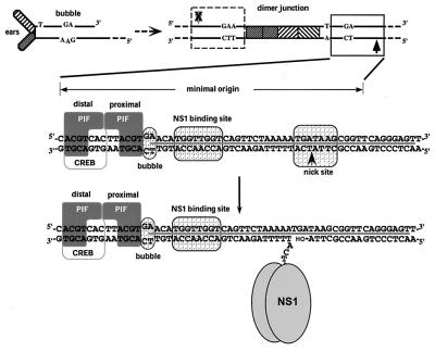
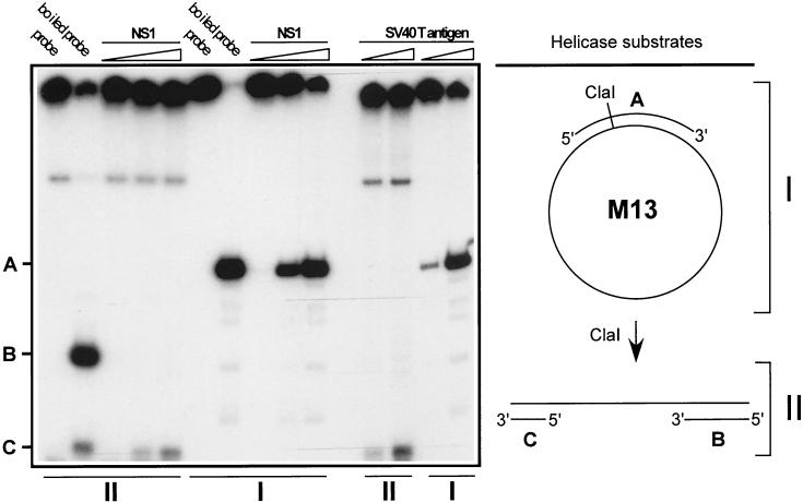
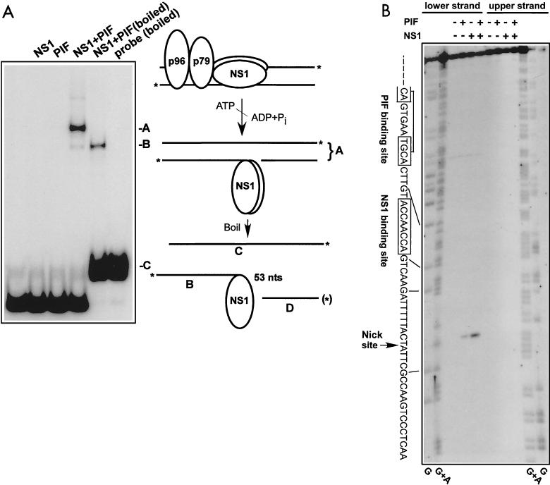
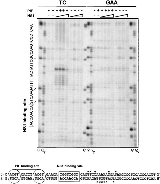
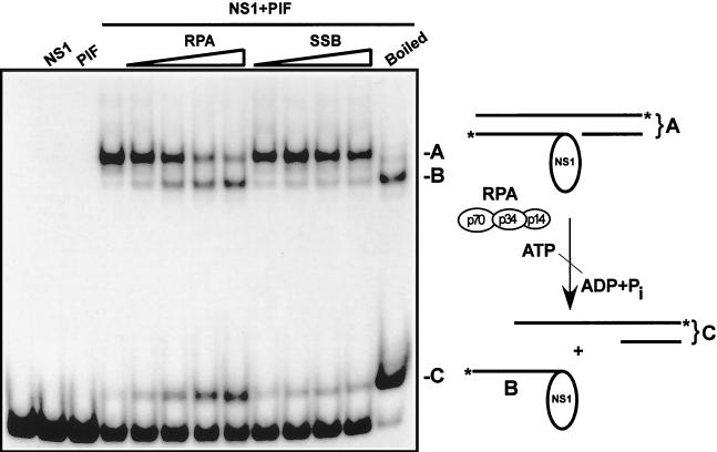
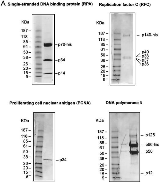
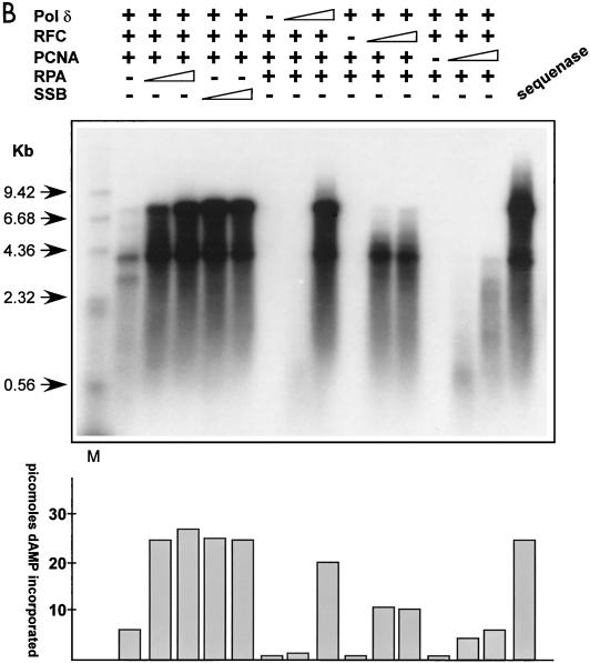
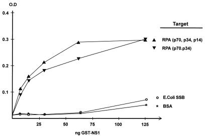
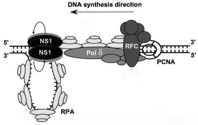
We show here that the DNA helicase activity of the parvoviral initiator protein NS1 is highly directional, binding to the single strand at a recessed 5' end and displacing the other strand while progressing in a 3'-to-5' direction on the bound strand. NS1 and a cellular site-specific DNA binding factor, PIF, also known as glucocorticoid modulating element binding protein, bind to the left-end minimal replication origin of minute virus of mice, forming a ternary complex. In this complex, NS1 is activated to nick one DNA strand, becoming covalently attached to the 5' end of the nick in the process and providing a 3' OH for priming DNA synthesis. In this situation, the helicase activity of NS1 did not displace the nicked strand, but the origin duplex was distorted by the NS1-PIF complex, as assayed by its sensitivity to KMnO(4) oxidation, and a stretch of about 14 nucleotides on both strands of the nicked origin underwent limited unwinding. Addition of Escherichia coli single-stranded DNA binding protein (SSB) did not lead to further unwinding. However, addition of recombinant human single-stranded DNA binding protein (RPA) to the initiation reaction catalyzed extensive unwinding of the nicked origin, suggesting that RPA may be required to form a functional replication fork. Accordingly, the unwinding mediated by NS1 and RPA promoted processive leading-strand synthesis catalyzed by recombinant human DNA polymerase delta, PCNA, and RFC, using the minimal left-end origin cloned in a plasmid as a template. The requirement for RPA, rather than SSB, in the unwinding reaction indicated that specific NS1-RPA protein interactions were formed. NS1 was tested by enzyme-linked immunosorbent assay for binding to two- or three-subunit RPA complexes expressed from recombinant baculoviruses. NS1 efficiently bound each of the baculovirus-expressed complexes, indicating that the small subunit of RPA is not involved in specific NS1 binding. No NS1 interactions were observed with E. coli SSB or other proteins included as controls.


Figures

FIG. 1.
FIG. 1.
Formation and organization of MVM oriL. In the upper left is the structure of the left-end hairpin (oriLH) showing the 3′ hydroxyl group used for priming replication and the mismatched bubble sequence as present in the parental single-stranded viral genome. In the upper right is the organization of the left-end hairpin sequences within the duplex dimer junction generated by replication through the hairpin. The hatched boxes represent the palindromic sequences that were originally folded to give the ears of the hairpin form. The boxed sequence (expanded below) represents the minimum active replication origin on the outboard arm, oriLTC, while the sequence in the dashed box represents the corresponding origin on the inboard arm, oriLGAA, which is inactive. The arrows denote the potential nick sites on each side of the junction, solid for active and X-ed out shaded for inactive. The middle diagram illustrates the sequence of oriLTC, showing the different elements involved in replication. The PIF binding site (see the text) overlaps a consensus binding site for the CREB/ATF family of host transcription factors. The two tetranucleotide motifs bound by PIF are labeled “distal” and “proximal” to indicate their positions relative to the NS1 binding site. The other boxes indicate sequences involved in the bubble dinucleotide (or trinucleotide) spacer element, the ACCA2 NS1 binding site, and the nick site, a specific sequence required for nicking and covalent attachment of NS1. The heavy line between the DNA strands indicates sequences protected by NS1 from DNase I digestion. The position of the nick reflects the new determination described in this report. The bottom diagram shows the nicking and covalent attachment of an oligomer of NS1 via a phosphotyrosine bond. The nicking reaction liberates a 3′ hydroxyl group, which primes DNA synthesis mediated by host cell DNA polymerases.
FIG. 2.
FIG. 2.
Determination of NS1 helicase directionality. The preparation and 3′-end labeling of helicase substrates were done as described in Materials and Methods. Lanes I represent the circular helicase substrate before ClaI digestion, diagrammed on the upper right, which serve as a positive control. Lanes II contained the ClaI-digested helicase substrate, diagrammed on the lower right, used to determine directionality. As indicated at the top of the gel, the different helicase substrates were incubated with increasing amounts of GST-NS1 (10, 30, or 90 ng) or purified SV40 large TAg (25 and 75 ng). Helicase assays were carried out as described in Materials and Methods. A, 341-base HaeIII fragment derived from double-stranded M13 DNA; B and C, linear substrate with 143- (C) and 202-bp (B) duplexes connected by a stretch of single-stranded M13 DNA in which each of the 3′ ends is labeled (directional substrate).
FIG. 3.
FIG. 3.
Characterization of the nick site in the minimal left-end origin. (A) A DNA fragment containing the oriLTC sequence was 32P labeled at its 3′ ends as described in Materials and Methods; incubated in the absence or presence of NS1 (100 ng), PIF (25 ng), and ATP (3 mM); and analyzed on a neutral SDS-polyacrylamide gel. Only covalent NS1-DNA complexes are significantly retarded in this gel system. For lane NS1+PIF (boiled), the nicking reaction mixture was denatured by boiling immediately before electrophoresis. A schematic model of the nicking reaction is depicted to the right of the gel autoradiograph. The asterisks indicate the positions of the 32P label at the 3′ end of each strand of the origin DNA. The letters A, B, and C correlate the DNA or DNA-protein structures produced in the reaction with bands on the gel. The fragment D labeled with an asterisk in parentheses indicates the 5′-end-labeled fragment analyzed in panel B. (B) DNA fragments containing the oriLTC sequence, separately labeled at the 5′ end of either the upper or lower strand (as depicted in panel A), were incubated with 3 mM ATP in the presence (+) or absence (−) of NS1 (100 ng) and PIF (25 ng) as indicated at the top of the gel. The samples were digested with proteinase K before analysis on a 6% denaturing polyacrylamide gel. Lanes G and G+A contain the products of G- and GA-specific chemical sequencing reactions of each labeled substrate and were used for aligning the position of the nick site. The sequence of the lower strand, depicted in panel A, is shown on the left side of the gel, and the lines indicate the position of this sequence in relation to the chemical sequencing reaction. An arrow indicates the putative nick site, representing the position of migration of fragment D in panel A. The boxed sequences show the positions of the specific NS1 and PIF binding sites.
FIG. 4.
FIG. 4.
PIF and NS1 nicking of the minimal left-end origin causes local unwinding. DNA fragments containing either the oriLTC or oriLGAA sequence were 32P labeled at the 3′ end of the lower strand (Fig. 1) and incubated with 3 mM ATP in the presence (+) or absence (−) of NS1 (increasing amounts of purified GST-tagged NS1 [25, 50, 100, and 225 ng]) and PIF (50 ng of purified PIF p79-p96 complex) as indicated at the top of the gel. The samples were exposed to KMnO4 and subjected to alkaline hydrolysis before analysis on a 6% denaturing sequencing polyacrylamide gel. TC and GAA indicate the form of oriL that was used as a substrate in the left and right sets of KMnO4 assays, respectively. Lanes G and GA contain the products of G- and GA-specific chemical sequencing reactions of each labeled substrate and were used to align the positions of KMnO4-sensitive residues. The sequence of the relevant strand of the origin is shown on the left, and the lines indicate the alignment of this sequence to the chemical sequencing reaction. The boxed sequence shows the position of the specific NS1 binding site. At the bottom is a summary of the positions of KMnO4-sensitive thymidine residues (labeled with asterisks) detected in both strands of the origin.
FIG. 5.
FIG. 5.
RPA catalyzes unwinding of the nicked minimal left-end origin. A DNA fragment containing the oriLTC sequence, 32P labeled at its 3′ ends, was incubated with 3 mM ATP in the presence or absence of NS1 (100 ng) and PIF (25 ng) and increasing amounts of either recombinant RPA (0.125, 0.25, 0.5, and 1 μg) or E. coli SSB (0.125, 0.25, 0.5, and 1 μg), as indicated at the top of the gel. For lane NS1+PIF (boiled), the nicking reaction mixture was boiled immediately before electrophoresis in an SDS-polyacrylamide gel. Note that only covalent NS1-DNA complexes are significantly retarded in this assay. A schematic model of the nicking-coupled unwinding reaction is depicted at the right of the gel autoradiograph. The asterisks indicate the position of the 32P label on each strand of the origin DNA.
FIG. 6.
FIG. 6.
Recombinant RPA/SSB, RFC, PCNA, and Pol δ replicate primed M13 DNA. (A) SDS-PAGE analyses of the purified human recombinant RPA, RFC, PCNA, and Pol δ preparations used in replication and protein-protein interaction assays (see Materials and Methods for details). The positions of the relevant purified proteins or subunits are indicated on the right of each gel, and the positions of a standard set of molecular mass markers run in the first lane of each gel are shown on the left. The gels were stained with Coomassie brilliant blue. (B) Autoradiograph of an M13 singly primed extension assay analyzed by alkaline agarose gel electrophoresis. Singly primed M13 DNA (7.3 kb) was incubated with deoxynucleoside triphosphates, including [32P]dATP and ATP, in the presence (+) or absence (−) of titrated amounts of purified recombinant RFC, PCNA, Pol δ, and RPA or SSB. For reactions including RPA or E. coli SSB, the amounts added were 0.5 or 1.5 μg, respectively, and elsewhere + represents addition of 1.5 μg of either protein. RFC titrations were 5 and 25 ng, and elsewhere + represents 125 ng of protein added. PCNA titrations contained 10 or 25 ng, and elsewhere + indicates 100 ng of protein added. For Pol δ titrations, 5 or 25 ng was added to the assay, and elsewhere + represents 125 ng of protein added. The first lane contains a 32P-labeled, HindIII-digested λ phage DNA marker (M), while the arrows on the left indicate the sizes of individual fragments. The amount of dAMP (in picomoles) incorporated into each replication product is indicated in the histogram at the bottom.
FIG. 6.
FIG. 6.
Recombinant RPA/SSB, RFC, PCNA, and Pol δ replicate primed M13 DNA. (A) SDS-PAGE analyses of the purified human recombinant RPA, RFC, PCNA, and Pol δ preparations used in replication and protein-protein interaction assays (see Materials and Methods for details). The positions of the relevant purified proteins or subunits are indicated on the right of each gel, and the positions of a standard set of molecular mass markers run in the first lane of each gel are shown on the left. The gels were stained with Coomassie brilliant blue. (B) Autoradiograph of an M13 singly primed extension assay analyzed by alkaline agarose gel electrophoresis. Singly primed M13 DNA (7.3 kb) was incubated with deoxynucleoside triphosphates, including [32P]dATP and ATP, in the presence (+) or absence (−) of titrated amounts of purified recombinant RFC, PCNA, Pol δ, and RPA or SSB. For reactions including RPA or E. coli SSB, the amounts added were 0.5 or 1.5 μg, respectively, and elsewhere + represents addition of 1.5 μg of either protein. RFC titrations were 5 and 25 ng, and elsewhere + represents 125 ng of protein added. PCNA titrations contained 10 or 25 ng, and elsewhere + indicates 100 ng of protein added. For Pol δ titrations, 5 or 25 ng was added to the assay, and elsewhere + represents 125 ng of protein added. The first lane contains a 32P-labeled, HindIII-digested λ phage DNA marker (M), while the arrows on the left indicate the sizes of individual fragments. The amount of dAMP (in picomoles) incorporated into each replication product is indicated in the histogram at the bottom.
FIG. 7.
FIG. 7.
Reconstitution of replication initiated from the minimal left-end origin. Replication reaction mixtures were assembled using combinations of purified recombinant His-tagged NS1, PIF p79-p96 complex, RPA or E. coli SSB, RFC, PCNA Pol δ, or previously described phosphocellulose fractions of 293 cell extracts as positive controls (10) as indicated above the gel. Plasmid pL1-2 TC containing the minimal active left-end origin was the replication-positive template, and pL1-2 GAA was used as a negative control. The different purified proteins were titrated to achieve optimal replication. For PIF, RFC, PCNA, and Pol δ, constant amounts of 50, 125, 100, and 100 ng, respectively, were used in the assay. The increasing amounts of NS1, as indicated at the top of the gel, were 90 and 270 ng. RPA or E. coli SSB was added (+) in 0.5- or 1.5-μg amounts. Replicated DNA was digested sequentially with proteinase K and HindIII as described in Materials and Methods and then analyzed by agarose gel electrophoresis, along with the 32P-labeled λ phage DNA markers described in the legend to Fig. 6B. The amount of dAMP (in picomoles) incorporated into each replication product is indicated in the histogram at the bottom of the autoradiograph. A schematic model of the replication reaction is at the right.
FIG. 8.
FIG. 8.
Binding of NS1 to purified RPA. Purified RPA complex comprising three (p70, p34, p14) or two (p70, p34) subunits or E. coli SSB was immobilized on ELISA wells and incubated with increasing amounts of purified GST-NS1. The different target proteins are indicated at the right. In each ELISA, bound NS1 was detected by a rabbit polyclonal antiserum directed against its carboxyl terminus, followed by horseradish peroxidase-coupled anti-rabbit immunoglobulin G antibody and a chromogenic substrate. ELISA wells coated with BSA served as negative controls. Inclusion of DNase I or micrococcal nuclease in these assays did not quantitatively or qualitatively affect the observed interactions. O.D., optical density.
FIG. 9.
FIG. 9.
Model of a minimal parvovirus replication fork. The model is based partly on the data described in this report and partly on the body of published data describing the interactions of RPA, RFC, PCNA, and Pol δ (6, 23, 24, 45, 46, 54). See the text for additional description.

References

    1. Astell, C. R., M. B. Chow, and D. C. Ward. 1985. Sequence analysis of the termini of virion and replicative forms of minute virus of mice DNA suggests a modified rolling hairpin model for autonomous parvovirus DNA replication. J. Virol. 54:171-177. - PMC - PubMed
    1. Astell, C. R., Q. Liu, C. E. Harris, J. Brunstein, H. K. Jindal, and P. Tam. 1996. Minute virus of mice cis-acting sequences required for genome replication and the role of the trans-acting viral protein, NS-1. Prog. Nucleic Acid Res. Mol. Biol. 55:245-285. - PubMed
    1. Baldauf, A. Q., K. Willwand, E. Mumtsidu, J. P. Nuesch, and J. Rommelaere. 1997. Specific initiation of replication at the right-end telomere of the closed species of minute virus of mice replicative-form DNA. J. Virol. 71:971-980. - PMC - PubMed
    1. Bashir, T., R. Horlein, J. Rommelaere, and K. Willwand. 2000. Cyclin A activates the DNA polymerase delta-dependent elongation machinery in vitro: a parvovirus DNA replication model. Proc. Natl. Acad. Sci. USA 97:5522-5527. - PMC - PubMed
    1. Borowiec, J. A., F. B. Dean, P. A. Bullock, and J. Hurwitz. 1990. Binding and unwinding—how T antigen engages the SV40 origin of DNA replication. Cell J. 60:181-184. - PubMed

Publication types

MeSH terms

LinkOut - more resources