. 2002 Jul;76(13):6618-35.
doi: 10.1128/jvi.76.13.6618-6635.2002.

Critical role for glial cells in the propagation and spread of lymphocytic choriomeningitis virus in the developing rat brain

Affiliations

Critical role for glial cells in the propagation and spread of lymphocytic choriomeningitis virus in the developing rat brain

Daniel J Bonthius et al. J Virol. 2002 Jul.

Abstract

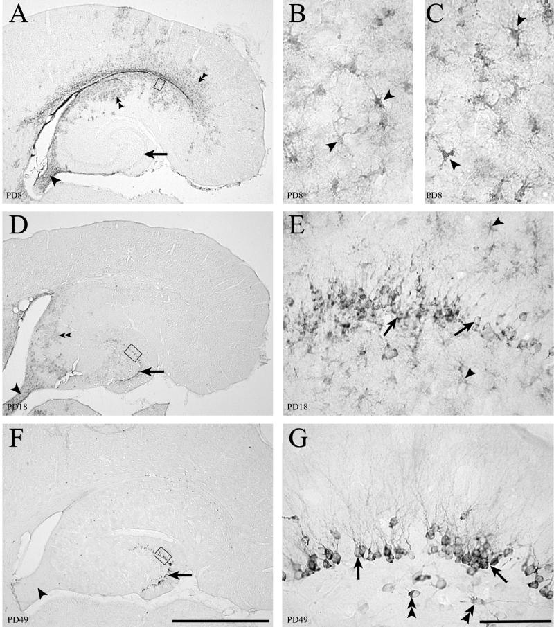
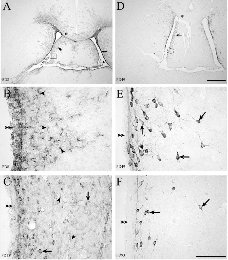
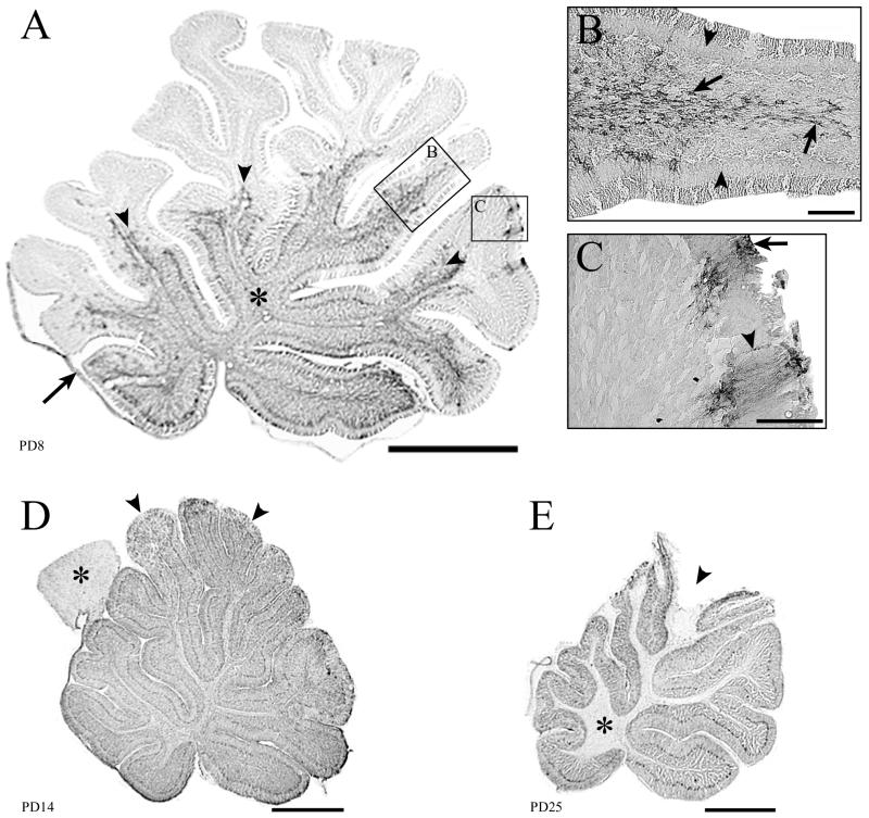
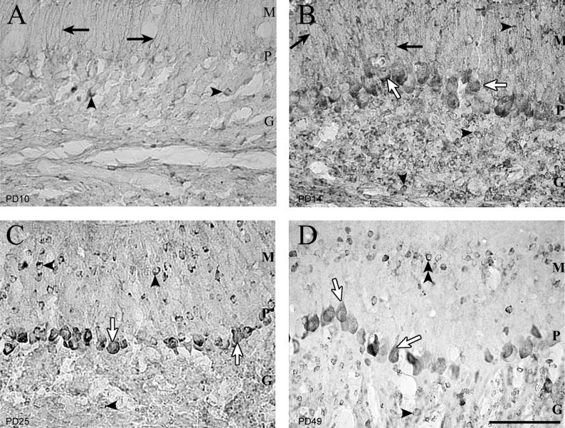
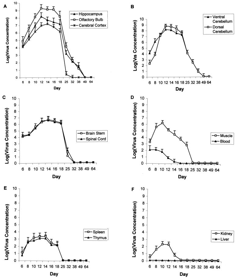
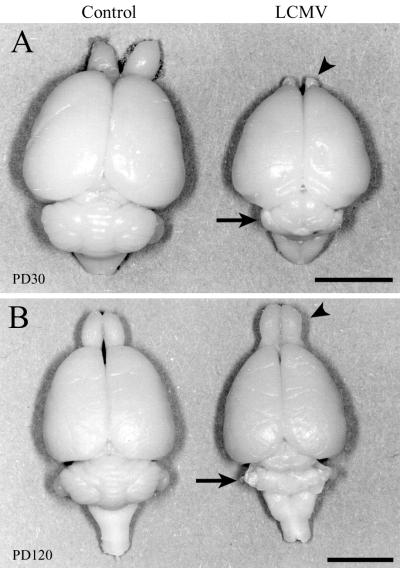
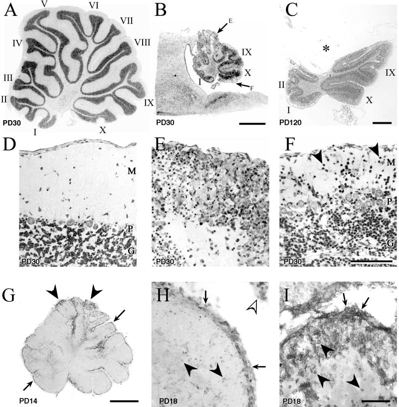
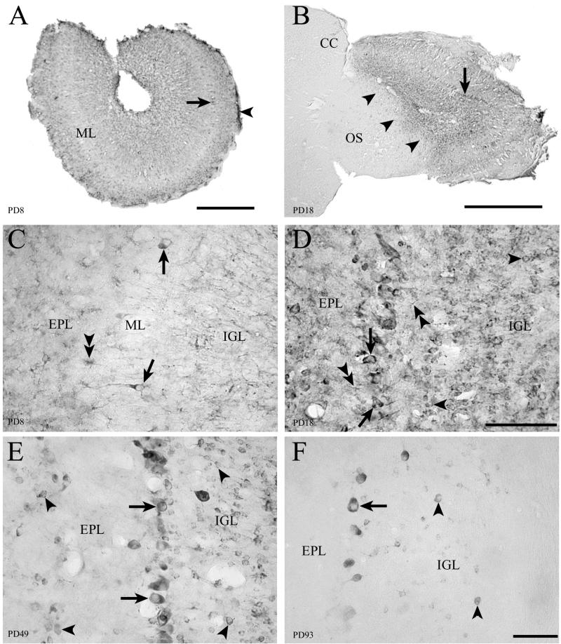
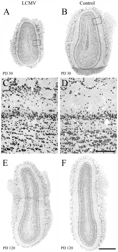
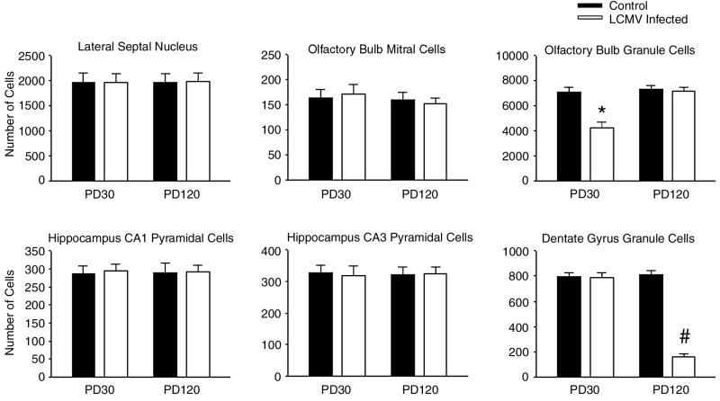
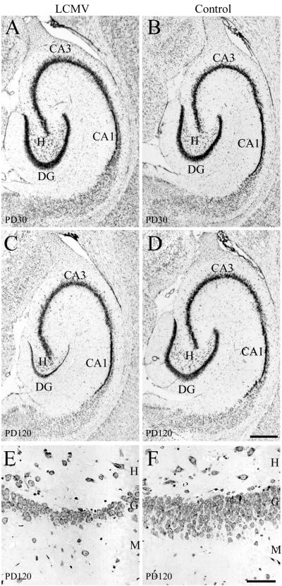
Inoculation of the neonatal rat with lymphocytic choriomeningitis virus (LCMV) results in the selective infection of several neuronal populations and in focal pathological changes. However, the pathway by which LCMV reaches the susceptible neurons has not been described, and the nature and time course of the pathological changes induced by the infection are largely unknown. This study examined the sequential migration of LCMV in the developing rat brain and compared the pathological changes among infected brain regions. The results demonstrate that astrocytes and Bergmann glia cells are the first cells of the brain parenchyma infected with LCMV and that the virus spreads across the brain principally via contiguous glial cells. The virus then spreads from glial cells into neurons. However, not all neurons are susceptible to infection. LCMV infects neurons in only four specific brain regions: the cerebellum, olfactory bulb, dentate gyrus, and periventricular region. The virus is then cleared from glial cells but persists in neurons. LCMV induces markedly different pathological changes in each of the four infected regions. The cerebellum undergoes an acute and permanent destruction, while the olfactory bulb is acutely hypoplastic but recovers fully with age. Neurons of the dentate gyrus are unaffected in the acute phase but undergo a delayed-onset mortality. In contrast, the periventricular region has neither acute nor late-onset cell loss. Thus, LCMV infects four specific brain regions in the developing brain by spreading from glial cells to neurons and then induces substantially different pathological changes with diverse time courses in each of the four infected regions.


Figures

FIG. 1.
FIG. 1.
Fifty-micrometer-thick sections through the hippocampal formation immunohistochemically stained for LCMV (panels A, B, D, E, F, and G) or GFAP (panel C). LCMV infection in the hippocampus initially involves astrocytes. The granule cells of the dentate gyrus become chronically infected. (A) Low-power image of hippocampal region at PD8 shows infection within the ependyma, parenchymal cells adjacent to the ependyma (double arrowheads), and fornix (arrowhead). The dentate gyrus (arrow) is not labeled. (B) Area beneath the ependyma, represented by the box in panel A, shows that the infected cells have the morphology of astrocytes (arrowheads). (C) A section adjacent to panel B immunohistochemically labeled for GFAP, a marker for astrocytes. The GFAP-positive cells in panel C have the same morphology as the LCMV-infected cells in panel B, indicating that the infected parenchymal cells on PD8 are astrocytes. (D) By PD18, infection in the hippocampal region is less intense but still present beneath the ependyma (double arrowheads) and in the fornix (arrowhead). The dentate gyrus stratum granulosum (arrow) now shows evidence of infection. (E) Higher magnification of box in panel D shows that granule cells of the dentate gyrus (arrows), and nearby astrocytes (arrowheads) are both infected with LCMV on PD18. (F) By PD49, infection has been cleared from the hippocampal region, including the fornix (arrowhead), except for granule cells of the dentate gyrus (arrow), where viral antigen persists. (G) Higher magnification of the box in panel F demonstrates that cell bodies and dendrites of dentate granule cells (arrows) and a few hilar neurons (double arrowheads) are still infected. The virus has been cleared from glial cells. Bars, 1 mm (A, D, and F); 20 μm (B and C); 100 μm (E and G).
FIG. 2.
FIG. 2.
Fifty-micrometer-thick sections through the septum immunohistochemically stained for LCMV. LCMV infection in the periventricular region of rat forebrain spreads from the ependyma and astrocytes to neurons. (A) On PD8 (4 days postinoculation), infected areas include the choroid plexus (arrowhead), ependymal lining of the ventricle (arrow), and cells of the septal region (double arrowheads) and corpus callosum (∗). (D) With the passage of time, infection is cleared from the corpus callosum (∗) and deeper portions of the septal region. Infection persists in cells adjacent to the ventricle (arrow). (B) High magnification of boxed area in panel A shows infection of the ependymal lining of the ventricle (double arrowheads) and of cells beneath the ependyma (arrowheads) whose morphology is consistent with astrocytes. (C) On PD18, infection of the ependymal cells (double arrowheads) and astrocytes (arrowheads) is less evident. However, LCMV is now apparent in neuronal cell bodies (arrows). (E) High magnification of the boxed area in panel D shows that the infected cells are no longer astrocytes but are neurons (arrows). The ependymal lining (double arrowheads) no longer shows any evidence of infection. (F) On PD93, viral antigen remains detectable in the cell bodies and processes of many neurons (arrows) near the ependymal wall (double arrowheads). Bars, 1 mm (A and D); 100 μm (B, C, E, and F).
FIG. 3.
FIG. 3.
Fifty-micrometer-thick midsagittal sections through the cerebellum immunohistochemically stained for LCMV. Infection of the cerebellum can spread via astrocytes in the white matter or via the leptomeninges and Bergmann glia. Infection is cleared from glia but persists in cortical neurons. (A) On PD8, the proximal white matter tracts (∗) and leptomeninges (arrow) are infected. The arrowheads point to the leading edge of infection, which is spreading from the proximal to the distal portions of the lobules. Boxed areas (B and C) demonstrate the two pathways by which the infection spreads through the cerebellum. (B) Spread of infection via astrocytes within a lobule. Note that the cortex is uninfected (arrowheads), but the white matter astrocytes (arrows) are heavily labeled. (C) Infection spreading into the cerebellum from the leptomeninges (arrow) and Bergmann glia (arrowhead). (D) By PD14, the entire cerebellum is heavily infected, including white and gray matter. In contrast to the cerebellum, the tectal plate (∗) of the brain stem has scattered infection of astrocytes only and is thus less intensely labeled. Early pathological changes are visible in the tips of the dorsal lobules (arrowheads), where tissue breakdown is beginning to occur. (E) By PD25, substantial pathology is evident in the dorsal lobules (arrowhead). Infection has been cleared from the white matter (∗) but persists in cortical neurons. Bars, 1 mm (A, D, and E); 100 μm (B and C).
FIG. 4.
FIG. 4.
Fifty-micrometer-thick sections of cerebellar cortex immunohistochemically stained for LCMV. The first cells in the cerebellar cortex infected with LCMV are the Bergmann glia. The virus persists in granule cells and Purkinje cells. (A) On PD10, the only infected cortical cells are the Bergmann glia (arrows) and occasional granule cells (arrowheads). Purkinje cell bodies are not yet infected. (B) Four days later, Purkinje cell bodies (open arrows), granule cells (arrowheads), and Bergmann glia (arrows) are infected. (C) By PD25, the Bergmann glia are no longer infected. Purkinje cell bodies (open arrows) and granule cells (arrowheads) remain infected. (D) On PD49, Purkinje cell bodies and proximal apical dendrites (open arrows) are infected. Granule cells, which have failed to migrate to the granular layer (double arrowheads), remain infected in the molecular layer. Granule cells within the internal granule cell layer (arrowheads) are also persistently infected. M, molecular layer; P, Purkinje cell layer; G, internal granule cell layer. Bar, 100 μm.
FIG. 5.
FIG. 5.
Log of viral concentrations (per gram of tissue) versus postnatal age following LCMV inoculation on PD4. (A) The olfactory bulb has the heaviest viral burden among all tissues. Viral load is lower in cerebral cortex than in hippocampus or olfactory bulb. There is a large reduction in viral load between PD18 and PD25 in all three brain regions. However, in the cerebral cortex, virus titers fall to near zero on PD25, while virus titers remain substantially greater than zero in the hippocampus and olfactory bulb at this age. Immunohistochemistry demonstrates that reduction in virus titer between PD18 and PD25 reflects clearance of virus from glial cells. The continued presence of infectious virus in the olfactory bulb and hippocampus on PD25 and beyond reflects the persistent infection of neurons. (B) During the initial days following inoculation, virus titers are higher in ventral cerebellum than in dorsal cerebellum. This reflects the ventral-to-dorsal spread of LCMV through the cerebellar lobules. At the peak of infection, the dorsal cerebellum has a higher virus titer than the ventral cerebellum. (C) LCMV infects brain stem and spinal cord but at lower levels than in hippocampus, olfactory bulb, or cerebellum. The nearly total clearance of virus from these tissues by PD25 reflects the fact that only glial cells, not neurons, are infected in these tissues. (D) Following intracerebral inoculation of LCMV, there is a transient low-level viremia. Muscle is more heavily infected than any other extracerebral tissue. (E) Spleen and thymus both contain infectious LCMV particles, though the concentrations do not approach those found in the brain. (F) Low levels of LCMV infection transiently occur in the kidney, but infectious LCMV is never isolated in any quantity from the liver.
FIG. 6.
FIG. 6.
Dorsal views of control and LCMV-infected rat brains one month (A) and 4 months (B) postinoculation. The effect of LCMV infection on brain growth is region and age dependent. LCMV infection induces a temporary hypoplasia of the olfactory bulbs and a permanent destruction of the cerebellum. (A) On PD30, a substantial microencephaly induced by LCMV infection is evident. While brain growth is globally disrupted, the cerebellum (arrow) and olfactory bulbs (arrowhead) are particularly affected. (B) By PD120, the previously hypoplastic olfactory bulb has rebounded to a normal size (arrowhead), but the cerebellum remains permanently and severely affected (arrow). Bars, 7 mm (A); 7.5 mm (B).
FIG. 7.
FIG. 7.
Midsagittal sections through the cerebellar vermis of control (A and D) and LCMV-infected (B, C, and E to I) littermate rats, demonstrating the acute and long-term pathological changes induced by LCMV infection. The ages of the animals are noted on each panel. Panels A to F are Nissl-stained 2-μm-thick sections, while panels G to I are 50-μm-thick sections stained immunohistochemically for CD8a antigen. Within the cerebellum, LCMV induces a destructive process that is region dependent and permanent and that involves the infiltration of CD8+ lymphocytes. LCMV infection destroys the cytoarchitecture of some lobules and induces a migrational defect in others. (A) The 10 lobules of the control (uninfected) cerebellum are labeled according to the system of Larsell (22). (B) Midsagittal section through the cerebellum of an LCMV-infected rat on PD30, photographed at the same magnification as the control cerebellum in panel A. The severe reduction in the size of the cerebellum induced by LCMV is obvious. Lobules I, IX, and X, which are among the ventral lobules, contain less severe pathological change than do the dorsal lobules. (C) LCMV-infected cerebellum 4 months postinoculation. The dorsal lobules have been obliterated and are now absent. The asterisk (*) denotes the position where the dorsal lobules would normally reside. In contrast, the ventral lobules (I, II, IX, and X) are relatively intact. (D) Control cerebellar cortex demonstrating the normal trilaminar architecture of the cortex. (E) Enlargement of boxed area E from panel B demonstrating the pathological changes within the cerebellar cortex of a dorsal lobule. The cortical architecture has been completely disrupted. No Purkinje cells can be identified, and the molecular layer has been completely destroyed. (F) Enlargement of boxed area F from panel B. In the ventral lobules, all layers of the cortex can be clearly identified and contain many healthy-appearing cells. However, note the abnormal continued presence of granule cells in the molecular layer (arrowheads), indicating a migrational defect. (G) LCMV-infected cerebellum on PD14 immunohistochemically stained for CD8+ cells. Labeling of CD8+ cells is evident along the entire extent of the ependymal and meningeal surfaces of the cerebellum (arrows). In addition, CD8+ cells are prominently present within cerebellar parenchyma at the tips of the dorsal lobules (arrowheads). (H) Lobule I (a ventral lobule) on PD18 has a dense labeling of CD8+ cells over the meningeal surface of the lobule (arrows) and within the choroid plexus (open arrowhead) but relatively few labeled cells within the cerebellar parenchyma (arrowheads). (I) In contrast, a dorsal lobule from the same cerebellum as in panel H has a dense infiltration of CD8+ cells both on the meningeal surface (arrows) and within the parenchyma (arrowheads) of the lobule. M, molecular layer; P, Purkinje cell layer; G, granule cell layer. Bars, 1 mm (A, B, C, and G); 100 μm (D, E, F, H, and I).
FIG. 8.
FIG. 8.
Fifty-micrometer-thick sections through the olfactory bulb immunohistochemically labeled for LCMV. In the olfactory bulb, infection of LCMV spreads from astrocytes to neurons. Mitral cells are the first neuronal population of the brain to become infected. (A) Low-power image on PD8 shows infection of the leptomeninges (arrowhead) and mitral cell bodies (arrow) as well as parenchyma deep to the mitral cell layer (ML). (B) Arrowheads show a clear demarcation of infection in this parasagittal section of the olfactory bulb on PD18. The olfactory bulb is selectively and heavily infected, while the adjacent olfactory stalk (OS) and cerebral cortex (CC) are spared. Mitral cell bodies are clearly infected (arrow). (C) On PD8, infection principally involves glial cells (double arrowheads). However, a few mitral cells (arrows) are also infected. Mitral cells are the first neuronal population of the brain infected with LCMV. (D) On PD18, glial cells remain infected (double arrowheads). However, the infection has spread beyond glial cells and into large numbers of neurons. Granule cells are now clearly infected (arrowheads), and the proportion of mitral cells (arrows) that are infected has increased. (E) By PD49, the infection has been cleared from glia. However, granule cells (arrowheads) and mitral cells (arrows) remain heavily infected. (F) By adulthood, mitral cells remain heavily infected (arrow) and viral antigen persists in some granule cells (arrowheads). CC, cerebral cortex; EPL, external plexiform layer; IGL, internal granule cell layer; ML, mitral cell layer; OS, olfactory stalk. Bars, 300 μm (A); 1 mm (B); 100 μm (C and D); 80 μm (E and F).
FIG. 9.
FIG. 9.
Two-micrometer-thick Nissl-stained coronal sections through the olfactory bulb of control and LCMV-infected rats at 1 month and 4 months postinoculation. In the olfactory bulb, LCMV infection induces a temporary hypoplasia. At the 1-month time point, the olfactory bulb of the LCMV-infected rat (A) is hypoplastic, relative to that of the control rat (B). C and D are enlargements of the boxed areas from panels A and B, respectively. The cytoarchitecture of the infected olfactory bulb (C) does not differ from that of the uninfected olfactory bulb (D). By 4 months postinoculation, the previously infected olfactory bulb (E) has recovered and does not differ substantially in size from the control olfactory bulb (F). Thus, unlike the cerebellum, the olfactory bulb infected with LCMV does not undergo an acute destructive process. Bars, 500 μm (A, B, E, and F); 100 μm (C and D).
FIG. 10.
FIG. 10.
LCMV infection induces neuronal deficits that are region and age dependent. Cell counts were made from single 2-μm-thick sections through the septum, olfactory bulb, and hippocampal formation. Six neuronal populations within these three regions were quantified in LCMV-infected and control rats on PD30 and on PD120. Within the olfactory bulb, only the granule cells were substantially reduced in number, and the cellular deficits were temporary. By PD120, the number of olfactory granule cells in LCMV-infected animals did not differ from that in controls. In contrast, in the hippocampal formation, the number of dentate granule cells in LCMV-infected animals was normal on PD30 but was significantly reduced by PD120. In further contrast, the number of olfactory mitral cells, hippocampal CA1 and CA3 pyramidal cells, and neurons of the lateral septal nucleus was never significantly affected by LCMV infection. ∗, significantly different from controls at PD30 and from controls and LCMV-infected animals at PD120 (P < 0.0001); #, significantly different from controls and LCMV-infected animals at PD30 and from controls at PD120 (P < 0.0001).
FIG. 11.
FIG. 11.
LCMV infection induces a delayed-onset selective dropout of dentate granule cells. Panels A to D are Nissl-stained 50-μm-thick sections cut in the horizontal plane through the hippocampal formation at the midtemporal level of control and LCMV-infected rats 1 month and 4 months postinoculation. At the 1-month time point, the hippocampal formation of infected rats (A) does not appear histologically different from that of controls (B). A full complement of pyramidal and granule cells is present despite a selective and heavy infection of the granule cells. However, at 4 months postinoculation (C), the granule cell layer of the dentate gyrus is markedly reduced in width relative to the 4-month-old control (D). The LCMV-induced cell loss is selective to the dentate gyrus, as the CA1 and CA3 pyramidal cell populations are unaffected. Panels E and F are 2-μm-thick cresyl violet-stained horizontal sections through the dentate gyrus from LCMV-infected (E) and control (F) rats 4 months postinoculation. In the LCMV-infected rats, the substantially reduced width of the granule cell layer is due to a massive loss of granule cells. Whereas the granule cell layer of the dentate gyrus is normally 10 to 12 cells thick (F), in theLCMV-infected animal, the dentate gyrus has been reduced to 1 to 3 cells in thickness (E). M, molecular layer; G, granule cell layer; H, hilus; CA1, CA1 pyramidal cells; CA3, CA3 pyramidal cells; DG, dentate gyrus granule cells. Bars, 500 μm (A to D); 50 μm (E and F).

References

    1. Altman, J. 1969. Autoradiographic and histological studies of postnatal neurogenesis. III. Dating the time of production and onset of differentiation of cerebellar microneurons in rats. J. Comp. Neurol. 136:269-294. - PubMed
    1. Altman, J. 1972. Postnatal development of the cerebellar cortex in the rat. II. Phases in the maturation of Purkinje cells and of the molecular layer. J. Comp. Neurol. 145:399-464. - PubMed
    1. Altman, J., and S. A. Bayer. 1997. Development of the cerebellar system, p. 498-523. CRC Press, New York, N.Y.
    1. Baldridge, J. R., B. D. Pearce, B. S. Parekh, and M. J. Buchmeier. 1993. Teratogenic effects of neonatal arenavirus infection on the developing rat cerebellum are abrogated by passive immunotherapy. J. Virol. 197:669-677. - PubMed
    1. Barton, L. L., S. C. Budd, W. S. Morfitt, C. J. Peters, T. G. Ksiazek, R. F. Schindler, and M. T. Yoshino. 1993. Congenital lymphocytic choriomeningitis virus infection in twins. Pediatr. Infect. Dis. J. 12:942-946. - PubMed

Publication types

MeSH terms

LinkOut - more resources