. 2002 Jul;76(13):6815-24.
doi: 10.1128/jvi.76.13.6815-6824.2002.

P0 of beet Western yellows virus is a suppressor of posttranscriptional gene silencing

Affiliations

P0 of beet Western yellows virus is a suppressor of posttranscriptional gene silencing

S Pfeffer et al. J Virol. 2002 Jul.

Retraction in

Abstract

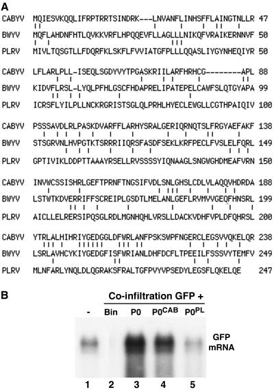
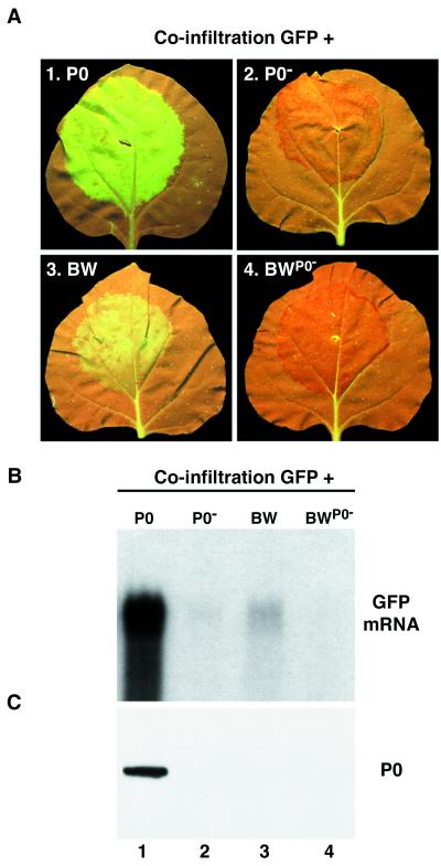
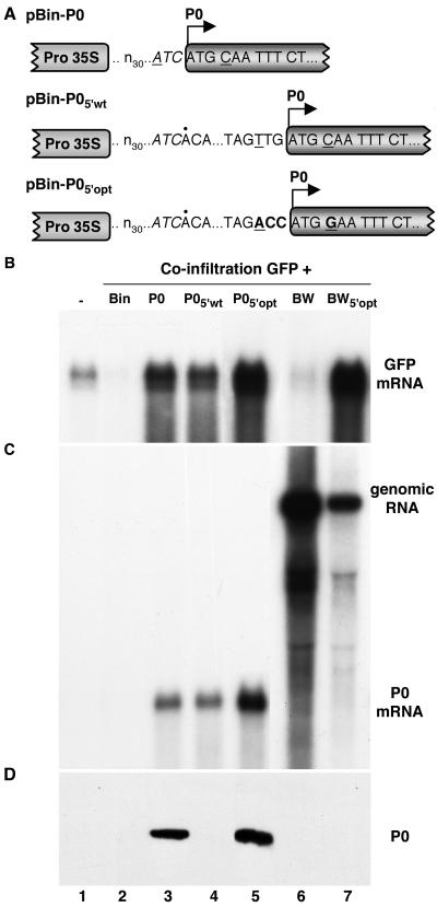
Higher plants employ a homology-dependent RNA-degradation system known as posttranscriptional gene silencing (PTGS) as a defense against virus infection. Several plant viruses are known to encode proteins that can suppress PTGS. Here we show that P0 of beet western yellows virus (BWYV) displays strong silencing suppressor activity in a transient expression assay based upon its ability to inhibit PTGS of green fluorescent protein (GFP) when expressed in agro-infiltrated leaves of Nicotiana benthamiana containing a GFP transgene. PTGS suppressor activity was also observed for the P0s of two other poleroviruses, cucurbit aphid-borne yellows virus and potato leafroll virus. P0 is encoded by the 5'-proximal gene in BWYV RNA but does not accumulate to detectable levels when expressed from the genome-length RNA during infection. The low accumulation of P0 and the resulting low PTGS suppressor activity are in part a consequence of the suboptimal translation initiation context of the P0 start codon in viral RNA, although other factors, probably related to the viral replication process, also play a role. A mutation to optimize the P0 translation initiation efficiency in BWYV RNA was not stable during virus multiplication in planta. Instead, the P0 initiation codon in the progeny was frequently replaced by a less efficient initiation codon such as ACG, GTG, or ATA, indicating that there is selection against overexpression of P0 from the viral genome.


Figures

FIG. 1.
FIG. 1.
Schematic representation of the BWYV genome and of some of the clones used in the study. (A) Genome organization of BWYV RNA. Numbered rectangles represent ORFs. The ORF for P0 is shaded. The diagonal arrow corresponds to the approximate position of translation frameshift to synthesize the P1-2 fusion protein mentioned in the text. The subgenomic RNA (sgRNA) which is used for translation of 3′-proximal genes is represented by a horizontal arrow below the map. (B) Structure of PVX-P0 chimera RNA. The white rectangles (not to scale) represent PVX genes, and the shaded rectangle represents the BWYV P0 gene. The black vertical lines correspond to the duplicated PVX subgenomic RNA promoter sequences, and horizontal arrows below indicate major subgenomic RNAs. The arrow above the P0 gene indicates the position of the frameshift (fs) mutation in PVX-P0fs. (C) Structures of the agro-infection vectors used for expression of different proteins in plants. Only the transcription cassette of each vector is shown with the cauliflower mosaic virus 35S transcription promoter and termination sequences indicated.
FIG. 2.
FIG. 2.
A PVX-P0 chimera is hypervirulent on N. benthamiana. (A) N. benthamiana plants which have been mock inoculated (panel 1) or inoculated with wild-type PVX transcript (panel 2), PVX-P0 (panel 3), or PVX-P0fs (panel 4). In panel 3, the arrow indicates an inoculated leaf and the arrowhead indicates an upper leaf with severe systemic necrosis. Photographs were taken 15 days postinfection. (B) Northern (RNA) blot analysis of viral RNA extracted from upper leaves of the plants shown in panel A and probed with either a PVX RNA-specific probe (lanes 1 to 4) or a probe specific for the BWYV P0 coding region (lanes 5 to 8). The upper band in each lane corresponds to genomic RNA (g). The lower bands in lanes 2 to 4 are the 3′-proximal subgenomic RNAs sg1, sg2, and sg3 (see Fig. 1B). The extra sg1 band in lane 4 compared to lane 3 indicates that a short deletion occurred in a fraction of the PVX-P0fs progeny RNA in this experiment.
FIG. 3.
FIG. 3.
Expression of BWYV P0 in N. benthamiana line 16c suppresses PTGS of the GFP transgene. (A) Leaves of line 16c plants agro-infiltrated with bacteria mixtures harboring pBin-GFP plus one of the following: empty vector pBin61 (panel 1), pBin-HCPro (panel 2), pBin-P0 (panel 3), or pBin-P0 (panel 4). Photographs were taken with long-wavelength UV light 5 days p.i. (B) Northern (RNA) blot analysis of high-molecular-weight RNA (upper panel) and low-molecular-weight RNA (lower panel) extracted from the agro-infiltrated zone (patch) of 16c leaves such as those shown in panel A. The patches had received bacterial mixtures harboring pBin-GFP plus one of the following: empty vector pBin61 (lane 2), pBin-HCPro (lane 3), pBin-P0 (lane 4), or pBin-P0 (lane 5). The RNA loaded in lane 1 came from a noninfiltrated 16c leaf. The blots were hybridized with a probe specific for GFP mRNA. In the lower panel, the mobilities of 32P-labeled oligodeoxynucleotides of the indicated length are shown to the left. (C) Line 16c plants 15 days p.i. of bacterial mixtures harboring pBin-GFP plus one of the following: pBin61 (panel 1), pBin-HCPro (panel 2), or pBin-P0 (panel 3). Upper leaves in panels 1 and 3 show vein-proximal silencing of the GFP signal. The inserts show an agro-infiltrated leaf from each plant harvested at the same time.
FIG. 4.
FIG. 4.
The P0s of the poleroviruses CABYV and PLRV have silencing suppressor activity. (A) Sequence similarity between the different P0s. The P0 sequences of CABYV and PLRV were aligned pairwise to the BWYV P0 sequence by using the MultAlign interface. (B) Northern (RNA) blot analysis of high-molecular-weight RNA extracted from the infiltrated patch of 16c leaves which had received a mixture of bacteria harboring pBin-GFP plus one of the following: empty vector pBin61 (lane 2), pBin-P0 (lane 3), pBin-P0CAB (lane 4), or pBin-P0PL (lane 5). The RNA loaded in lane 1 came from a noninfiltrated 16c leaf. The blot was hybridized with a probe specific for GFP mRNA.
FIG. 5.
FIG. 5.
Down-regulation of P0 levels and silencing suppressor activity from genome-length BWYV RNA. (A) Leaves of 16c plants agro-infiltrated with bacteria mixtures harboring pBin-GFP plus one of the following: empty vector pBin-P0 (panel 1), pBin-P0 (panel 2), pBin-BW (panel 3), or Pbin-BWP0− (panel 4). Photographs were taken with long-wavelength UV light 5 days p.i. (B) Northern (RNA) blot analysis of high-molecular-weight RNA extracted from the agro-infiltrated patches of the leaves shown in panel A. The blot was hybridized with a probe specific for GFP mRNA. (C) Western (immunoblot) analysis of total protein from the infiltrated patches of the leaves shown in panel A. The blot was probed with a polyclonal antiserum specific for BWYV P0. The position of P0 is indicated to the right.
FIG. 6.
FIG. 6.
Effect of initiation codon context on expression of P0. (A) Sequence of the region immediately surrounding the P0 initiation codon in the constructs. The first 33 residues of each transcript (designated n30ATC) is derived from the vector. The first residue of the 31-nt viral 5′-noncoding region in pBin-P05′wt and pBin-P05′opt is indicated by a dot above the sequence. Underlined residues correspond to positions −3 and +4 relative to the A residue in the P0 initiation codon. Residues modified in pBin-P05′opt are shown in bold. (B) Northern (RNA) blot analysis of high-molecular-weight RNA extracted from agro-infiltrated patches of 16c leaves which had received bacterial mixtures harboring pBin-GFP plus one of the following: empty vector pBin61 (lane 2), pBin-P0 (lane 3), pBin-P05′wt (lane 4), pBin-P05′opt (lane 5), pBin-BW (lane 6), or pBin-BW5′opt (lane 7). The RNA loaded in lane 1 came from a noninfiltrated 16c leaf. The blot was hybridized with a probe specific for GFP mRNA. (C) Northern (RNA) blot analysis of the same samples shown in panel B, using a probe specific for the P0 coding region. The bands corresponding to 5.7-kb BWYV RNA (genomic RNA) and 0.8-kb P0 mRNA are labeled to the right. (D) Western (immunoblot) analysis of total protein from the infiltrated patches. The blot was probed with a polyclonal antiserum specific for BWYV P0. The position of P0 is indicated to the right. Lane 5 was loaded with one-third the amount of protein extract loaded in the other lanes.
FIG. 7.
FIG. 7.
BWYV RNA in which the P0 translation initiation codon context has been optimized undergoes second-site mutations in the initiation codon. (A) Northern (RNA) blot analysis of viral genome-length RNA in N. benthamiana 16c plants agro-infiltrated with pBin-BW (lanes 2 and 4) or pBin-BW5′opt (lanes 3 and 5). The RNA shown in lane 1 was extracted from a healthy leaf. High-molecular-weight RNA was isolated from patches 5 days after infiltration (lanes 2 and 3) or from upper leaves 21 days after infiltration (lanes 4 and 5). The blot was hybridized with a radioactive probe complementary to the 3′-terminal 196 nt of BWYV RNA. The positions of the genomic RNA and a subgenomic RNA (sgRNA) are shown to the left. An autoradiographic exposure time of 1 h was used to visualize lanes 1 to 3, and an exposure time of 16 h was used for lanes 4 and 5. The subgenomic RNA was difficult to detect in lanes 4 and 5 due to rRNA interference (30). (B) Sequence in the vicinity of the P0 initiation codon for cloned RT-PCR products obtained from viral progeny RNA in the upper leaves of three plants agro-infiltrated with pBin-BW5′opt. The number of clones containing the indicated sequence relative to the number of clones analyzed is shown to the right. Positions modified to optimize codon context are underlined, and second-site mutations in the progeny RNA are in bold type.

References

    1. Anandalakshmi, R., G. J. Pruss, X. Ge, R. Marathe, T. H. Smith, and V. B. Vance. 1998. A viral suppressor of gene silencing in plants. Proc. Natl. Acad. Sci. USA 95:13079-13084. - PMC - PubMed
    1. Baulcombe, D. C., S. N. Chapman, and S. Santa Cruz. 1995. Jellyfish green fluorescent protein as a reporter for virus infections. Plant J. 7:1045-1053. - PubMed
    1. Béclin, C., R. Berthome, J. C. Palauqui, M. Tepfer, and H. Vaucheret. 1998. Infection of tobacco or Arabidopsis plants by CMV counteracts systemic post-transcriptional silencing of nonviral (trans)genes. Virology 252:313-317. - PubMed
    1. Bosher, J. M., and M. Labouesse. 2000. RNA interference: genetic wand and genetic watchdog. Nat. Cell Biol. 2:E31-E36. - PubMed
    1. Brault, V., J. F. J. M. van den Heuvel, M. Verbeek, V. Ziegler-Graff, A. Reutenauer, E. Herrbach, J. C. Garaud, H. Guilley, K. Richards, and G. Jonard. 1995. Aphid transmission of beet western yellows luteovirus requires the minor capsid readthrough protein P74. EMBO J. 14:650-659. - PMC - PubMed

Publication types

MeSH terms