. 2002 May 24:2:9.
doi: 10.1186/1471-2334-2-9.

Dimethyl sulfoxide blocks herpes simplex virus-1 productive infection in vitro acting at different stages with positive cooperativity. Application of micro-array analysis

Affiliations

Dimethyl sulfoxide blocks herpes simplex virus-1 productive infection in vitro acting at different stages with positive cooperativity. Application of micro-array analysis

J S Aguilar et al. BMC Infect Dis. .

Abstract

Background: Dimethyl sulfoxide (DMSO) is frequently used at a concentration of up to 95% in the formulation of antiherpetic agents because of its properties as a skin penetration enhancer. Here, we have analyzed the effect of DMSO on several parameters of Herpes Simplex Virus replication.

Methods: Productive infection levels of HSV-1 were determined by plaque assay or by reporter gene activity, and its DNA replication was estimated by PCR. Transcript levels were evaluated with HSV-specific DNA micro-arrays.

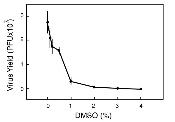
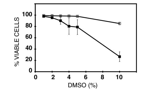
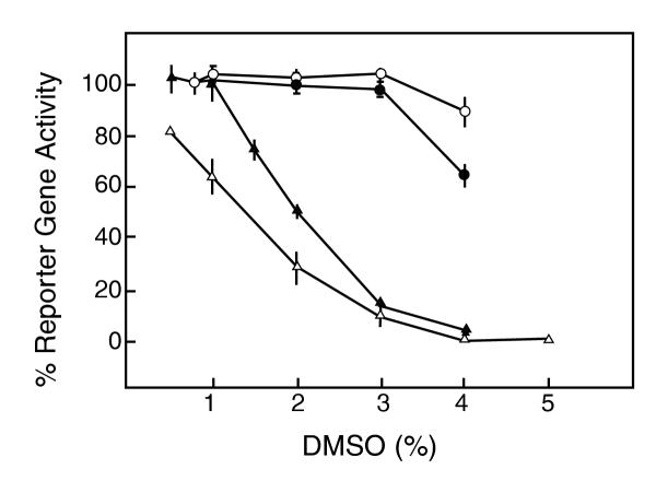
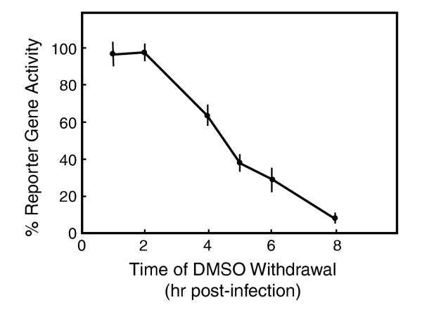
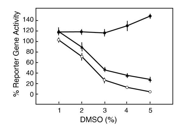
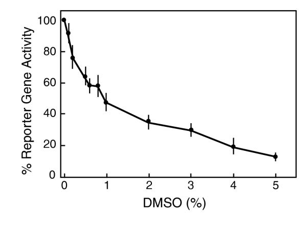
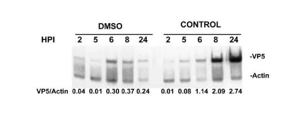
Results: DMSO blocks productive infection in vitro in different cell types with a 50% inhibitory concentration (IC50) from 0.7 to 2% depending upon the multiplicity of infection. The concentration dependence exhibits a Hill coefficient greater than 1, indicating that DMSO blocks productive infection by acting at multiple different points (mechanisms of action) with positive cooperativity. Consistently, we identified at least three distinct temporal target mechanisms for inhibition of virus growth by DMSO. At late stages of infection, DMSO reduces virion infectivity, and markedly inhibits viral DNA replication. A third mode of action was revealed using an oligonucleotide-based DNA microarray system for HSV. These experiments showed that DMSO reduced the transcript levels of many HSV-1 genes; including several genes coding for proteins involved in forming and assembling the virion. Also, DMSO markedly inhibited some but not all early transcripts indicating a previously unknown mode for inhibiting the early phase of HSV transcription-replication cycle.

Conclusion: These observations suggest that DMSO itself may have a role in the anti-herpetic activity of formulations utilizing it as a dispersant.


Figures

Figure 1
Figure 1
Effect of DMSO in the productive infection of HSV-1. Cultured Vero cells (105 cells in 24-well plates) were infected with 1 million PFU of the dUTPase/LAT recombinant virus. After 1 hr adsorption period, overlay medium was added containing the indicated concentrations of DMSO. At 24 hr post-infection, the cells were harvested and the virus yield was determined by plaque assay. Data are the average ± SD of 3 duplicate determinations.
Figure 2
Figure 2
Cell toxicity of DMSO. Confluent cultures of Vero cells in 96-well plates were treated with the indicated concentrations of DMSO for either 24 h (filled circles) or for 8 hr (open circles) and cytotoxicity determined 28 hrs after the initiation of the treatment. Results are average ± SD of 4 duplicated determinations.
Figure 3
Figure 3
Effect of DMSO treatment on productive infection at different phases of infection. Cultured Vero cells (105 cells in 24-well plates) were infected with 1000 pfu of the dUTPase/LAT recombinant virus. Treatments were as follows: DMSO at the indicated concentrations was present for 24 h before adsorption (filled circles), during the adsorption phase (open circles) or for 23 hr after the adsorption period (filled triangles). Also included is the effect of DMSO present for 23 hr after adsorption on HFF cells infected under similar conditions (open triangles). Reporter gene activity was determined at 24 hr following infection. Data are the average ± SD of 3–6 duplicate determinations.
Figure 4
Figure 4
Time course dependency of DMSO blockade of productive infection. Cultured Vero cells (105 cells in 24-well plates) were infected with 1000 PFU of the dUTPase/LAT recombinant virus. After the adsorption phase, 4% DMSO was added in the ovelay medium and kept for the indicated time periods. At those times, the media were replaced with fresh overlay medium without DMSO and the incubation continued 24 hr. Reporter gene activity was determined at 24 hr post-infection. Data are the average ± SD of 3–6 duplicate determinations.
Figure 5
Figure 5
Effect of 8 hr treatment of DMSO at different phases of infection. Cultured Vero cells (105 cells in 24-well plates) were infected with 1000 PFU of the dUTPase/LAT recombinant virus. Treatments were as follows: DMSO at the indicated concentrations were present for 8 h before adsorption (filled circles), 8 h immediately after adsorption (open circles) and 16 to 24 hr (triangles). Reporter gene activity was determined at 24 pi. Data are the averages ± S.D. of 3–6 duplicate determinations.
Figure 6
Figure 6
Inactivation of HSV-1 virion infectivity by DMSO. The dUTPase/LAT recombinant virus was suspended in PBS-glucose medium at 2000 PFU/ml and incubated at 37°C for 7 hr. Aliquots of 0.5 ml of the suspension were then adsorbed to Vero cells (105 cells in 24-well plates) for 1 hr. The virus suspension was then replaced with overlay medium, and reporter gene activity was determined at 24 hr. Data are the averages ± S.D of 4 duplicate determinations.
Figure 7
Figure 7
PCR analysis of HSV-1 DNA replication in the presence or absence of DMSO. Total DNA was isolated from cultures of Vero cells infected with 1000 PFU of the dUTPase/LAT recombinant and the indicated hrs post-infection and amplified in the conditions described in Material and Methods. Aliquots of the amplified products were fractionated in 6% polyacrylamide gels in Tris-borate-EDTA and evaluated by densitometry.

Similar articles

Cited by

References

    1. Whitley RJ, Soong SJ, Dolin R, Galasso GJ, Chien LT, Alford CA. Adenine arabinoside therapy of biopsy-proved herpes simplex encephalitis. National Institute of Allergy and Infectious Diseases collaborative antiviral study. N. 1977;297:289–294. - PubMed
    1. Wagner EK, Bloom DC. Experimental investigation of herpes simplex virus latency. Clin Microbiol Rev. 1997;Jul 10:419–443. - PMC - PubMed
    1. Corey L, Spear PG. Infectious with herpes simplex viruses (1). N. 1986;314:686–691. - PubMed
    1. Corey L, Spear PG. Infectious with herpes simplex viruses (2). N. 1986;314:749–757. - PubMed
    1. Nesburn AB. "Report of the Corneal Disease Panel: Vision Research-A National Plan 1983–1987". 1986.

Publication types

Substances