. 2002 Jul;22(13):4587-97.
doi: 10.1128/MCB.22.13.4587-4597.2002.

Cell attachment to the extracellular matrix induces proteasomal degradation of p21(CIP1) via Cdc42/Rac1 signaling

Affiliations

Cell attachment to the extracellular matrix induces proteasomal degradation of p21(CIP1) via Cdc42/Rac1 signaling

Wenjie Bao et al. Mol Cell Biol. 2002 Jul.

Abstract

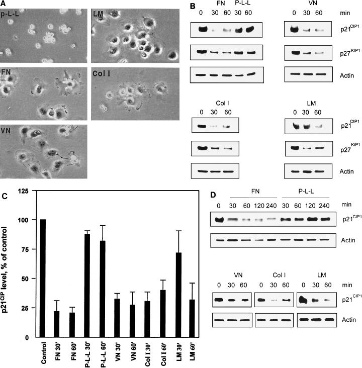
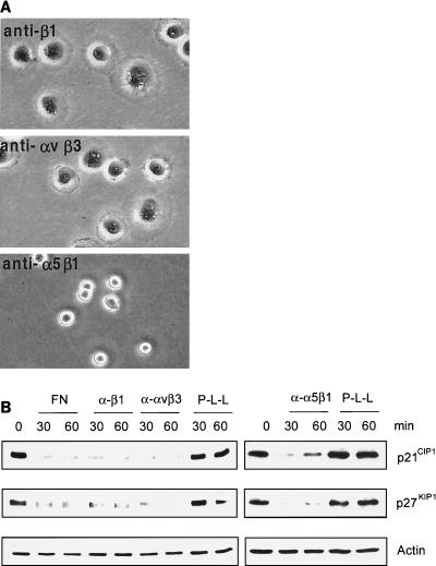
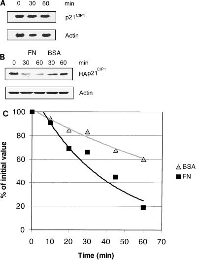
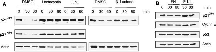
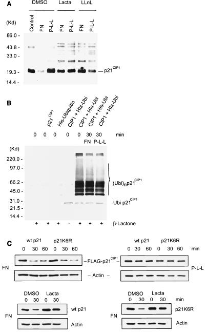
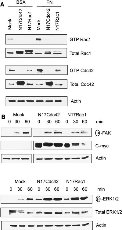
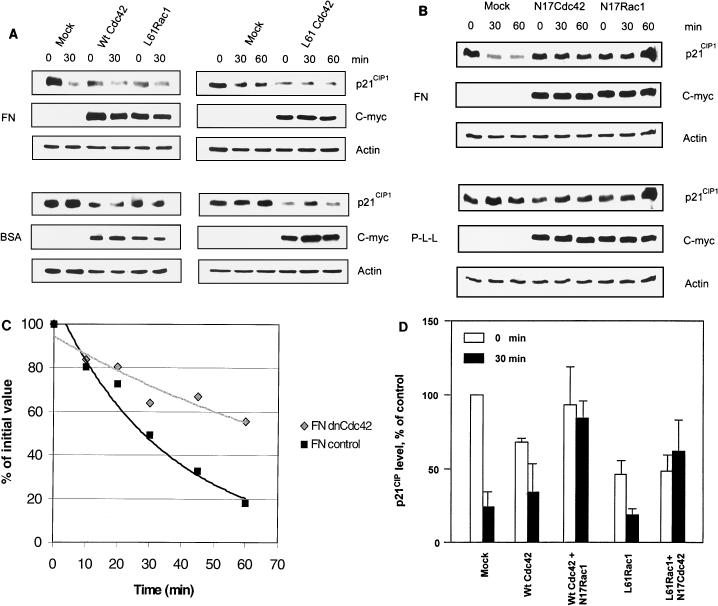
The cyclin-dependent kinase 2 (Cdk2) inhibitors p21(CIP1) and p27(KIP1) are negatively regulated by anchorage during cell proliferation, but it is unclear how integrin signaling may affect these Cdk2 inhibitors. Here, we demonstrate that integrin ligation led to rapid reduction of p21(CIP1) and p27(KIP1) protein levels in three distinct cell types upon attachment to various extracellular matrix (ECM) proteins, including fibronectin (FN), or to immobilized agonistic anti-integrin monoclonal antibodies. Cell attachment to FN did not rapidly influence p21(CIP1) mRNA levels, while the protein stability of p21(CIP1) was decreased. Importantly, the down-regulation of p21(CIP1) and p27(KIP1) was completely blocked by three distinct proteasome inhibitors, demonstrating that integrin ligation induced proteasomal degradation of these Cdk2 inhibitors. Interestingly, ECM-induced proteasomal proteolysis of a ubiquitination-deficient p21(CIP1) mutant (p21K6R) also occurred, showing that the proteasomal degradation of p21(CIP1) was ubiquitin independent. Concomitant with our finding that the small GTPases Cdc42 and Rac1 were activated by attachment to FN, constitutively active (ca) Cdc42 and ca Rac1 promoted down-regulation of p21(CIP1). However, dominant negative (dn) Cdc42 and dn Rac1 mutants blocked the anchorage-induced degradation of p21(CIP1), suggesting that an integrin-induced Cdc42/Rac1 signaling pathway activates proteasomal degradation of p21(CIP1). Our results indicate that integrin-regulated proteasomal proteolysis might contribute to anchorage-dependent cell cycle control.


Figures

FIG. 1.
FIG. 1.
Down-regulation of the Cdk2 inhibitors p21CIP1 and p27 KIP1 upon attachment to ECM components. (A) Human bladder carcinoma ECV 304 cells were allowed to attach to dishes precoated with FN, VN, LM, ColI, or P-L-L under serum-free conditions. Photographs (20× objective) show the representative morphology of ECV 304 cells after attachment to the distinct ECM proteins for 30 min. (B) p21CIP1, p27KIP1, and actin protein levels in ECV 304 cell lysates were analyzed by Western blotting after attachment for the indicated times to different ECM components or P-L-L under serum-free conditions in order to analyze the exclusive influence of integrin ligation without intervening growth factor signaling. Actin levels were detected as a loading control. (C) Estimation of p21CIP1 levels in ECV 304 cells by densitometry of Western blots. Bars represent mean values of three independent experiments ± the standard error of the mean. The level at time zero is defined as 100% (control). (D) HUVECs were allowed to attach to FN, P-L-L, VN, ColI, or LM for various times. Cell lysates were analyzed for p21CIP1, p27KIP1, and actin protein levels by Western blotting.
FIG. 2.
FIG. 2.
Integrin ligation specifically down-regulates p21CIP1 and p27KIP1. (A) ECV 304 cells were plated on immobilized anti-β1 (P4C10), anti-αvβ3 (LM609), or anti-α5β1 (JBS5) for 30 min and photographed with a 20× objective. (B) ECV 304 cells attached to immobilized anti-β1, anti-αvβ3, or anti-α5β1 MAb were analyzed for p21CIP1, p27KIP1, and actin protein levels by Western blotting.
FIG. 3.
FIG. 3.
Cell attachment to FN affects p21CIP1 protein stability. (A) Total mRNA isolated from ECV 304 cells previously plated on FN for the indicated times was analyzed for p21CIP1 mRNA levels by Northern blotting. β-Actin mRNA levels were measured as a loading control. (B) ECV 304 cells transiently transfected with HA-tagged p21CIP1 were allowed to attach to FN or suspended upon BSA, and then HA-p21CIP1 levels were analyzed by Western blotting. Actin levels were used as a loading control. (C) ECV 304 cells transfected with HA-p21CIP1 were plated on FN or kept in suspension (BSA), and then the stability of HA-p21CIP1 was detected by pulse-chase analysis with incorporated [35S]methionine. The values shown are the means of two or three independent experiments.
FIG. 4.
FIG. 4.
Proteasome inhibitors block FN-induced down-regulation of p21CIP1 and p27KIP1. (A) ECV 304 cells were pretreated with lactacystin, β-lactone, or LLnL or with the dimethyl sulfoxide (DMSO) vehicle as a control and then plated on FN for the indicated times. p21CIP1, p27 KIP1, and actin protein levels were analyzed by Western blotting. (B) ECV 304 cells were allowed to attach to FN or P-L-L, and then the levels of cyclin E and p53 were examined by Western blotting. The levels of p21CIP1 were used as a comparison and determined with anti-p21CIP1 MAb SX118.
FIG. 5.
FIG. 5.
Ubiquitination of p21CIP1 upon cell attachment. (A) ECV 304 cells were treated with the proteasome inhibitor lactacystin (Lacta) or LLnL and then plated on FN or P-L-L for 30 min. Cell lysates were analyzed by Western blotting with anti-p21CIP1 PAb (Ab5; Oncogene). DMSO, dimethyl sulfoxide; MW, molecular mass; Kd, kilodaltons. (B) ECV 304 cells transiently mock transfected or transfected with p21CIP1 or His-ubiquitin or cotransfected with p21CIP1 and His-ubiquitin were treated with or without the proteasome inhibitor β-lactone, as indicated. Cells cotransfected with p21CIP1 and His-ubiquitin were allowed to attach to FN or P-L-L, and then His-ubiquitin was trapped by an Ni2+ column and ubiquitinated p21CIP1 was detected with anti-p21CIP1 MAb SX118 by Western blotting. (C, upper portion) ECV 304 cells transiently transfected with FLAG-tagged wild-type (wt) p21 or with the ubiquitination-deficient FLAG-tagged p21K6R mutant were plated on FN or P-L-L for the times indicated. The levels of overexpressed p21CIP1 were detected by Western blotting with anti-FLAG MAb (M2; Sigma). Actin levels were analyzed as a loading control. (C, lower portion) ECV 304 cells transiently transfected with FLAG-tagged wild-type p21 or the FLAG-tagged p21K6R mutant were treated with the specific proteasome inhibitor lactacystin or with the dimethyl sulfoxide vehicle and plated on FN for 30 min. The levels of wild-type p21 and p21K6R were determined by Western blotting with anti-FLAG MAb (M2).
FIG. 6.
FIG. 6.
Cell attachment to FN activates a Cdc42/Rac1 signaling pathway. (A) ECV 304 cells transiently transfected with dn N17Cdc42 or dn N17Rac1 cDNA or mock transfected were plated on FN for 20 min. Cell lysates were analyzed for active GTPases by binding to a GST-PAK-CRIB domain fusion protein, followed by Western blotting with anti-Rac1 or anti-Cdc42 antibodies. Total levels of Rac1, Cdc42, and actin were analyzed by Western blotting of original cell lysates. (B) ECV 304 cells transfected with Cdc42 or dn Rac1 cDNA or mock transfected were allowed to attach to FN for the indicated times, and then activated FAK and ERK1/2 were analyzed by Western blotting with anti-phospho-FAKY397 PAb or anti-phospho-ERK1/2 PAb. The levels of c-myc were detected with anti-c-Myc MAb 9E10. Actin levels were analyzed as a loading control.
FIG. 7.
FIG. 7.
Anchorage-dependent Cdc42/Rac1 signaling regulates proteolysis of p21CIP1. (A) ECV 304 cells transiently transfected with c-myc-tagged wild-type (Wt) Cdc42 or c-myc-tagged mutant ca Cdc42 or ca L61Rac1 were plated on FN- or BSA-coated plates for the indicated times. Levels of p21CIP1, c-myc, and actin were analyzed by Western blotting. Levels of c-myc were analyzed for transfection efficiency with anti-c Myc MAb 9E10, and actin was used as a loading control. (B) ECV 304 cells transiently overexpressing c-myc-tagged mutant dn N17Cdc42 or dn N17Rac1 were plated on FN or P-L-L. p21CIP1, c-myc, and actin levels were analyzed by Western blotting. (C) ECV 304 cells transiently transfected with HA-p21CIP1 or cotransfected with HA-p21CIP1 and dn Cdc42 were allowed to attach to FN for the times indicated, and then the stability of HA-p21CIP1 was examined by pulse-chase analysis. (D) Quantitative estimations of p21CIP1 levels in ECV 304 cells by densitometry of Western blots from single mock transfections or transfections with L61Rac1 or wild-type Cdc42 or cotransfections with ca L61Rac1 and dn N17Cdc42 or wild-type Cdc42 and dn N17Rac1, respectively, before and 30 min after plating onto FN. Bars represent mean values of three independent experiments ± the standard error of the mean.

References

    1. Blagosklonny, M. V., G. S. Wu, S. Omurand, and W. S. El-Deiry. 1996. Proteasome-dependent regulation of p21WAF1/CIP1 expression. Biochem. Biophys. Res. Commun. 227:564-569. - PubMed
    1. Bottazzi, M. E., X. Y. Zhu, R. M. Böhmer, and R. K. Assoian. 1999. Regulation of p21cip1 expression by growth factors and the extracellular matrix reveals a role for transient ERK activity in G1 phase. J. Biol. 146:1255-1264. - PMC - PubMed
    1. Boyer, S. N., D. E. Wazer, and V. Band. 1996. E7 protein of human papilloma virus-16 induces degradation of retinoblastoma protein through the ubiquitin-proteasome pathway. Cancer Res. 56:4620-4624. - PubMed
    1. Brown, J., S. J. Reading, S. Jones, C. J. Fitchett, J. Howl, A. Martin, C. Longland, F. Michelangeli, Y. E. Dubrova, and C. A. Brown. 2000. Critical evaluation of ECV 304 as a human endothelial cell model defined by genetic analysis and functional responses: a comparison with the human bladder cancer derived epithelial cell line T24/83. Lab. Investig. 80:37-45. - PubMed
    1. Chen, C. S., M. Marksich, S. Huang, G. M. Whitesides, and D. E. Ingber. 1997. Geometric control of cell life and death. Science 276:1425-1428. - PubMed

Publication types

MeSH terms

Substances

LinkOut - more resources