. 2002 Jul;22(13):4863-75.
doi: 10.1128/MCB.22.13.4863-4875.2002.

Cyclin D and cdk4 are required for normal development beyond the blastula stage in sea urchin embryos

Affiliations

Cyclin D and cdk4 are required for normal development beyond the blastula stage in sea urchin embryos

Jennifer C Moore et al. Mol Cell Biol. 2002 Jul.

Abstract

cdk4 mRNA and protein are constitutively expressed in sea urchin eggs and throughout embryonic development. In contrast, cyclin D mRNA is barely detectable in eggs and early embryos, when the cell cycles consist of alternating S and M phases. Cyclin D mRNA increases dramatically in embryos at the early blastula stage and remains at a constant level throughout embryogenesis. An increase in cdk4 kinase activity occurs concomitantly with the increase in cyclin D mRNA. Ectopic expression of cyclin D mRNA in eggs arrests development before the 16-cell stage and causes eventual embryonic death, suggesting that activation of cyclin D/cdk4 in cleavage cell cycles is lethal to the embryo. In contrast, blocking cyclin D or cdk4 expression with morpholino antisense oligonucleotides results in normal development of early gastrula-stage embryos but abnormal, asymmetric larvae. These results suggest that in sea urchins, cyclin D and cdk4 are required for normal development and perhaps the patterning of the developing embryo, but may not be directly involved in regulating entry into the cell cycle.


Figures

FIG. 1.
FIG. 1.
Sea urchin cdk4 and cyclin D. The protein sequences of sea urchin S. purpuratus cdk4 (panel A) and cyclin D (panel B) are compared to the sequences of cyclin D1 from vertebrates (Homo sapiens, Mus musculus, and Xenopus laevis) and cyclin D from D. melanogaster. In panel A, the amino acid sequences used to construct primers for PCR to identify the cdk genes are underlined. The QMALT sequence characteristic of the vertebrate cdk4/cdk6 genes is boxed. In panel B, the asterisk indicates the threonine identified as the phosphorylation site that determines cyclin D half-life (6). The diamonds indicate the Rb binding motif. Dark shading indicates identical residues, and light shading indicates similar residues.
FIG. 2.
FIG. 2.
Sea urchin cdk4 interacts with human cyclin D and p16, The S. purpuratus cdk4 open reading frame was fused to the Gal4 DNA binding domain and transformed into yeast cells expressing the Gal4 activation domain (sector 2) or the Gal4 activation domain fused to human p16 (sector 4), human cyclin D2 (sector 5), or human cyclin D1 (sector 6). Sector 1 is the HF7c yeast strain with no vectors, and sector 3 is yeast cells expressing the human cdk4 open reading frame fused to the Gal4 DNA binding domain transformed with human cyclin D1. The yeast cells were grown on plates lacking leucine and tryptophan and then streaked onto plates lacking leucine, tryptophan, and histidine. The colonies were transferred to nitrocellulose and then assayed for β-galactosidase activity.
FIG. 3.
FIG. 3.
Sea urchin cyclin D and cdk4 form a complex in reticulocyte lysates. (A) Synthetic mRNAs encoding S. purpuratus cyclin D and cdk4 were mixed and translated in a rabbit reticulocyte lysate containing [35S]methionine. Equal amounts of lysate were analyzed directly (lane 1) or precipitated with the anti-cdk4 antibody in the absence (lane 2) or presence (lane 3) of competing antigenic peptide or the antibody to the C terminus of cyclin E (lane 4). (B) mRNAs encoding S. purpuratus cdk4 and cyclin D (lanes 1 and 2), cdk4 and cyclin E (lanes 3 and 4), cyclin E and cdk2 (lanes 5 and 6), or cyclin D and cdk2 (lanes 7 and 8) were cotranslated in a rabbit reticulocyte lysate system containing [35S]methionine. The resulting proteins were analyzed directly (lanes 1, 3, 5, and 7) or after precipitation with α-cdk4 (lanes 2 and 4) or α-cdk2 (lanes 6 and 8). The cyclin E and cdk2 were analyzed directly or after precipitation with α-cyclin E (lane 4). (C) mRNAs encoding S. purpuratus cdk2 and cyclin D were cotranslated in the presence of [35S]methionine (lane 1). The resulting proteins were immunoprecipitated with α-cdk4 (lane 2).
FIG. 4.
FIG. 4.
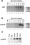
Expression of cdk4 and cyclin D mRNA during development. Total RNA was prepared from S. purpuratus eggs and embryos at the indicated times after fertilization and culturing at 15°C. Equal amounts of total RNA were resolved by Northern blotting, transferred to nitrocellulose, and probed with random primed labeled cdk4 cDNA (A) or cyclin D cDNA (B and C). Eggs are shown in lane 1, and embryos at the indicated times of development are shown in lanes 2 to 8 of panel B and lanes 2 to 5 of panel C. The 12-h embryos in panel B were at early blastula stage.
FIG. 5.
FIG. 5.
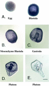
Expression of cyclin D mRNA during embryogenesis by in situ hybridization. L. variegatus embryos at different stages were fixed and analyzed for cyclin D mRNA by in situ hybridization. Eggs (left) and early embryos (A) have no detectable signal until blastula stage (embryo at right), when the signal appears uniform throughout the embryo. At mesenchyme blastula (B), the mRNA is concentrated in the vegetal plate and the future oral ectoderm. This ectodermal regionalization persists through gastrulation (C) to the larval stage (D), and cyclin D mRNA remains concentrated in regions of the ciliary band. Endoderm labeling also increases during gastrulation to maximal levels in plutei. Panel E is a pluteus larva hybridized with a sense strand probe. The slight purple color in the egg is due to the pigment in the egg and not to hybridization.
FIG. 6.
FIG. 6.
Expression of cdk4 protein during development. (A) A coupled transcription-translation reaction in a reticulocyte lysate programmed with a plasmid expressing cdk4 mRNA (lane 1) or a plasmid (lane 2) was analyzed by Western blotting with the anti-cdk4 antibody. IVT, in vitro translation. (B) Extracts were prepared from S. purpuratus eggs (lane 1) and embryos at various times of development. In panel B, embryos were harvested at the indicated time throughout the first two cell cycles. The embryos divided between 110 and 120 min. (C) Embryos were harvested at the indicated time after fertilization. Equal amounts of total protein were resolved by SDS gel electrophoresis, transferred to nitrocellulose, and analyzed by Western blotting with the anti-cdk4 antibody.
FIG. 7.
FIG. 7.
Activity of cdk4 kinase during development. (A) Extracts were prepared from S. purpuratus embryos cultured at 18°C 10 h after fertilization (early blastula). The extracts were immunoprecipitated with α-cdk4 in the absence (lane 1) or presence (lane 2) of antigenic peptide. The precipitates were assayed for kinase activity with GST-pRb as a substrate (52). (B) Extracts were prepared from a single batch of embryos at the indicated times after fertilization and culturing at 18°C. Equal amounts of total protein were immunoprecipitated with α-cdk4 (lanes 1 to 5) or α-cyclin E (lanes 6 to 10), and the immunoprecipitates were assayed for kinase activity. The 10-h embryos were at early blastula (the same stage as the 12-h embryos in Fig. 4).
FIG. 8.
FIG. 8.
Premature expression of cyclin D mRNA is lethal to sea urchin embryos. Fertilized L. variegatus sea urchin eggs were injected with cyclin D (top) or cdk4 (bottom) mRNA and photographed 2.5 and 5 h after fertilization.
FIG. 9.
FIG. 9.
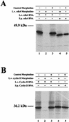
Morpholino oligonucleotides inhibit in vitro translation of cdk4 mRNA (A) and cyclin D mRNA (B). (A) Synthetic L. variegatus (L.v., lanes 1 to 3) or S. purpuratus (S.p., lanes 4 and 5) cdk4 mRNAs were translated in a reticulocyte lysate. The L. variegatus cdk4 antisense morpholino oligonucleotide was included in lanes 2 and 4, and the control morpholino was included in lanes 3 and 5. (B) Synthetic L. variegatus (lanes 1 to 3) or S. purpuratus (lanes 4 and 5) cyclin D mRNAs were translated in a reticulocyte lysate. The L. variegatus cyclin D antisense morpholino oligonucleotide was included in lanes 2 and 4, and the control morpholino was included in lanes 3 and 5.
FIG. 10.
FIG. 10.
Cyclin D and cdk4 are required for the normal development of sea urchin larvae. Fertilized L. variegatus sea urchin eggs were injected with a morpholino antisense control oligonucleotide, a morpholino antisense oligonucleotide to cyclin D, a morpholino antisense oligonucleotide to cdk4, or a mixture of the antisense oligonucleotide and a synthetic S. purpuratus cdk4 mRNA. Embryos were photographed 24 and 48 h after fertilization. Two examples of the embryos injected with antisense cyclin D or antisense cdk4 are shown.

References

    1. Audic, Y., B. Boyle, M. Slevin, and R. S. Hartley. 2001. Cyclin E morpholino delays embryogenesis in Xenopus. Genesis 30:107-109. - PubMed
    1. Ceol, C. J., and H. R. Horvitz. 2001. dpl-1 DP and elf-1 E2F act with lin-35 Rb to antagonize ras signaling in C. elegans vulval development. Mol. Cell 3:461-474. - PubMed
    1. Datar, S. A., H. W. Jacobs, A. F. De la Cruz, C. F. Lehner, and B. A. Edgar. 2000. The D. melanogaster cyclin D-Cdk4 complex promotes cellular growth. EMBO J. 19:4543-4554. - PMC - PubMed
    1. De Nooij, J. C., M. A. Letendre, and I. K. Hariharan. 1996. A cyclin-dependent kinase inhibitor, dacapo, is necessary for timely exit from the cell cycle during D. melanogaster embryogenesis. Cell 87:1237-1247. - PubMed
    1. Diehl, J. A., M. G. Cheng, M. F. Roussel, and C. J. Sherr. 1998. Glycogen synthase kinase 3β regulates cyclin D1 proteolysis and subcellular localization. Genes Dev. 12:3499-3511. - PMC - PubMed

Publication types

MeSH terms

Associated data

LinkOut - more resources