Comparative Study
. 2002 May 10;318(4):941-50.
doi: 10.1016/S0022-2836(02)00172-9.

Intrinsic membrane targeting of the flagellar export ATPase FliI: interaction with acidic phospholipids and FliH

Affiliations
Comparative Study

Intrinsic membrane targeting of the flagellar export ATPase FliI: interaction with acidic phospholipids and FliH

Frédéric Auvray et al. J Mol Biol. .

Abstract

The specialised ATPase FliI is central to export of flagellar axial protein subunits during flagellum assembly. We establish the normal cellular location of FliI and its regulatory accessory protein FliH in motile Salmonella typhimurium, and ascertain the regions involved in FliH(2)/FliI heterotrimerisation. Both FliI and FliH localised to the cytoplasmic membrane in the presence and in the absence of proteins making up the flagellar export machinery and basal body. Membrane association was tight, and FliI and FliH interacted with Escherichia coli phospholipids in vitro, both separately and as the preformed FliH(2)/FliI complex, in the presence or in the absence of ATP. Yeast two-hybrid analysis and pull-down assays revealed that the C-terminal half of FliH (H105-235) directs FliH homodimerisation, and interacts with the N-terminal region of FliI (I1-155), which in turn has an intra-molecular interaction with the remainder of the protein (I156-456) containing the ATPase domain. The FliH105-235 interaction with FliI was sufficient to exert the FliH-mediated down-regulation of ATPase activity. The basal ATPase activity of isolated FliI was stimulated tenfold by bacterial (acidic) phospholipids, such that activity was 100-fold higher than when bound by FliH in the absence of phospholipids. The results indicate similarities between FliI and the well-characterised SecA ATPase that energises general protein secretion. They suggest that FliI and FliH are intrinsically targeted to the inner membrane before contacting the flagellar secretion machinery, with both FliH105-235 and membrane phospholipids interacting with FliI to couple ATP hydrolysis to flagellum assembly.


Figures

Figure 1
Figure 1
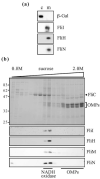
FliI and FliH localisation in wild-type S. typhimurium. (a) Cell lysates of the flagellated S. typhimurium SJW1103 strain expressing β-galactosidase from plasmid pOZ172 were fractionated. Proteins from cytoplasmic (c), and membrane (m) fractions were analysed by SDS-12.5% PAGE and immunoblotting with anti-β-galactosidase, anti-FliI, anti-FliH, and anti-FliN antisera. (b) Membrane fractions were separated on a 0.8 M-2.0 M sucrose gradient. Proteins were resolved by SDS-12.5% PAGE and either stained with Coomassie blue (top panel, molecular mass markers are in kDa), or immunoblotted with anti-FliI, anti-FliH, anti-FliN and anti-FliM antisera (bottom panels). Positions of co-separated inner membrane NADH oxidase and outer membrane proteins (OMPs) are indicated.
Figure 2
Figure 2
FliI and FliH localisation in S. typhimurium flagellar mutant strains. Cell lysates from SJW2702 (fliI mutant), SJW1616 (flhA), SJW1467 (flhB), SJW192 (fliOPQR), and SJW1684 (fliF) were fractionated. Proteins from cytoplasmic (c), and membrane (m) fractions were analysed by SDS-12.5% PAGE and immunoblotting with anti-FliI, anti-FliH and anti-FliN antisera.
Figure 3
Figure 3
Localisation of FliI and FliH artificially expressed in a flhDC mutant. (a) Cell lysates from the non-flagellated S. typhimurium SJW1368 flhDC mutant expressing either FliI or FliH from plasmids pBADFliI or pBADFliH were fractionated. Proteins from cytoplasmic (c), and membrane (m) fractions were analysed by SDS-12.5% PAGE and immunoblotting with anti-FliI or anti-FliH antisera. (b) Membrane fractions were separated on a 0.8 M-2.0 M sucrose gradient and analysed as above, using inner membrane NADH oxidase and outer membrane proteins (OMPs) as markers.
Figure 4
Figure 4
Extraction of membrane-asssociated FliI and FliH. Membrane pellets from the S. typhimurium SJW1368 flhDC mutant expressing FliI or FliH were either sonicated for 30 seconds, or resuspended in one of the solutions indicated. Soluble (s) and pellet (p) material was separated by centrifugation, analysed by SDS-12.5% PAGE and immunoblotting with anti-FliI or anti-FliH antisera.
Figure 5
Figure 5
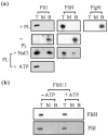
Flotation of purified FliI and FliH with liposomes. (a) Purified proteins FliI, FliH and (negative control) FlgN were added to E. coli phospholipid (PL) vesicles and mixed with 55% (w/v) sucrose, overlaid with 40% (w/v) sucrose and allowed to float through the density-gradient during centrifugation (75,000g, 16 hours). Flotations were also performed with PL in the presence of 1 M NaCl or 20 mM ATP, or in the absence of PL, as indicated. Top (T), middle (M) and bottom (B) fractions were analysed by SDS-12.5% PAGE and immunoblotting with anti-FliI, -FliH or -FlgN antisera. (b) Purified FliH2/FliI complex was mixed with E. coli PL in the absence or presence of 20 mM ATP and analysed as above.
Figure 6
Figure 6
Complex formation by FliH and FliI truncated derivatives. (a) Plasmid constructs co-expressing His-tagged FliH and FliI, or their deletion derivatives. The fliH and fliI genes, represented by grey and black bars, respectively, are co-transcribed in an operon. The phage T7 promoter (broken arrow) and histidine-tag (hatched bar) are represented 5′ of the FliH coding sequence. (b) Pull-down assay (Ni-NTA affinity chromatography) using the protein pairs shown in (a). Lysates of IPTG uninduced (-) and induced (+) cell cultures, and proteins eluted from the nickel column (eluate) were analysed by SDS-12.5% (1, 3 and 5) or 15% (2 and 4) PAGE and Coomassie blue staining. Molecular mass markers were the same in each gel, as indicated.
Figure 7
Figure 7
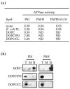
Role of acidic phospholipids in liposome binding and activation of FliI. (a) ATP hydrolysis (±10% nmol phosphate min.-1 μg-1 FliI) was assayed at 37 °C with FliI (10 μg ml-1), FliH/FliI or FliH105-235/FliI complex (30 μg ml-1), in the presence or absence of phospholipids (PL) (10 μg ml-1). We used E. coli PL, dioleoyl phosphatidylcholine (DOPC) alone, or mixtures (5:1 ratio) of DOPC/phosphatidylglycerol (PG) and DOPC/cardiolipin (CL). Released inorganic phosphate was measured using the malachite green assay. ND, not determined. (b) Flotation of purified FliI or FliH to DOPC, DOPC/PG, or DOPC/CL, as measured by density-gradient centrifugation as in Figure 5.

References

    1. Macnab RM. Flagella and motility. In: Neidhardt FC, editor. Escherichia coli and Salmonella typhimurium: Cellular and Molecular Biology. 2nd edit. Washington DC: American Society for Microbiology; 1996. pp. 123–145.
    1. Namba K, Vonderviszt F. Molecular architecture of the bacterial flagellum. Quart. Rev. Biophys. 1997;30:1–65. - PubMed
    1. Macnab RM. Reversible rotary propellor and type III export apparatus. J. Bacteriol. 1999;181:7149–7153. - PMC - PubMed
    1. Aizawa SI. Bacterial flagella and type III secretion systems. FEMS Microbiol. Letters. 2001;202:157–164. - PubMed
    1. Minamino T, Macnab RM. Components of the Salmonella flagellar export apparatus and classification of export substrates. J. Bacteriol. 1999;181:1388–1394. - PMC - PubMed

Publication types

MeSH terms

LinkOut - more resources