. 2002 Jun;122(7):1967-77.
doi: 10.1053/gast.2002.33661.

Norwalk virus binds to histo-blood group antigens present on gastroduodenal epithelial cells of secretor individuals

Affiliations

Norwalk virus binds to histo-blood group antigens present on gastroduodenal epithelial cells of secretor individuals

Severine Marionneau et al. Gastroenterology. 2002 Jun.

Abstract

Background & aims: Norwalk Virus (NV) is a member of the Caliciviridae family, which causes acute epidemic gastroenteritis in humans of all ages and its cellular receptors have not yet been characterized. Another calicivirus, Rabbit Hemorrhagic Disease Virus, attaches to H type 2 histo-blood group oligosaccharide present on rabbit epithelial cells. Our aim was to test if, by analogy, recombinant NV-like particles (rNV VLPs) use carbohydrates present on human gastroduodenal epithelial cells as ligands.

Methods: Attachment of rNV VLPs was tested on tissue sections of the gastroduodenal junction and on saliva from individuals of known ABO, Lewis, and secretor phenotypes. It was also tested on human Caco-2 cells and on animal cell lines transfected with glycosyltransferases complementary DNA (cDNA). Competition experiments were performed with synthetic oligosaccharides and anticarbohydrate antibodies. Internalization was monitored by confocal microscopy.

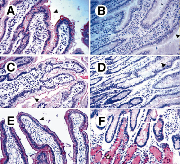
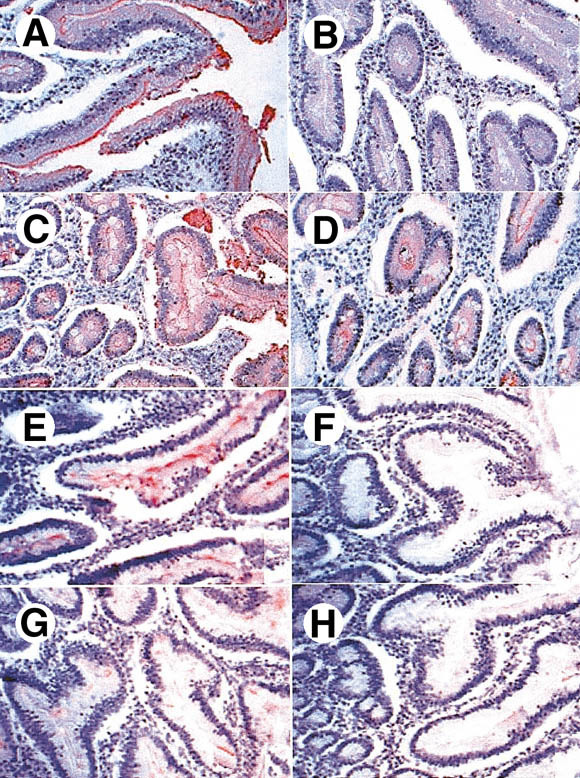
Results: Attachment of rNV VLPs to surface epithelial cells of the gastroduodenal junction as well as to saliva was detected, yet only from secretor donors. It was abolished by alpha1,2fucosidase treatment, and by competition with the H types 1 and 3 trisaccharides or with anti-H type 1 and anti-H types (3/4) antibodies. Transfection of CHO and TS/A cells with an alpha1,2fucosyltransferase cDNA allowed attachment of VLPs. These transfectants as well as differentiated Caco-2 cells expressing H type 1 structures internalized the bound particles.

Conclusions: rNV VLPs use H type 1 and/or H types (3/4) as ligands on gastroduodenal epithelial cells of secretor individuals.


Figures

Fig. 1
Fig. 1
(A–D) Binding of rNV VLPs to duodenal tissue sections. Tissue sections from secretor individuals were incubated with VLPs and the binding was detected as described in the Materials and Methods section. Before incubation with VLPs, sections were treated with (B) 10 mmol/L periodate or with fucosidase II, either (C) heat inactivated or (D) not. rNV VLPs binding to the villi epithelial cell surface are indicated by arrowheads. No binding was detected on tissue sections from nonsecretors (not shown). No staining of Brünner's glands was detectable, irrespective of the secretor status (not shown). Immunostaining of a duodenal section from a secretor individual by the (E) anti-H type 1 BG-4 and from (F) a nonsecretor individual by the anti-H type 2 19-OLE. (F) Staining of Brünner's glands by the anti-H type 2 is shown by a star.
Fig. 2
Fig. 2
Inhibition of rNV VLPs binding to duodenal tissue sections from secretor individuals by (A–D) anti-H mAbs or by (E–H) synthetic oligosaccharides. Sections were first incubated with the anti-H mAbs and then with VLPs. Binding of the VLPs was detected as described in the Materials and Methods section. (A) No preincubated antibody, (B) preincubated anti-H type 1 BG-4, (C) preincubated anti-H type 2 19-OLE, and (D) preincubated anti-H types 3 and 4 MBr1. Inhibitions with oligosaccharides were performed by preincubating the VLPs with the following trisaccharides: (E) Lewis a, (F) H type 1, (G) H type 2, and (H) H type 3.
Fig. 3
Fig. 3
(A) Binding of rNV VLPs to serially diluted saliva samples from either secretor (n = 4) or nonsecretor individuals (n = 4) coated to an enzyme-linked immunosorbent assay plate. Three of the secretors were homozygotes, and 1 was heterozygote. Binding was detected by using the polyclonal rabbit anti-NV VLPs as described in the Materials and Methods section. The optical densities at 405 nm are plotted against the log2 of reciprocal dilutions of saliva. (B) Inhibition of rNV VLPs binding to saliva from a secretor individual by synthetic oligosaccharides. VLPs were preincubated with oligosaccharides at a final concentration of 1.8 mmol/L before incubation on saliva coated at a dilution of 1/4000.
Fig. 4
Fig. 4
Cytofluorimetric analysis of the expression of A and H histo-blood group antigens and of the rNV VLPs attachment to glycosyltransferases-transfected CHO and TS/A cells. Both cell types were transfected with empty vectors (controls), with the rat FTB α1,2fucosyltransferase cDNA (FTB transfectants), or with both the rat FTB α1,2fucosyltransferase and the rat A enzyme cDNAs (FTB + A transfectants). Cells were incubated with either the anti-H type 1 BG-4, the anti-H type 2 19 OLE, the anti-H types 3 and 4 MBr1, the anti-A 3-3A, or with rNV VLPs. The log of fluorescence intensities in arbitrary units is plotted against the cell number.
Fig. 5
Fig. 5
Confocal microscopy analysis of the attachment and internalization of rNV VLPs to transfected CHO cells and to differentiated Caco-2 cells. CHO cells transfected with the FTB cDNA and expressing H structures (H+) or mock transfected (CONTROL) were incubated at 4°C or 37°C for 2 hours with rNV VLPs. Before incubation at 4°C, cells were fixed to avoid any internalization. After incubation at 37°C, cells were permeabilized and fixed. VLPs were detected by using mAb 9C3 followed by FITC-conjugated secondary antibody (green). Nuclei were stained with propidium iodide (red). Confocal images were taken at the (A) top, (B) middle, and (C) bottom of cells. rNV VLPs were incubated at either 4°C or 37°C on differentiated Caco-2 cells grown on Transwell filters. Their presence was detected as described earlier for CHO cells. Confocal xz sections show the surface labeling after incubation at 4°C and the intracellular labeling after incubation at 37°C. Caco-2 cells grown on glass slides were incubated with rNV VLPs at 4°C and binding was detected as described earlier. Confocal xy sections were recorded. When grown on glass, differentiated Caco-2 cells form cysts as shown in D, E, and F. Before incubation on cells, VLPs were preincubated with either (D) a nonsecretor saliva or (E) a secretor saliva, or cells were treated with (F) fucosidase II. Images obtained in absence of preincubation with saliva or after treatment with inactivated fucosidase were similar to that shown in D.

References

    1. Thiel HJ, König M. Caliciviruses: an overview. Vet Microbiol. 1999;69:55–62. - PubMed
    1. Green K, Ando T, Balayan MS, Berke T, Clarke IN, Estes MK, Matson DO, Nakata S, Neill JD, Studdert MJ, Thiel HJ. Taxonomy of the caliciviruses. J Infect Dis. 2000;181:S322–S330. - PubMed
    1. Shaw RD. Viral infections of the gastrointestinal tract. Curr Opin Gastroenterol. 2000;16:12–17. - PubMed
    1. Glass RI, Noel J, Ando T, Fankhauser R, Belliot G, Mounts A, Parashar UD, Bresee JS, Monroe SS. The epidemiology of enteric caliciviruses from humans: a reassessment using new diagnostics. J Infect Dis. 2000;181:S254–S261. - PubMed
    1. Koopmans M, Vinjé J, de Witt M, Leenen I, van der Poel W, van Duynhoven Y. Molecular epidemiology of human enteric caliciviruses in the Netherlands. J Infect Dis. 2000;181:S262–S269. - PubMed

Publication types