Bacterial cholangitis causing secondary sclerosing cholangitis: a case report
- PMID: 12057011
- PMCID: PMC116430
- DOI: 10.1186/1471-230x-2-14
Bacterial cholangitis causing secondary sclerosing cholangitis: a case report
Abstract
Background: Although bacterial cholangitis is frequently mentioned as a cause of secondary sclerosing cholangitis, it appears to be extremely rare, with only one documented case ever reported.
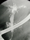
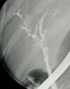
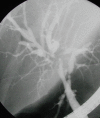
Case presentation: A 48-year-old woman presented with an episode of acute biliary pancreatitis that was complicated by pancreatic abcess formation. After 3 months she had an episode of severe pyogenic (E. Coli) cholangitis that recurred over the subsequent 7 months on a further two occasions. Initially, cholangiography suggested the presence of extra-biliary intrahepatic abcesses while repeated investigations demonstrated development of multiple segmental biliary duct strictures. After maintenance antibiotic treatment was started, no episodes of cholangitis occurred over a 14-month period.
Conclusions: Sclerosing cholangitis can rapidly develop after an episode of bacterial cholangitis. Extra-biliary involvement of the hepatic parenchyma with abcess formation may be a risk factor for developing this rare but particularly severe complication.
Figures


References
-
- Sherlock S, Dooley J. Diseases of the Liver and Biliary System. 9th edn London: Blackwell Scientific Publications. 1994. pp. 237–48.
-
- Vleggaar FP, van Buuren HR, Lameris JS. Bile duct lesions in portal vein thrombosis. Ned Tijdsch Geneesk. 1999;143:2057–2062. - PubMed
-
- Tanaka Y, Koshiyama H, Nakao K, Makita Y, Kobayashi Y, Yoshida Y, Kimura M, Adachi Y. Rapid progress of acute suppurative cholangitis to secondary sclerosing cholangitis sequentially followed-up by endoscopic retrograde cholangiography. Endoscopy. 2001;33:633–5. doi: 10.1055/s-2001-15325. - DOI - PubMed
-
- Maluenda F, Csendes A, Burdiles P, Diaz J. Bacteriological study of choledochal bile in patients with common bile duct stones, with or without acute suppurative cholangitis. Hepatogastroenterology. 1989;36:132–5. - PubMed
Publication types
MeSH terms
LinkOut - more resources
Full Text Sources
Medical

