. 2002 Apr 26:1:5.
doi: 10.1186/1475-2875-1-5.

Characterisation and expression of a PP1 serine/threonine protein phosphatase (PfPP1) from the malaria parasite, Plasmodium falciparum: demonstration of its essential role using RNA interference

Affiliations

Characterisation and expression of a PP1 serine/threonine protein phosphatase (PfPP1) from the malaria parasite, Plasmodium falciparum: demonstration of its essential role using RNA interference

Rajinder Kumar et al. Malar J. .

Abstract

Background: Reversible protein phosphorylation is relatively unexplored in the intracellular protozoa of the Apicomplexa family that includes the genus Plasmodium, to which belong the causative agents of malaria. Members of the PP1 family represent the most highly conserved protein phosphatase sequences in phylogeny and play essential regulatory roles in various cellular pathways. Previous evidence suggested a PP1-like activity in Plasmodium falciparum, not yet identified at the molecular level.

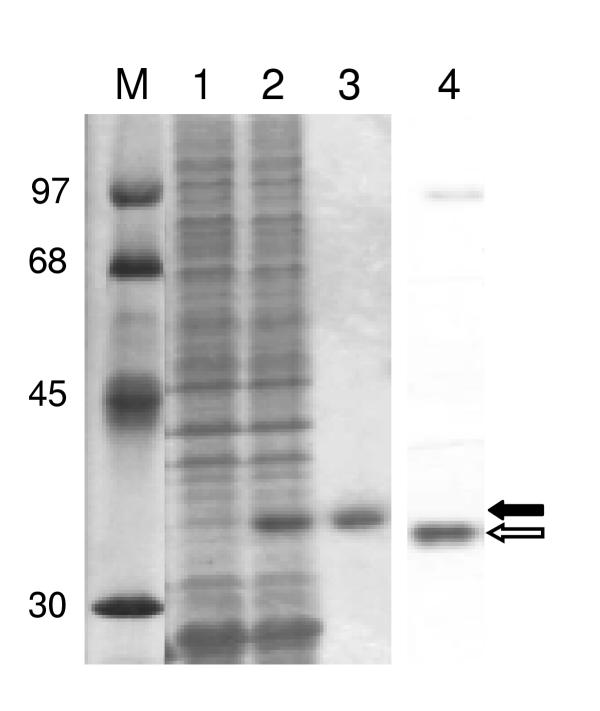
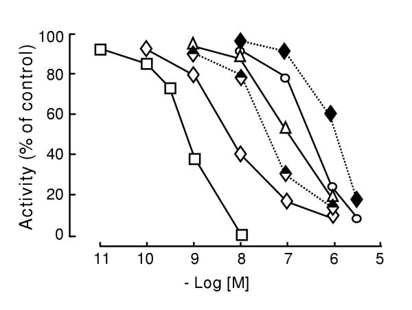
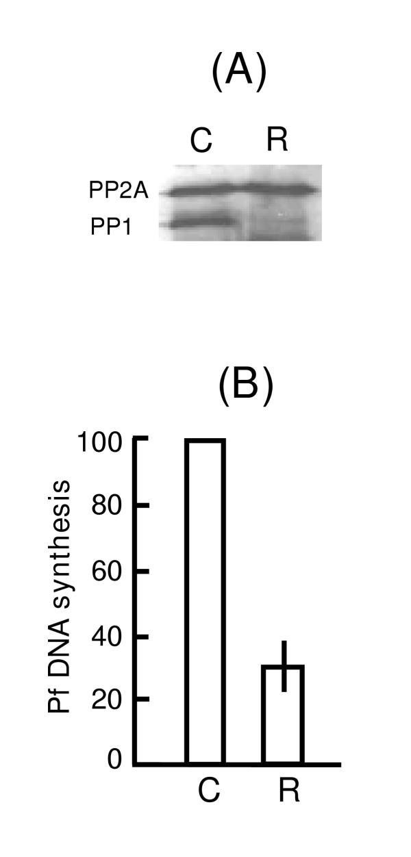
Results: We have identified a PP1 catalytic subunit from P. falciparum and named it PfPP1. The predicted primary structure of the 304-amino acid long protein was highly similar to PP1 sequences of other species, and showed conservation of all the signature motifs. The purified recombinant protein exhibited potent phosphatase activity in vitro. Its sensitivity to specific phosphatase inhibitors was characteristic of the PP1 class. The authenticity of the PfPP1 cDNA was further confirmed by mutational analysis of strategic amino acid residues important in catalysis. The protein was expressed in all erythrocytic stages of the parasite. Abrogation of PP1 expression by synthetic short interfering RNA (siRNA) led to inhibition of parasite DNA synthesis.

Conclusions: The high sequence similarity of PfPP1 with other PP1 members suggests conservation of function. Phenotypic gene knockdown studies using siRNA confirmed its essential role in the parasite. Detailed studies of PfPP1 and its regulation may unravel the role of reversible protein phosphorylation in the signalling pathways of the parasite, including glucose metabolism and parasitic cell division. The use of siRNA could be an important tool in the functional analysis of Apicomplexan genes.


Figures

Figure 1
Figure 1
PfPP1 gene structure. The exon and intron sequences of PfPP1 gene are shown in capital and small letters, respectively. Underlined primers were used in RT-PCR to amplify the PP1 ORF, and have been described under Results. The amino acid sequence of PfPP1 is in single-letter codes below the coding sequence.
Figure 2
Figure 2
PfPP1 sequence comparison. The predicted sequences of Plasmodium PP1 (this study) and human PP1 alpha (P08129) catalytic subunits were aligned using the CLUSTALW program at the European Bioinformatics Institute (EMBL) server, and later refined by visual inspection. The amino acid residue numbers are shown on the right. Residues are marked as: non-conservative replacement (.); conservative replacement (:), and identical (*). Residues important in I-2 interaction are highlighted in gray: E52, E54; D164, E165, and K166.
Figure 3
Figure 3
Recombinant expression of PfPP1 in bacteria. The following proteins / extracts were analyzed by SDS-PAGE followed by staining with Coomassie Brilliant Blue R250: approximately 30 μg (protein) of total extract [14] of IPTG-induced E. coli BL21(DE3) containing the RIG plasmid and pET-15-PfPP1 (lane 2) or pET-15 without insert (lane 1); 4 μg of the purified recombinant (His)8-tagged PfPP1 (lane 3). Lane 4 shows an immunoblot in which 80 μg of 100,000 × g extract of Pf [10] was probed using a PP1 antibody described under Materials and Methods. Parasitic PfPP1 and the recombinant His-tagged PfPP1 bands are indicated by open and closed arrowheads, respectively. Protein markers (lane M) are indicated by Mr in thousands.
Figure 4
Figure 4
PfPP1 dose response to inhibitors. Inhibition assays for recombinant PfPP1 were performed using 32P-phosphorylase a as substrate essentially as described [14]. The inhibitors and the symbols are: tautomycin (square), inhibitor-2 (diamond), OA (triangle), and I-1 (circle). The half-closed and fully closed diamonds represent I-2 against the double mutant (E52A/E54A) and triple mutant (D164A/E165A/K166A) enzymes, respectively. Activities are expressed as percentage of the inhibitor-free reaction. As shown, the X-axis represents negative logarithm of molar concentration of the inhibitors.
Figure 5
Figure 5
Constitutive expression of parasitic PfPP1. (A) Western blot: Total protein (80 μg) from the ring (R), Trophozoite (T), schizont (S), and early (G1) and late (G2) sexual stages of Pf were probed with a mixture of anti-PP1 and anti-Pfg27 antibodies as described [14]. (B) Microcystin-sepharose chromatography: About 500 μg of the following extracts was subjected to microcystin affinity chromatography and the bound proteins analyzed by Western blot using PP1 antibody: extract of uninfected RBC processed identically (lane 1); Pf extract (lane 2); Pf extract pre-incubated with 1 μM microcystin-LR at room temperature for 5 min (lane 3); unbound fraction (a double-pass flow-through from the column) (lane 4). Recombinant His-tagged PfPP1 is displayed in lane 5 for comparison. The native and recombinant PP1 bands are marked by open and closed arrows, respectively. Sizes of protein standards are indicated on the left. Two non-PP1 proteins are also seen in the blot in panel B. The ~25 kDa band (common in lanes 1, 2, and 3) is evidently a RBC protein. The ~40 kDa band (lanes 2, 3), on the other hand, is a Plasmodium protein, since it is absent in the RBC fraction. We speculate that these proteins non-specifically bound to the Sepharose matrix, since they could not be competed out by microcystin (lane 3).
Figure 6
Figure 6
Inhibition of parasite growth and abrogation of PfPP1 expression by interfering dsRNA. The dsRNA sequences, and electroporation procedure have been described under Materials and Methods. Infected RBC were transfected with dsRNA against PfPP1 (R) or luciferase (C). Western blot (A) shows loss of PfPP1 by the dsRNA, but no effect on the control PfPP2A. The PP1 monoclonal and PP2A peptide antibodies have been described in Materials and Methods. Hypoxanthine incorporation assay (B) shows an approximately 70% inhibition of parasitic DNA synthesis in PP1-depleted cells, compared to the luciferase antisense-treated control.

References

    1. Graves JD, Krebs EG. Protein phosphorylation and signal transduction. Pharmacol Ther. 1999;82:111–121. doi: 10.1016/S0163-7258(98)00056-4. - DOI - PubMed
    1. Cohen PT. Novel protein serine/threonine phosphatases: variety is the spice of life. Trends Biochem Sci. 1997;22:245–251. doi: 10.1016/S0968-0004(97)01060-8. - DOI - PubMed
    1. Wera S, Hemmings BA. Serine/threonine protein phosphatases. Biochem J. 1995;311:17–29. - PMC - PubMed
    1. Fischer EH. Cell signaling by protein tyrosine phosphorylation. Adv Enzyme Regul. 1999;39:359–369. doi: 10.1016/S0065-2571(98)00014-4. - DOI - PubMed
    1. Guan KL, Dixon JE. Bacterial and viral protein tyrosine phosphatases. Semin Cell Biol. 1993;4:389–396. doi: 10.1006/scel.1993.1046. - DOI - PubMed

Publication types

MeSH terms

LinkOut - more resources