Comparative Study
. 2002 Jun;160(6):1973-80.
doi: 10.1016/S0002-9440(10)61147-9.

An invasion-independent pathway of blood-borne metastasis: a new murine mammary tumor model

Affiliations
Comparative Study

An invasion-independent pathway of blood-borne metastasis: a new murine mammary tumor model

Takashi Sugino et al. Am J Pathol. 2002 Jun.

Abstract

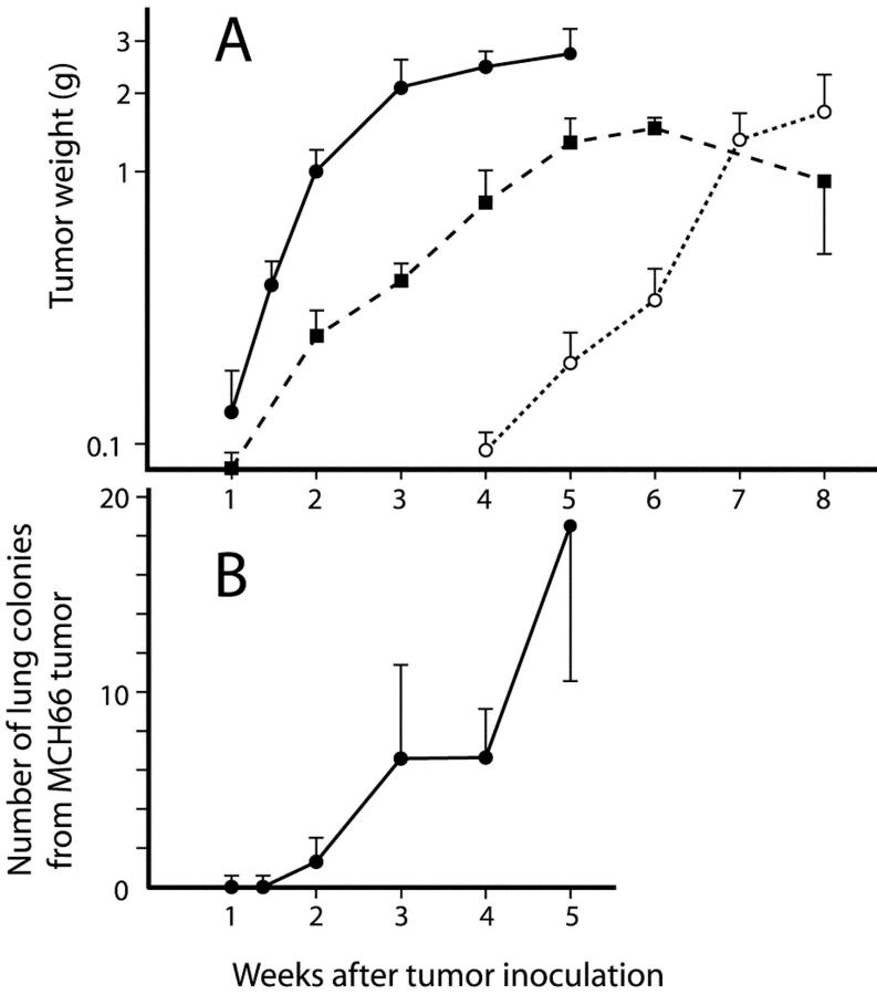
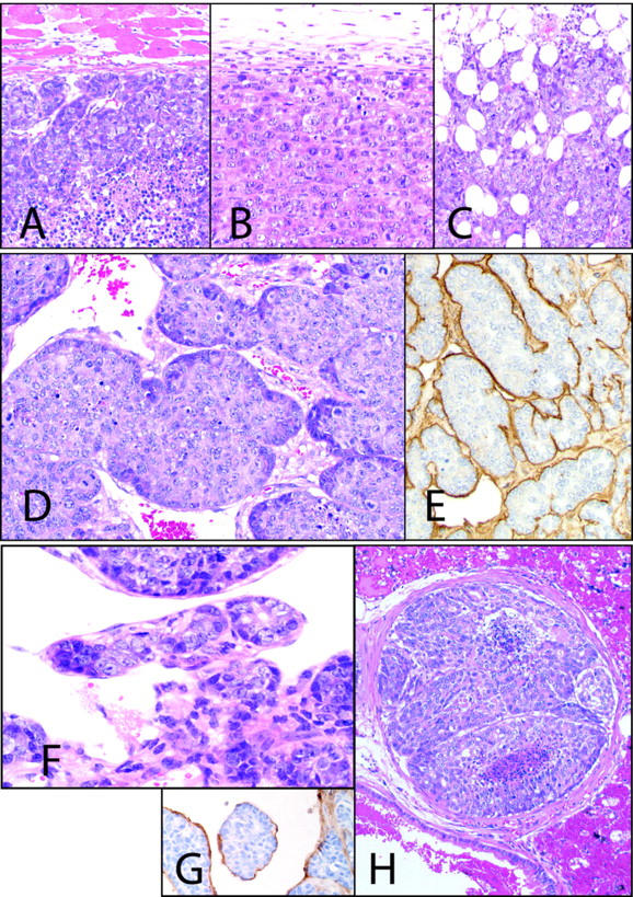
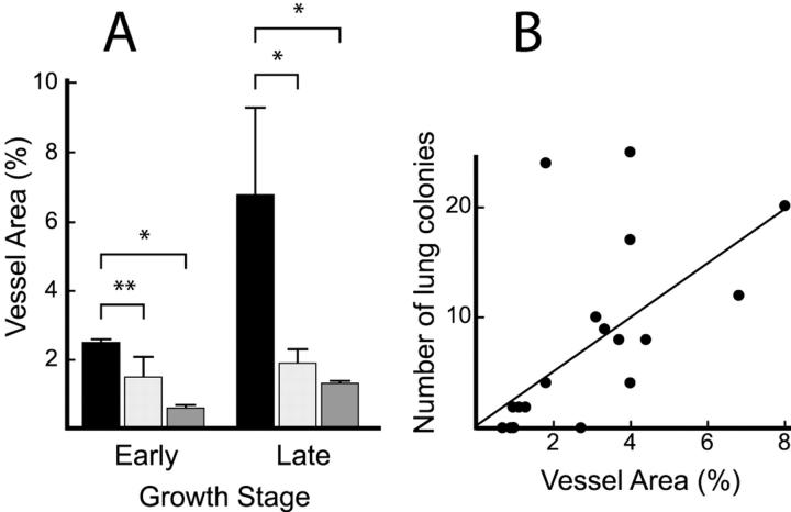
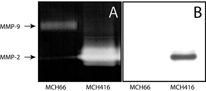
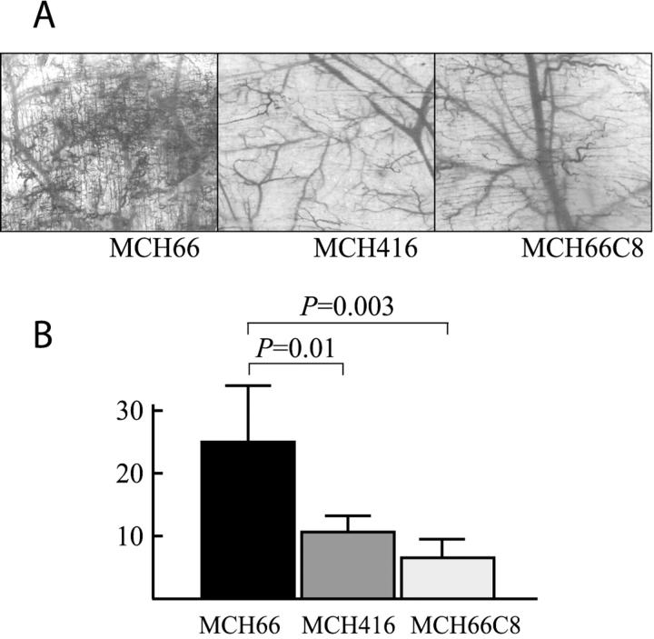
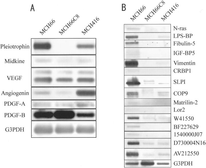
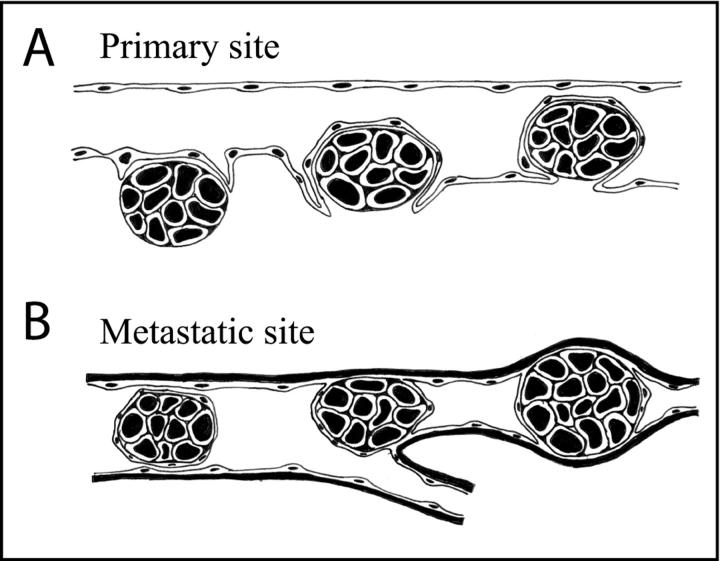
It is generally believed that active invasion by cancer cells is essential to the metastatic process. In this report, we describe a murine mammary tumor (MCH66) model of metastasis that does not require invasion into the vascular wall of both the primary tumor and the target organ, in this case, the lung. The process involves intravasation of tumor nests surrounded by sinusoidal blood vessels, followed by intravascular tumor growth in the lung, without penetration of the vascular wall during the process. Comparative studies using a nonmetastatic MCH66 clone (MCH66C8) and another highly invasive metastatic cell line (MCH416) suggested that high angiogenic activity and sinusoidal remodeling of tumor blood vessels were prerequisites for MCH66 metastasis. Differential cDNA analysis identified several genes that were overexpressed by MCH66, including genes for the angiogenesis factor pleiotrophin, and extracellular matrix-associated molecules that may modulate the microenvironment toward neovascularization. Our analyses suggest that tumor angiogenesis plays a role in the induction of invasion-independent metastasis. This model should prove useful in screening and development of new therapeutic agents for cancer metastasis.


Figures

Figure 1.
Figure 1.
In vivo growth properties and generation of pulmonary metastasis. A: Tumor growth curves for the subcutaneously inoculated cell lines. Filled circle, MCH66; filled square, MCH66C8; open circle, MCH416. Each point represents the mean ± SEM tumor weight. B: Time course of the number of lung metastatic colonies from MCH66 tumors. No colonies were detected in MCH416- or MCH66C8-inoculated mice until 8 weeks after inoculation.
Figure 2.
Figure 2.
Cell line histology. MCH66 (A) and MCH66C8 (B) tumors grew expansively, whereas invasive growth of MCH416 (C) was evident. D: MCH66 tumors at 7 days after inoculation formed small nests surrounded by fibrous stroma. Dilated sinusoidal vessels were observed around the nests. E: Immunohistochemistry for laminin shows continuous basement membrane around the MCH66 tumor nests. F: MCH66 tumors at 21 days after inoculation. Tumor nest was involved in the developed sinusoidal vessels. G: Tumor nest in the blood was surrounded by a vascular endothelial layer that immunostained with anti-CD31 antibody. H: Intravascular MCH66 tumor growth subsequent to embolization in the pulmonary arteriole. For laminin and CD31 immunohistochemistry, counterstain is hematoxylin. All others are H&E. Original magnifications: ×100 (A–C); ×200 (D–H).
Figure 3.
Figure 3.
Intratumoral vascularity. A: The proportion of vascular area within tumors at an early growth stage (days 12 to 14 of MCH66 and MCH66C8, and day 35 of MCH416) and at a late growth stage (day 35 of MCH66 and days 56 to 70 of MCH416). Filled square, MCH66; light gray square, MCH66C8; dark gray square, MCH416. Results are means ± SEM, n = 15 microscopic fields for each group. *, P < 0.01; **, P < 0.05. B: Correlation between the number of pulmonary metastatic colonies in mice inoculated with MCH66 and the proportion of vessel area in the tumors. P = 0.017.
Figure 4.
Figure 4.
Activities of gelatin degradation and MMP-2 expression. A: Gelatin zymography of conditioned media from cultured cells. Molecular weights were calculated using protein standards. B: Immunoblot analysis with an MMP-2 antibody confirmed the zymography results.
Figure 5.
Figure 5.
Angiogenesis activities of cell lines using dorsal air sac assay. A: Representative views with a dissecting microscopy of the dorsal skin of mice implanted with chambers filled with MCH66, MCH416, or MCH66C8. B: The number of vessels per 1-mm width of vertical histological section was estimated using an image analyzer. Results are means ± SEM.
Figure 6.
Figure 6.
Analysis of differentially expressed MCH66 genes. A: Semiquantitative RT-PCR and Southern blot analysis of angiogenesis factors. Basic fibroblast growth factor (bFGF) or hepatocyte growth factor (HGF) expression was not detected in any of the cell lines. VEGF, vascular endothelial growth factor; PDGF, platelet-derived growth factor. B: Virtual Northern blot analysis for cDNA clones of known genes from MCH66 libraries subtracted from MCH66C8 and MCH416. LPS-BP, lipopolysaccharide-binding protein; IGFb-BP5, insulin-like growth factor-binding protein-5; CRBP1, cellular retinol binding protein-1; SLPI, secretory leukocyte protease inhibitor.
Figure 7.
Figure 7.
Schematic showing the process of blood-borne metastasis of MCH66 tumors. A: Intravasation of a tumor nest enveloped by vascular endothelial cells. B: A tumor embolus, conserving its tissue organization and endothelial covering, is mechanically arrested in an arteriole and then intravascularly proliferates. During this metastatic process, the tumor cells do not pass through vascular walls.

Comment in

References

    1. Sugino T, Kawaguchi T, Suzuki T: Sequential process of blood-borne lung metastases of spontaneous mammary carcinoma in C3H mice. Int J Cancer 1993, 55:141-147 - PubMed
    1. Itoh T, Tanioka M, Yoshida H, Yoshioka T, Nishimoto H, Itohara S: Reduced angiogenesis and tumor progression in gelatinase A-deficient mice. Cancer Res 1998, 58:1048-1051 - PubMed
    1. Mohler KM, Butler LD: Quantitation of cytokine mRNA levels utilizing the reverse transcriptase-polymerase chain reaction following primary antigen-specific sensitization in vivo. I. Verification of linearity, reproducibility and specificity. Mol Immunol 1991, 28:437-447 - PubMed
    1. Fidler IJ: The relationship of embolic homogeneity, number, size and viability to the incidence of experimental metastasis. Eur J Cancer 1973, 9:223-227 - PubMed
    1. Liotta LA, Saidel GM, Kleinerman J: The significance of hematogenous tumor cell clumps in the metastasis process. Cancer Res 1979, 36:889-894 - PubMed

Publication types

MeSH terms