. 2002 Jun;160(6):2001-8.
doi: 10.1016/S0002-9440(10)61150-9.

Renal fibrosis. Extracellular matrix microenvironment regulates migratory behavior of activated tubular epithelial cells

Affiliations

Renal fibrosis. Extracellular matrix microenvironment regulates migratory behavior of activated tubular epithelial cells

Michael Zeisberg et al. Am J Pathol. 2002 Jun.

Abstract

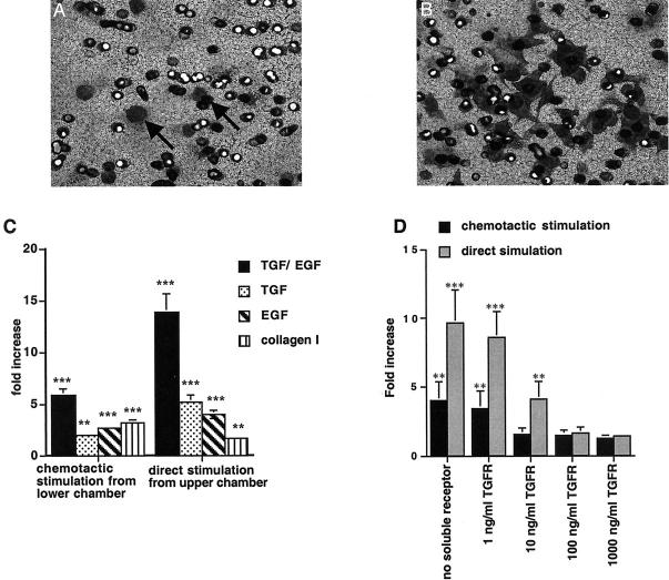
During progression of chronic renal disease, qualitative and quantitative changes in the composition of tubular basement membranes (TBMs) and interstitial matrix occur. Transforming growth factor (TGF)-beta(1)-mediated activation of tubular epithelial cells (TECs) is speculated to be a key contributor to the progression of tubulointerstitial fibrosis. To further understand the pathogenesis associated with renal fibrosis, we developed an in vitro Boyden chamber system using renal basement membranes that partially mimics in vivo conditions of TECs during health and disease. Direct stimulation of TECs with TGF-beta(1)/epithelial growth factor results in an increased migratory capacity across bovine TBM preparations. This is associated with increased matrix metalloproteinase (MMP) production, namely MMP-2 and MMP-9. Indirect chemotactic stimulation by TGF-beta(1)/EGF or collagen type I was insufficient in inducing migration of untreated TECs across bovine TBM preparation, suggesting that basement membrane integrity and composition play an important role in protecting TECs from interstitial fibrotic stimuli. Additionally, neutralization of MMPs by COL-3 inhibitor dramatically decreases the capacity of TGF-beta(1)-stimulated TECs to migrate through bovine TBM preparation. Collectively, these results demonstrate that basement membrane structure, integrity, and composition play an important role in determining interstitial influences on TECs and subsequent impact on potential aberrant cell-matrix interactions.


Figures

Figure 1.
Figure 1.
Two-compartment Boyden chamber system mimics in vivo conditions. A: Normal mouse kidney, PAS-stained. B: Schematic illustration of normal kidney representing tubuli (T), surrounded by TBM, which separates them from the interstitium that contains fibroblasts (F) and capillaries (C). C: Kidney specimen from α3 type IV collagen knockout mouse with severe tubulointerstitial fibrosis, Masson trichrome staining. D: Schematic illustration of kidney with tubulointerstitial fibrosis, a widened interstitium that contains deposited type I collagen (Col1), an increased number of fibroblasts, fibroblast-like cells, and inflammatory cells (I). E: The two-compartment Boyden chamber mimics the tubular compartment in the upper chamber and the interstitial compartment in the lower chamber, separated by a polycarbonate membrane coated with TBM. F: Direct stimulation by TGF-β1/EGF in the upper chamber or chemotactic stimulation by TGF-β1/EGF or type I collagen leads to enhanced migration in the lower chamber. G: Double staining of human kidney from a patient with membranous nephropathy for laminin (fluorescein isothiocyanate, green) and α-smooth muscle actin (Cy5, red). In areas that had basement membranes intact, α-smooth muscle actin was not detectable. H: Disruption of TBM is associated with EMT. The arrows indicate areas where tubular cells express α-smooth muscle actin around areas of TBM disruption. Original magnifications: ×200 (G); ×400 (A, C, H).
Figure 2.
Figure 2.
Migration of TECs. A: TECs migrate into the lower chamber and are visible on the lower side of the polycarbonate membrane when TGF-β1 and EGF were used as chemotactic stimuli. B: Cells pretreated with TGF-β1/EGF displayed an increased migratory capacity and continued to be responsive to TGF-β1 and EGF. C, left: Stimulation of TECs with TGF-β1/EGF from the lower chamber led to a 5.8 ± 0.62-fold increase of migration compared to unstimulated control cells. Incubation with TGF-β1 led to a 2.0 ± 0.17-fold increase and incubation with EGF led to 3.66 ± 0.17-fold increase compared to nonstimulated control. C, right: Direct stimulation of MCT cells by TGF-β1 and EGF in the upper chamber led to a 13.8 ± 1.82-fold increase compared to the control. Direct stimulation with TGF-β1 led to a 5.2 ± 0.32-fold increase in migration compared to untreated control and direct stimulation with EGF led to a 3.96 ± 0.36-fold increase. The addition of type I collagen in the lower chamber induced migration of unstimulated TECs by 3.2 ± 0.31-fold, whereas the addition of type I collagen into the upper chamber had no significant effect. D: Addition of soluble TGF-β receptor to cells in the upper chamber inhibited TGF-β1-induced migration. D, gray columns: Migration that was induced by direct stimulation with TGF-β1 (9.6 ± 2.43-fold increase), was reduced to 8.6 ± 1.94-fold by the addition of 1 ng/ml of TGF-β receptor, to 4.1 ± 1.28-fold by the addition of 10 ng/ml of TGF-β receptor, to 1.6 ± 0.42-fold by the addition of 100 ng/ml of TGF-β receptor, and to 1.4 ± 0.12-fold by the addition of 1000 ng/ml of TGF-β receptor. D, black columns: Inhibition of migration induced by chemotactic stimulation with TGF-β1 (4.0 ± 1.34-fold increase) was reduced to 3.39 (±1.3)-fold increase by the addition of 1 ng/ml of TGF-β receptor, to 1.52 (±0.47)-fold at a concentration of 10 ng/ml, to 1.47 (±0,37)-fold at 100 ng/ml, and to 1.25 (±0.25)-fold at 1000 ng/ml). ***, P < 0.0001; **, P < 0.001; *, P < 0.05. Original magnification: ×200 (B).
Figure 3.
Figure 3.
Regulation of migration by matrix microenvironment. A, left columns: Compared to chemotactic migration of MCT cells across an uncoated polycarbonate membrane, migration was significantly inhibited by coating the upper side of the membrane with type IV collagen [reduction by 44.6 (±2.5)% when compared to uncoated control]. Coating with bovine TBM preparation almost completely prevented chemotactic migrations of TECs in this setting [reduction by 88 (± 6.25)%]. A, right columns: Similarly, chemotactic migration of TECs across a membrane that was coated on the bottom side with type I collagen, was significantly inhibited by coating the upper side of the membrane with type IV collagen [reduction by 47.1 (±2.5)% when compared to control]. Also, TBM significantly inhibited chemotactic migrations of TECs [reduction by 92.2 (±6.3)%]. B, left columns: Migration of TECs across an uncoated membrane that was induced by direct stimulation with TGF-β1/EGF, was neither significantly inhibited by coating with type IV collagen, nor by coating with TBM. B, right columns: Migration of directly stimulated TECs across a membrane that was coated on the bottom side with type I collagen, was not significantly decreased by type IV collagen. Coating with bovine TBM preparation resulted in a decrease of migration in this setting, but was still permissive for migration of activated TECs [reduction by 49.3 (±1.3)% compared to uncoated control].
Figure 4.
Figure 4.
Activation of TECs with TGF-β1 and EGF leads to increased degradation of TBM via up-regulation of MMP-2 and MMP-9. A: Incubation of bovine TBM with active MMP-9 and MMP-2 generates NC1 domains and other type IV collagen degradation products (26 kd and 48 kd) as observed by Western blot. Incubation of TBM with supernatant derived from TGF-β1/EGF-treated TECs led to the generation of type IV collagen degradation products containing NC1 domains, whereas supernatant from untreated control TECs did not significantly degrade TBM. B: Supernatant from MCT cells treated with TGF-β1/EGF exhibited a significant up-regulation of MMP-2 and MMP-9 as observed by zymography assay using tissue culture supernatants. C and D summarize densitometric quantification of four independent experiments. *, P < 0.05.
Figure 5.
Figure 5.
Regulation of TEC migration. A: Inhibition of basement membrane-degrading MMP-2 and MMP-9 by COL-3, reduced migration that was induced by direct stimulation with TGF-β1/EGF across a polycarbonate membrane coated with bovine TBM preparation on the upper side and with type I collagen on the lower side in a dose-dependent manner [control 10.4 (±2.61)-fold stimulation, 8.91 (±1.98)-fold at 0.1 ng/ml, 4.57 (±1.48)-fold at 1 ng/ml, and 2.01 (±1.07)-fold at 10 ng/ml]. B: Migration induced by chemotactic stimulation with TGF-β1/EGF from the lower chamber, was not significantly inhibited by COL-3. ***, P < 0.0001; **, P < 0.001; *, P < 0.05.

References

    1. Stahl PJ, Felsen D: Transforming growth factor-beta, basement membrane, and epithelial-mesenchymal transdifferentiation: implications for fibrosis in kidney disease. Am J Pathol 2001, 159:1187-1192 - PMC - PubMed
    1. Muller GA, Zeisberg M, Strutz F: The importance of tubulointerstitial damage in progressive renal disease. Nephrol Dial Transplant 2000, 15:76-77 - PubMed
    1. Remuzzi G, Bertani T: Pathophysiology of progressive nephropathies. N Engl J Med 1998, 339:1448-1456 - PubMed
    1. Abrass CK, Berfield AK, Stehman-Breen C, Alpers CE, Davis CL: Unique changes in interstitial extracellular matrix composition are associated with rejection and cyclosporine toxicity in human renal allograft biopsies. Am J Kidney Dis 1999, 33:11-20 - PubMed
    1. Brito PL, Fioretto P, Drummond K, Kim Y, Steffes MW, Basgen JM, Sisson-Ross S, Mauer M: Proximal tubular basement membrane width in insulin-dependent diabetes mellitus. Kidney Int 1998, 53:754-761 - PubMed

Publication types

MeSH terms

Substances

LinkOut - more resources