. 2002 Jun;160(6):2045-53.
doi: 10.1016/S0002-9440(10)61154-6.

Signature tau neuropathology in gray and white matter of corticobasal degeneration

Affiliations

Signature tau neuropathology in gray and white matter of corticobasal degeneration

Mark S Forman et al. Am J Pathol. 2002 Jun.

Abstract

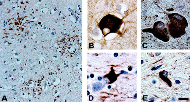
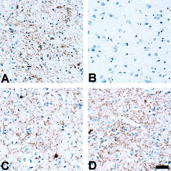
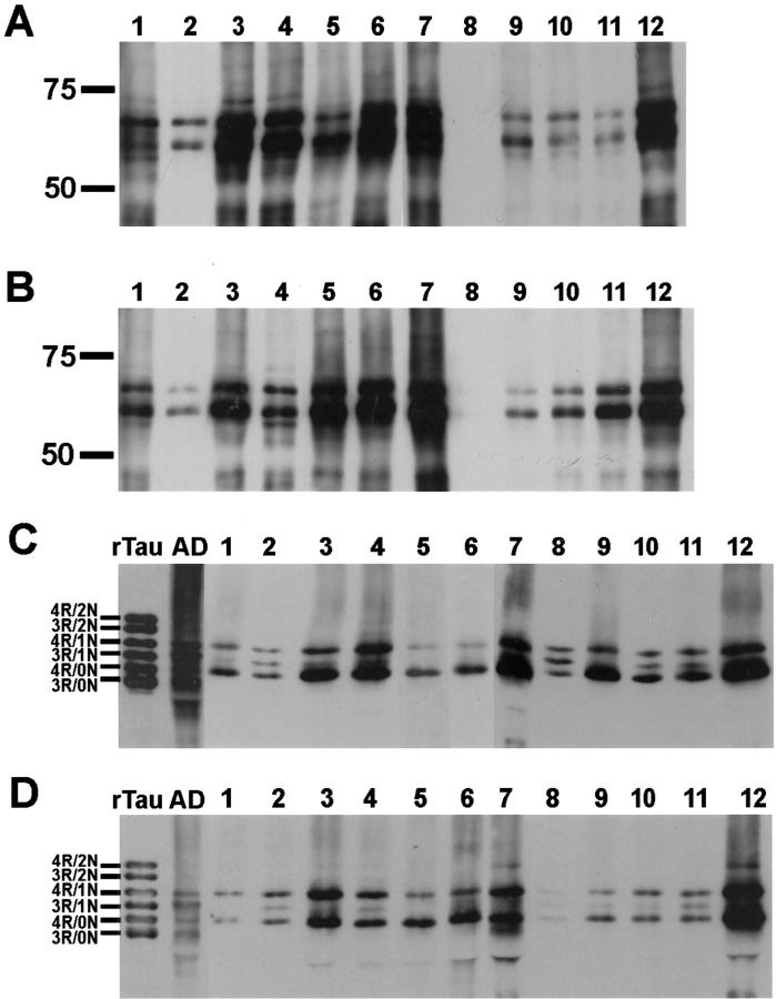
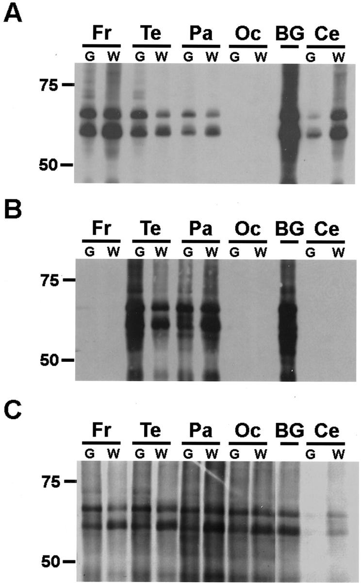
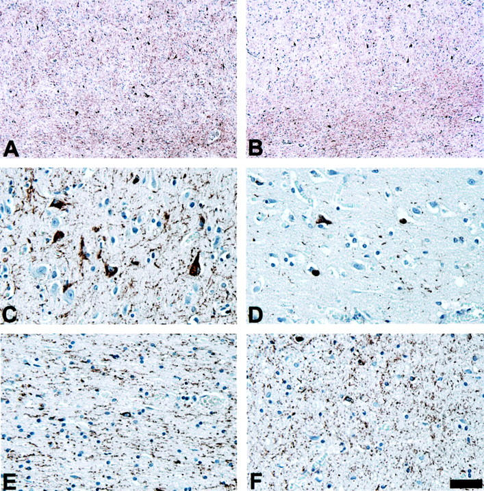
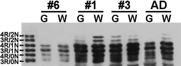
Corticobasal degeneration (CBD) is an adult-onset progressive neurodegenerative disorder characterized by L-dopa-resistant rigidity, focal cortical deficits, and variable dementia. The neuropathological hallmark of CBD is the deposition of filamentous inclusions in neurons and glia composed of hyperphosphorylated tau with only four microtubule-binding repeats (4R-tau). To characterize the regional burden of tau pathology in CBD, we studied 12 brains with the neuropathological diagnosis of CBD using biochemical and histochemical techniques. Eleven brain regions were evaluated including gray and white matter from frontal, parietal, temporal, and occipital lobes and cerebellum as well as basal ganglia. Although the distribution of tau pathology was variable, neuropathological and biochemical data showed a similar burden of tau abnormalities in frontal, temporal, and parietal lobes and basal ganglia of both hemispheres. This included abundant, sarkosyl-insoluble 4R-tau in both gray and white matter of two or more of these cortical regions and basal ganglia, and to a lesser extent, cerebellar white matter. The insoluble tau pathology in gray and white matter showed overlapping but distinct phosphorylated epitopes suggesting cell-type and subcellular localization (ie, cell bodies versus cell processes)-specific differences in tau phosphorylation. In contrast, soluble tau was composed of normal 4R/3R-tau ratios indicating no gross abnormality in tau splicing. Thus, although clinically heterogeneous, CBD is a distinct lobar and basal ganglionic tauopathy with selective aggregation of 4R-tau.


Figures

Figure 1.
Figure 1.
Characteristic brain lesions in CBD. A: Astrocytic plaque in neocortex (tau immunostain, PHF1). B: Balloon neuron in neocortex (neurofilament immunostain, RMO24). C: Neuronal cytoplasmic inclusions in nucleus basalis (tau immunostain, PHF1). D: Astrocytic cytoplasmic inclusion in neocortex (tau immunostain, PHF1). E: Oligodendroglial inclusion (coiled body) in subcortical white matter (tau immunostain, PHF1). Scale bars: 30 μm (A); 10 μm (B–E).
Figure 2.
Figure 2.
Topographic distribution of tau-immunoreactive pathology shows regional variability. A: Marked thread pathology in the basal ganglia of patient 10. B: In contrast, the occipital lobe of patient 10 is virtually devoid of tau pathology. C and D: Thread pathology in both the frontal lobe and occipital lobe of patient 7 who presented with a visual agnosia. Tau immunostains, PHF1. Scale bar, 80 μm.
Figure 3.
Figure 3.
Western blot analysis of insoluble tau in gray and white matter of the parietal lobe of CBD brains shows marked interpatient variability. Insoluble tau fractions extracted from gray (A) and white (B) matter of the parietal lobe of 12 CBD brains were resolved by SDS-PAGE and immunoblotted with PHF1. The insoluble tau protein is composed predominantly of two major proteins of 64 and 68 kd. Aliquots of the insoluble fractions were dephosphorylated with E. coli alkaline phosphatase, resolved by SDS-PAGE, and immunoblotted with T14 and T46 that recognize phosphorylation-independent tau epitopes on the amino- and carboxy terminus, respectively. On dephosphorylation, the insoluble tau fractions from gray (C) and white (D) matter co-migrate with 4R-tau; 3R-tau with one amino terminal insert could occasionally be detected in some of the cases. In contrast, in AD, dephosphorylated, insoluble tau fractions co-migrate with both 4R-tau and 3R-tau isoforms. Prolonged exposure of the immunoblots revealed at least mild tau pathology in all samples as reflected in Table 4 ▶ . Recombinant tau isoforms (rTau) are indicated to the left of C and D. Molecular weight standards are as indicated to the left of each panel here and in Figures 4 and 5 ▶ ▶ .
Figure 4.
Figure 4.
Western blot analysis of insoluble tau in CBD shows marked regional variability. Insoluble tau fractions from the brain regions indicated were resolved by SDS-PAGE and immunoblotted with PHF1. The insoluble tau protein from all brain regions is composed predominantly of two major proteins of 64 and 68 kd. Patient 2 (A), who presented clinically with an unclassified dementia, shows the most severe pathology in the frontal and temporal lobes. Patient 1 (B), who was diagnosed clinically with AD, shows the most severe pathology in the parietal and temporal lobes. Prolonged exposure of the immunoblot revealed mild tau pathology in frontal and occipital lobes as well as cerebellum as reflected in Table 4 ▶ . In contrast, patient 4 (C), who was diagnosed clinically with frontotemporal dementia, shows moderate pathology in all neocortical regions. Fr, frontal lobe; Te, temporal lobe; Pa, parietal lobe; Oc, occipital lobe; BG, basal ganglia; Ce, cerebellum; G, gray matter; W, white matter.
Figure 5.
Figure 5.
Western blot analysis of insoluble tau shows distinct phosphorylation profiles in white matter. Insoluble tau fractions from gray and white matter of the frontal lobe of CBD and AD patients were resolved by SDS-PAGE and immunoblotted with the epitope-specific, phosphorylation-dependent antibodies PHF1 and AT8 as indicated. PHF1 (B) detects similar patterns of phosphorylation in both gray and white matter. In contrast, AT8 (A), specific for phosphorylated Ser 202 and Thr 205, detects increased tau pathology in the white matter of the majority (10 of 12) of the cases. G, gray matter; W, white matter.
Figure 6.
Figure 6.
AT8 preferentially detects tau pathology in the white matter of CBD patients. Adjacent sections from the mid-frontal gyrus of patient 7 were immunostained with PHF1 (A, C, and E) or AT8 (B, D, and F). Although PHF1 detects abundant tau pathology in both the gray (C) and white (E) matter, AT8 preferentially detects the white matter pathology (F). A and B: Low-power photomicrographs of frontal lobe immunostained with PHF1 (A) and AT8 (B). C–F: High-power photomicrographs of cortex (C and D) and white matter (E and F) represented in A and B immunostained with PHF1 (C and E) and AT8 (D and F). Scale bars: 200 μm (A and B); 40 μm in (C–F).
Figure 7.
Figure 7.
Western blot analysis of soluble tau shows similar levels of tau isoform expression in gray and white matter. Soluble tau fractions from gray and white matter of CBD and AD patients were dephosphorylated with E. coli alkaline phosphatase, resolved by SDS-PAGE, and immunoblotted with T14. The soluble extracts showed similar levels of tau isoform expression in both gray and white matter, similar to that observed in AD patients. Recombinant tau isoforms (rTau) are as indicated.

References

    1. Rebeiz JJ, Kolodny EH, Richardson EP, Jr: Corticodentatonigral degeneration with neuronal achromasia. Arch Neurol 1968, 18:20-33 - PubMed
    1. Riley DE, Lang AE, Lewis A, Resch L, Ashby P, Hornykiewicz O, Black S: Cortical-basal ganglionic degeneration. Neurology 1990, 40:1203-1212 - PubMed
    1. Gibb WR, Luthert PJ, Marsden CD: Corticobasal degeneration. Brain 1989, 112:1171-1192 - PubMed
    1. Rinne JO, Lee MS, Thompson PD, Marsden CD: Corticobasal degeneration. A clinical study of 36 cases. Brain 1994, 117:1183-1196 - PubMed
    1. Wenning GK, Litvan I, Jankovic J, Granata R, Mangone CA, McKee A, Poewe W, Jellinger K, Ray CK, D’Olhaberriague L, Pearce RK: Natural history and survival of 14 patients with corticobasal degeneration confirmed at postmortem examination. J Neurol Neurosurg Psychiatry 1998, 64:184-189 - PMC - PubMed

Publication types

LinkOut - more resources