. 2002 Jun;160(6):2123-33.
doi: 10.1016/S0002-9440(10)61161-3.

E- and N-cadherin distribution in developing and functional human teeth under normal and pathological conditions

Affiliations

E- and N-cadherin distribution in developing and functional human teeth under normal and pathological conditions

Robert Heymann et al. Am J Pathol. 2002 Jun.

Abstract

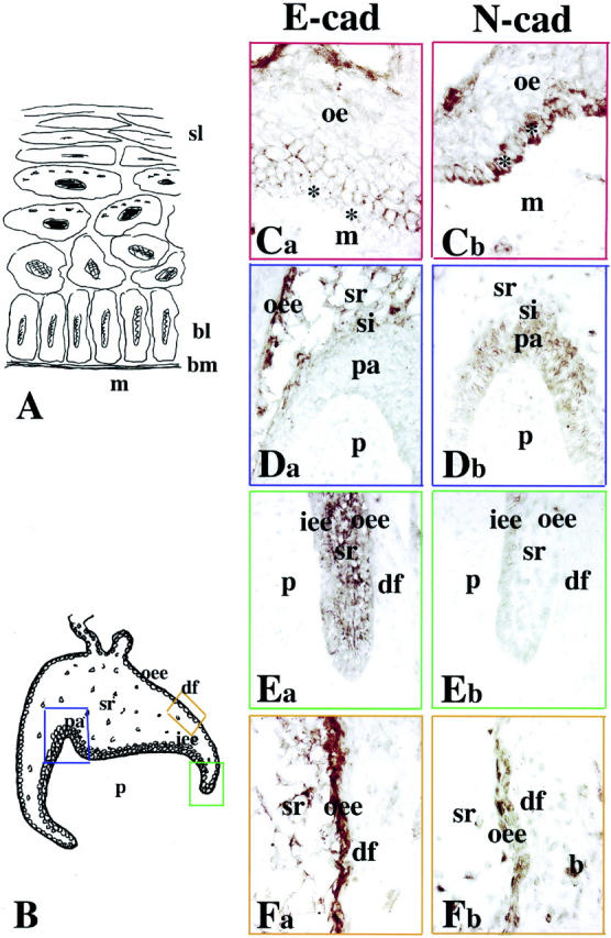
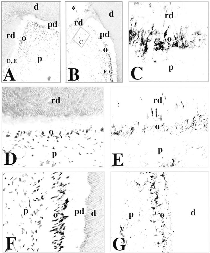
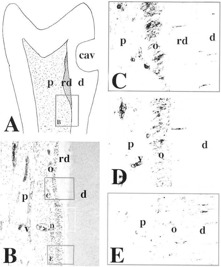
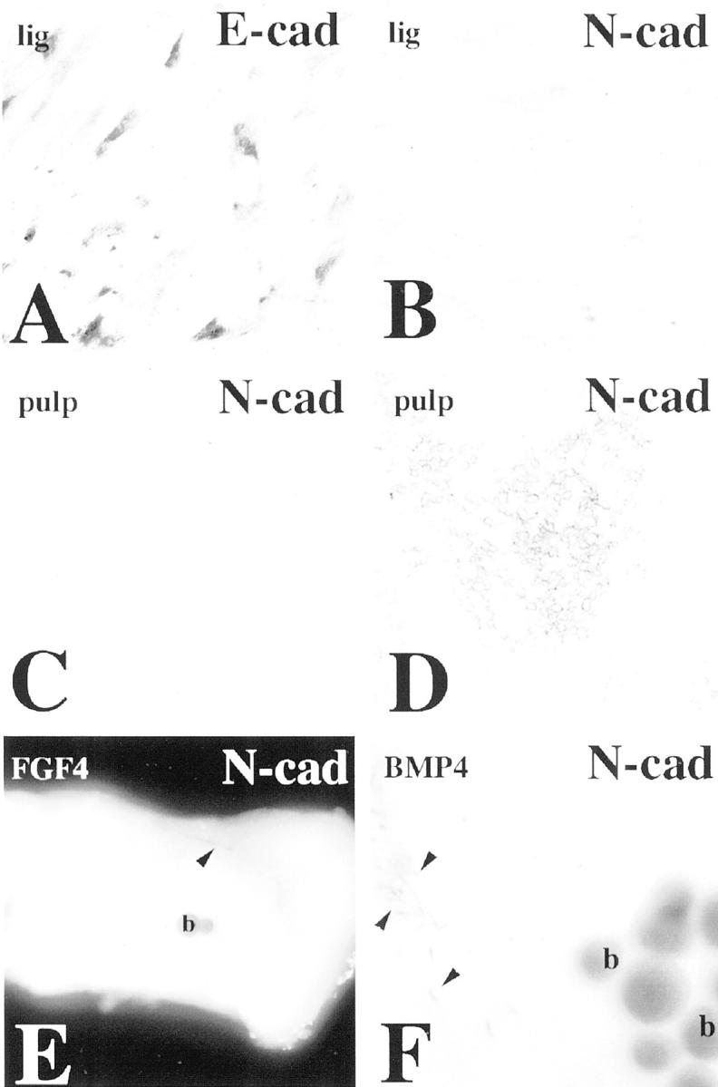
Cadherins are calcium-dependent cell adhesion molecules involved in the regulation of various biological processes such as cell recognition, intercellular communication, cell fate, cell polarity, boundary formation, and morphogenesis. Although previous studies have shown E-cadherin expression during rodent or human odontogenesis, there is no equivalent study available on N-cadherin expression in dental tissues. Here we examined and compared the expression patterns of E- and N-cadherins in both embryonic and adult (healthy, injured, carious) human teeth. Both proteins were expressed in the developing teeth during the cap and bell stages. E-cadherin expression in dental epithelium followed an apical-coronal gradient that was opposite to that observed for N-cadherin. E-cadherin was distributed in proliferating cells of the inner and outer enamel epithelia but not in differentiated cells such as ameloblasts, whereas N-cadherin expression was up-regulated in differentiated epithelial cells. By contrast to E-cadherin, N-cadherin was also expressed in mesenchymal cells that differentiate into odontoblasts and produce the hard tissue matrix of dentin. Although N-cadherin was not detected in permanent intact teeth, it was re-expressed during dentin repair processes in odontoblasts surrounding carious or traumatic sites. Similarly, N-cadherin re-expression was seen in vitro, in cultured primary pulp cells that differentiate into odontoblast-like cells. Taken together these results suggest that E- and N-cadherins may play a role during human tooth development and, moreover, indicate that N-cadherin is important for odontoblast function in normal development and under pathological conditions.


Figures

Figure 1.
Figure 1.
Comparison between the patterns of E- and N-cadherin immunostainings in oral epithelium and deciduous first molars. Ca, Da, Ea, and Fa: E-cadherin labeling; Cb, Db, Eb, and Fb: N-cadherin labeling. A: Schematic illustration of the oral epithelium. Note the presence of different epithelial cell layers. B: Schematic representation of a deciduous human first molar at the early bell stage of development (18 gestational weeks). The colored frames show the areas used for the comparative study. Ca and Cb: Oral epithelium (red frames). E-cadherin is distributed at the surface of cells overlying the proliferating N-cadherin-positive cells of the basal layer (asterisks). Da and Db: Cusp area (blue frames). Combinatory patterns of E- and N-cadherins: E-cadherin immunoreactivity is evident in outer enamel epithelium and stellate reticulum, whereas N-cadherin is observed in pre-ameloblasts and stratum intermedium. Note the weak N-cadherin staining in some dental pulp cells. Ea and Eb: Cervical loop area (green frames). Strong E-cadherin and faint N-cadherin staining in epithelial cells. Fa and Fb: Intermediate area (orange frames). Strong E-cadherin and weaker N-cadherin reactivity in outer enamel epithelium and stellate reticulum. Note also a positive staining in several mesenchymal cells surrounding the tooth germ. Abbreviations: b, bone forming cells; bl, basal layer; bm, basement membrane; de, dental epithelium; df, dental follicle; iee, inner enamel epithelium; m, mesenchyme; oe, oral epithelium; oee, outer enamel epithelium; p, dental papilla (pulp); pa, pre-ameloblasts; si, stratum intermedium; sl, squamus layer; sr, stellate reticulum.
Figure 2.
Figure 2.
Comparison between the E- and N-cadherin staining patterns in deciduous first incisors. Ca, Da, Ea, Fa, and Ga: E-cadherin labeling; B, Cb, Db, Eb, Fb, and Gb: N-cadherin labeling. A: Schematic illustration of a deciduous human first incisor at the late bell stage of development (30 gestational weeks). The colored frames represent the areas used for the comparative study. B: Mesenchymal part of the cusp area (blue frame). N-cadherin staining is present in odontoblasts and absent from pulp fibroblasts. Note that the gradient of N-cadherin staining in odontoblasts follows their maturation degree. Ca and Cb: Epithelial part of the cusp area (green frames). E-cadherin reactivity is restricted in outer enamel epithelium, whereas N-cadherin is detected in ameloblasts, stratum intermedium, and outer enamel epithelium. Note that in stellate reticulum the E- and N-cadherin immunoreactivities are weak. Da and Db: Intermediate area (red frames). Strong E-cadherin staining in outer enamel epithelium and weaker in pre-ameloblasts and inner enamel epithelium. Heavy N-cadherin labeling in pre-ameloblasts and weaker in outer enamal epithelium and inner enamel epithelium. Ea and Eb: Cervical loop area (orange frames). E- and N-cadherin reactivities in dental epithelial cells. Note a positive staining in mesenchymal cells forming the alveolar bone. Fa and Fb: Mesenchymal part of the cusp area (blue frames). Presence of N-cadherin and absence of E-cadherin staining in newly differentiated odontoblasts. Ga and Gb: Mesenchymal part of the cusp area (blue frames). Strong N-cadherin and very faint E-cadherin reactivities in functional odontoblasts. Abbreviations: a, ameloblasts; d, dentin; df, dental follicle; e, enamel; iee, inner enamel epithelium; o, odontoblast; oee, outer enamel epithelium; p, dental pulp; pa, pre-ameloblasts; si, stratum intermedium; sr, stellate reticulum.
Figure 3.
Figure 3.
Immunohistochemical localization of N-cadherin in sections of carious permanent human teeth. A and B: H&E staining. In carious teeth, tertiary or reparative dentin is seen near the sites of the carious lesions (A and B; asterisk in B). The frames represent the sites shown in the following panels. C: Higher magnification of the framed area in B. N-cadherin reactivity is found in odontoblasts involved in reparative dentin production. D and E: Higher magnification of the framed area in A (D), showing N-cadherin immunostaining in odontoblasts producing the reparative dentin (E). Note that the staining is distributed in both odontoblast cell bodies and processes. F and G: Higher magnification of the framed area in B (F), showing N-cadherin immunoreactivity in odontoblasts at a distance from the carious lesion (G). Note that the staining in odontoblasts is restricted to the odontoblast cell bodies. Cells in dilated blood vessels are also positive for N-cadherin. Abbreviations: d, dentin; o, odontoblasts; p, pulp; pd, predentin; rd, reparative dentin; v, vessel.
Figure 4.
Figure 4.
Immunohistochemical localization of N-cadherin in sections of permanent human premolars after cavity preparation. A: Schematic illustration of a premolar showing reparative dentin production, 9 weeks after class V cavity preparation. The frame represents the area shown in the B. B: H&E staining. The frames indicate the areas shown in C, D, and E. C: A strong N-cadherin labeling is observed in odontoblasts producing the reparative dentin. Note that the staining is detected in both odontoblast bodies and odontoblast processes. D: N-cadherin reactivity is decreased in odontoblasts near to the injury site, but not involved in reparative dentin synthesis. Note that blood vessels are N-cadherin-positive. E: N-cadherin staining is very faint in odontoblasts far away from the injury site. Note that the staining persists in some of the odontoblast processes. Abbreviations: cav, cavity; d, dentin; o, odontoblasts; p, pulp; rd, reparative dentin; v, vessel.
Figure 5.
Figure 5.
E- and N-cadherin immunoreactivities in human dental pulp explants, pulp cells, and periodontal ligament cells cultured in vitro. A: Confluent periodontal ligament cells exhibit E-cadherin staining. B: Confluent periodontal ligament cells are negative for N-cadherin. C: Confluent pulp cells cultured without β-glycerophosphate are negative for N-cadherin. D: A cell-surface staining for N-cadherin is observed in pulp cells implicated in the formation of the nodules after β-glycerophosphate treatment. E: FGF4 beads have no effect on N-cadherin expression in human dental pulp explants cultured for 14 hours. Note the positive N-cadherin staining in nerve fibers (arrowhead). F: N-cadherin immunoreactivity is not found in pulp cells surrounding the BMP4 beads. Note the positive staining of the nerve fibers (arrowheads). Abbreviation: b, bead.

References

    1. Peters H, Balling R: Teeth. Where and how to make them. Trends Genet 1999, 15:59-65 - PubMed
    1. Ruch JV, Lesot H, Begue-Kirn C: Odontoblast differentiation. Int J Dev Biol 1995, 39:51-68 - PubMed
    1. Thesleff I, Nieminen P: Tooth differentiation and cell differentiation. Curr Opin Cell Biol 1996, 8:844-850 - PubMed
    1. Thesleff I, Sharpe P: Signalling networks regulating dental development. Mech Dev 1997, 67:111-123 - PubMed
    1. Zeichner-David M, Diekwisch T, Fincham A, Lau E, MacDougall M, Moradian-Oldak J, Simmer J, Snead M, Slavkin HC: Control of ameloblast differentiation. Int J Dev Biol 1995, 39:69-92 - PubMed

Publication types

LinkOut - more resources