. 2002 Jun;160(6):2145-55.
doi: 10.1016/S0002-9440(10)61163-7.

Atherosclerotic lesions grow through recruitment and proliferation of circulating monocytes in a murine model

Affiliations

Atherosclerotic lesions grow through recruitment and proliferation of circulating monocytes in a murine model

Susan M Lessner et al. Am J Pathol. 2002 Jun.

Abstract

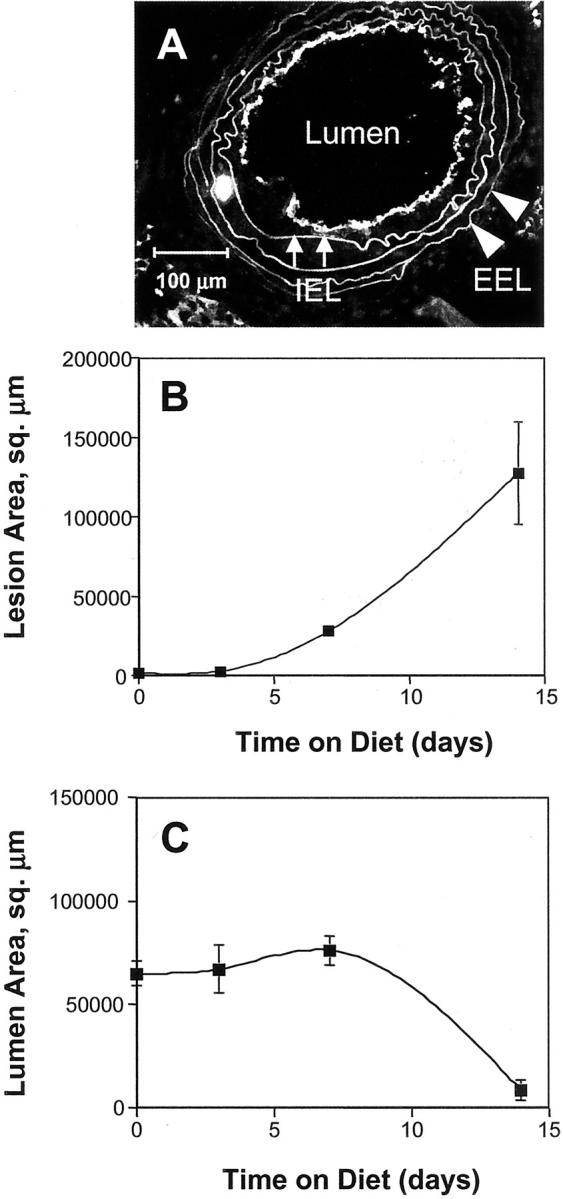
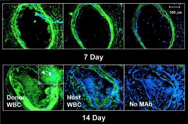
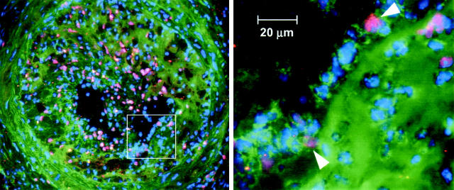
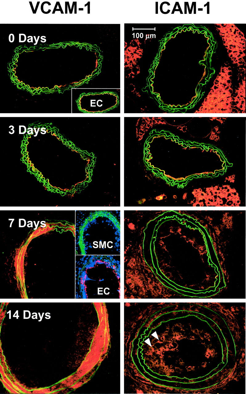
Macrophage-derived foam cells in developing atherosclerotic lesions may potentially originate either from recruitment of circulating monocytes or from migration of resident tissue macrophages. In this study, we have determined the source of intimal macrophages in the apoE-knockout mouse flow-cessation/hypercholesterolemia model of atherosclerosis using a bone marrow transplantation approach. We also examined the time course and spatial distribution of intercellular adhesion molecule-1 and vascular cell adhesion molecule-1 expression to assess whether endothelial adhesion molecules were involved in recruitment of either circulating monocytes or resident macrophages. We used allelic variants of the mouse common leukocyte antigen (CD45) to distinguish host-derived and donor-derived white blood cells (WBCs) both in blood and in macrophage-rich carotid lesions. We found that the distribution of CD45 isoforms in lesions is similar to that of circulating WBCs, whereas the host-type CD45 isoform is more prevalent in resident adventitial macrophages. These data indicate that macrophage-derived foam cells in the lesion derive mainly from circulating precursors rather than from resident macrophages. The corresponding time course of intercellular adhesion molecule-1 and vascular cell adhesion molecule-1 expression suggests that recruitment of circulating WBCs by endothelial adhesion molecules is likely to be more important during lesion initiation than during the later phase of rapid lesion growth.


Figures

Figure 1.
Figure 1.
Flow cytometric analysis of isolated peripheral WBCs demonstrates successful engraftment of CD45.2 donor bone marrow in CD45.1 recipient mice. A and B: Isolated peripheral WBCs stained with phycoerythrin-labeled anti-CD45.1 and fluorescein isothiocyanate-labeled anti-CD45.2 mAbs were analyzed by flow cytometry. A: In an apoE KO mouse homozygous for CD45.1, all peripheral WBCs stain positively for CD45.1. B: Six weeks after transplant with bone marrow from an apoE KO mouse homozygous for CD45.2, donor-type WBCs predominate in the circulation (cluster of points on x axis). Additional in vitro experiments with mixed populations of homozygous cells (data not shown) suggest that the double-labeled objects correspond to cell aggregates that account for 6% of gated events on average. These events have been excluded from the calculation of percentage engraftment.
Figure 2.
Figure 2.
Development of macrophage-rich lesions in ligated carotid arteries of ApoE KO mice that have undergone BMT leads to lumenal occlusion after 14 days on hypercholesterolemic diet. A: Representative frozen section used for morphometric analysis. Scale bar, 100 μm. Morphometric parameters were measured by image analysis of frozen sections immunostained with anti-CD31 to visualize the endothelium as a boundary for the vessel lumen. Elastin autofluorescence was used to identify the IEL (small arrows) and the EEL (arrowheads). Average values of morphometric parameters were calculated for groups of four to six mice sacrificed at the indicated time points after carotid ligation. Morphometric analysis of lesion area (B) and lumen area (C). Error bars, 1 SEM.
Figure 3.
Figure 3.
Donor-derived inflammatory cells predominate in mouse carotid lesions initiated by flow cessation/hypercholesterolemia. Serial frozen sections were stained without primary antibody (negative controls) or with mAbs to CD45.2 (donor WBCs) or CD45.1 (host WBCs). Sections were counterstained with Hoechst dye (blue) to visualize cell nuclei. Left: Numerous donor-derived (CD45.2) WBCs were evident in the neointima at both 7 and 14 days after ligation. Cells expressing CD45.2 surface antigen have distinct green borders (inset, arrowheads). Middle: In contrast, few host-derived (CD45.1-positive) WBCs were visible in lesions at either time point. Right: In negative controls, only elastin autofluorescence appears green. Scale bar, 100 μm.
Figure 4.
Figure 4.
Distribution and source (host versus donor) of macrophages in mouse carotid lesions throughout a 14-day time course. Frozen sections of carotid lesions were double labeled with mAbs to Mac-3 and CD45.2 to identify macrophages and donor-type WBCs, respectively. The corresponding lipid distribution was visualized by Nile Red staining. Top left: In mice that had undergone BMT, both host-type (red) and donor-type (yellow) macrophages were evident in the carotid adventitia before ligation (0 day). Top right: By 3 days after ligation, donor-type monocyte/macrophages (yellow) had begun to adhere to the endothelium, initiating neointima formation. Scale bars, 20 μm. Row 2, left: Macrophages occupied ∼35% of the lesion area (red staining) at 14 days after ligation. Row 2, right: CD45.2 mAb recognizes an antigen on the cell surface, resulting in strong labeling along leukocyte borders (green areas). Row 3, left: Superimposing the panels in row 2 produces an image in which donor-type macrophages appear as orange to yellow cells with green or yellow borders. Double labeling demonstrates that the majority of macrophages in the lesion are donor-derived. Row 3, right: Lipid distribution at 14 days after ligation. Scale bars, 100 μm. Controls (bottom left) Only host-type macrophages (red) were present in the adventitia of nontransplanted apoE KO mice homozygous for CD45.1. Right: No lesion is evident in the nonligated, contralateral vessel of transplanted mice. Both host-type (red) and donor-type (yellow) macrophages appear in the adventitia. Scale bar, 100 μm.
Figure 5.
Figure 5.
Donor-derived WBCs proliferate in the neointima of mouse carotid lesions. Frozen sections were immunostained with anti-BrdU mAb to identify proliferating cell nuclei (pink) and with anti-CD45.2 mAb to identify donor-derived WBCs (green outlines, arrowheads at right). Nuclei were counterstained with Hoechst 33258. Left: A cross-section of the entire vessel, with a boxed area indicating the region of the close-up highlighted at the right. Scale bar, 20 μm.
Figure 6.
Figure 6.
VCAM-1 and ICAM-1 expression in ligated mouse carotid arteries follow distinct time courses. Frozen sections were stained with mAb to either VCAM-1 (left) or ICAM-1 (right), followed by Rhodamine Red X-labeled secondary reagents. Before ligation and up to 3 days after ligation, VCAM-1 expression was confined to the luminal boundary of the vessel, where it co-localized with endothelial cells as demonstrated by CD31 staining (inset, top). By 7 days, VCAM-1 expression had disappeared from the endothelium (row 3, bottom inset) and had shifted to the media, where it co-localized with vascular SMCs, as demonstrated by staining for SM α-actin (green areas, top inset). This pattern continued up to 14 days after ligation (bottom). Before lesion initiation, ICAM-1 expression was confined to the luminal boundary of the artery (top right). By 3 days after ligation, some ICAM-1 staining could be detected in the adventitia as well. Adventitial staining became stronger at 7 and 14 days after ligation, coupled with a decrease in relative intensity of luminal staining. By 14 days after ligation, ICAM-1 was expressed by a subset of cells deeper within the neointima (arrowheads). Scale bar, 100 μm.

References

    1. Gerrity RG: The role of the monocyte in atherogenesis. I. Transition of blood-borne monocytes into foam cells in fatty lesions. Am J Pathol 1981, 103:181-190 - PMC - PubMed
    1. Joris I, Zand T, Nunnari JJ, Krolikowski FJ, Majno G: Studies on the pathogenesis of atherosclerosis. I. Adhesion and emigration of mononuclear cells in the aorta of hypercholesterolemic rats. Am J Pathol 1983, 113:341-358 - PMC - PubMed
    1. Stary HC: The intimal macrophage in atherosclerosis. Artery 1980, 8:205-207 - PubMed
    1. Miyata K, Shimokawa H, Kandabashi T, Higo T, Morishige K, Eto Y, Egashira K, Kaibuchi K, Takeshita A: Rho-kinase is involved in macrophage-mediated formation of coronary vascular lesions in pigs in vivo. Arterioscler Thromb Vasc Biol 2000, 20:2351-2358 - PubMed
    1. Rosenfeld ME, Tsukada T, Gown AM, Ross R: Fatty streak initiation in Watanabe heritable hyperlipidemic and comparably hypercholesterolemic fat-fed rabbits. Arteriosclerosis 1987, 7:9-23 - PubMed

Publication types

MeSH terms

Substances