Comparative Study
. 2002 Jun;160(6):2181-90.
doi: 10.1016/S0002-9440(10)61166-2.

Gene expression in Wilms' tumor mimics the earliest committed stage in the metanephric mesenchymal-epithelial transition

Affiliations
Comparative Study

Gene expression in Wilms' tumor mimics the earliest committed stage in the metanephric mesenchymal-epithelial transition

Chi-Ming Li et al. Am J Pathol. 2002 Jun.

Abstract

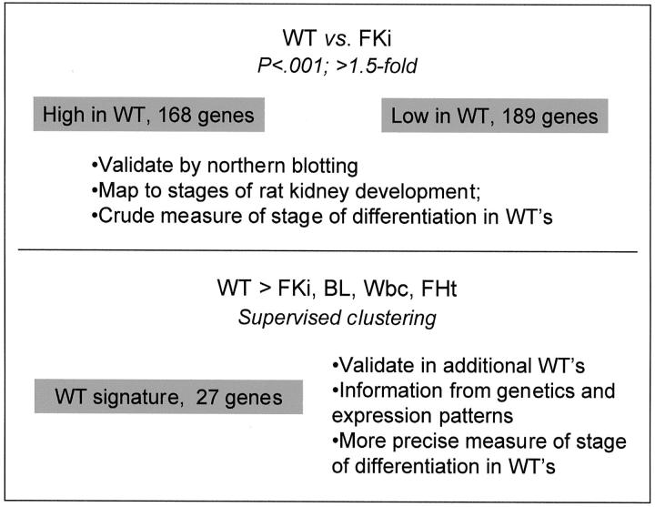
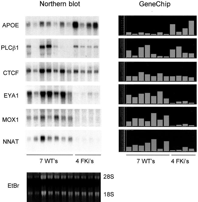
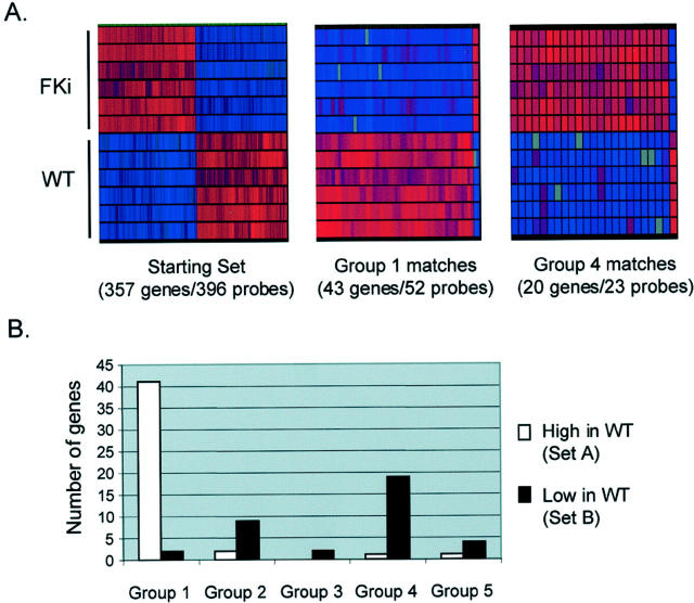
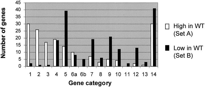
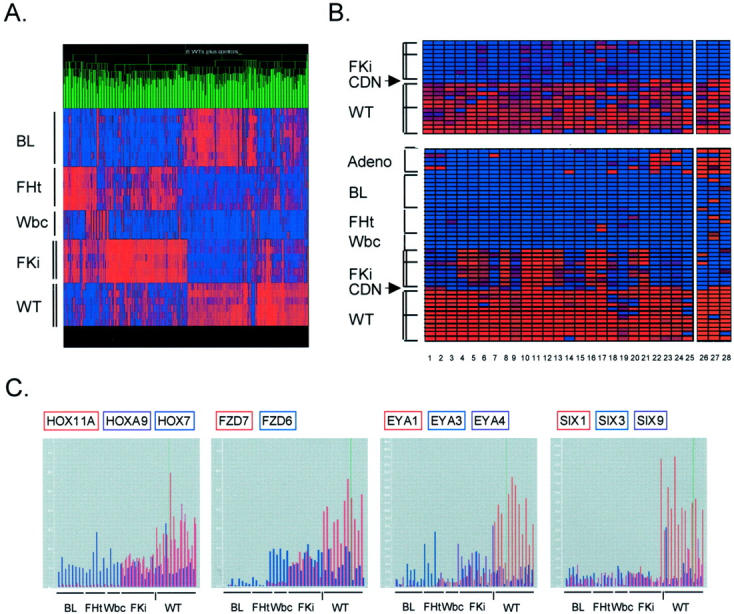
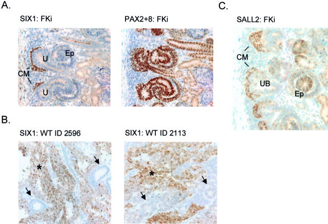
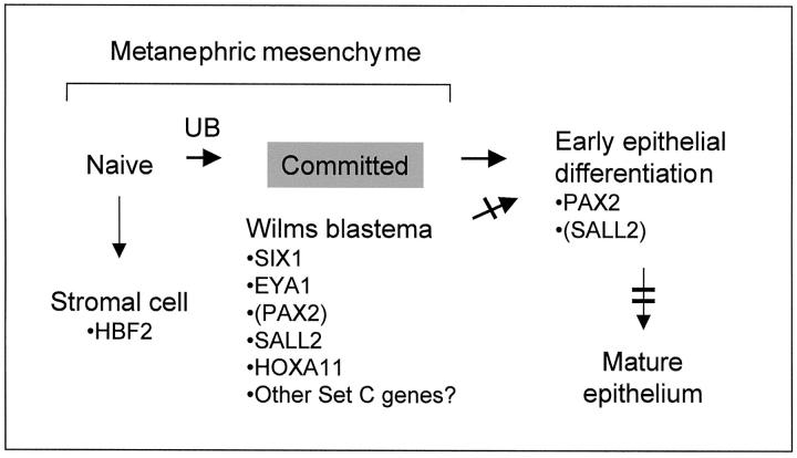
Wilms' tumor (WT) has been considered a prototype for arrested cellular differentiation in cancer, but previous studies have relied on selected markers. We have now performed an unbiased survey of gene expression in WTs using oligonucleotide microarrays. Statistical criteria identified 357 genes as differentially expressed between WTs and fetal kidneys. This set contained 124 matches to genes on a microarray used by Stuart and colleagues (Stuart RO, Bush KT, Nigam SK: Changes in global gene expression patterns during development and maturation of the rat kidney. Proc Natl Acad Sci USA 2001, 98:5649-5654) to establish genes with stage-specific expression in the developing rat kidney. Mapping between the two data sets showed that WTs systematically overexpressed genes corresponding to the earliest stage of metanephric development, and underexpressed genes corresponding to later stages. Automated clustering identified a smaller group of 27 genes that were highly expressed in WTs compared to fetal kidney and heterologous tumor and normal tissues. This signature set was enriched in genes encoding transcription factors. Four of these, PAX2, EYA1, HBF2, and HOXA11, are essential for cell survival and proliferation in early metanephric development, whereas others, including SIX1, MOX1, and SALL2, are predicted to act at this stage. SIX1 and SALL2 proteins were expressed in the condensing mesenchyme in normal human fetal kidneys, but were absent (SIX1) or reduced (SALL2) in cells at other developmental stages. These data imply that the blastema in WTs has progressed to the committed stage in the mesenchymal-epithelial transition, where it is partially arrested in differentiation. The WT-signature set also contained the Wnt receptor FZD7, the tumor antigen PRAME, the imprinted gene NNAT and the metastasis-associated transcription factor E1AF.


Figures

Figure 1.
Figure 1.
Strategy for analyzing gene expression in WTs. For a gene to be included in set A or set B, we first required a value of P < 0.001 by a chi-square test for differential expression between the WTs and the control fetal kidneys. We additionally required a normalized expression value of >1.5 relative to the experiment mean in at least three samples, either WTs or fetal kidneys.
Figure 2.
Figure 2.
Validation of the GeneChip data by comparisons with Northern blotting. Left: A single Northern blot was successively hybridized with cDNA probes for the indicated genes. Right: The GeneChip expression data (normalized to the experiment mean) are displayed with the same order of samples. EtBr, ethidium bromide stain of the Northern gel showing 28S and 18S ribosomal RNAs.
Figure 3.
Figure 3.
Correlation of gene expression in WTs with normal kidney development in the rat. A: Genes identified in the current study that mapped to genes identified by Stuart and colleagues. Genes that are more highly expressed in WTs than in whole fetal kidneys map predominantly to the earliest group of Stuart and colleagues, whereas genes that are expressed at lower levels in WTs than in fetal kidneys map to the later groups. B: Graphical representations (GeneTree function, GeneSpring) showing the uniformity of relative overexpression or underexpression across multiple genes and multiple samples. Genes are arrayed along the x axis; WT and fetal kidney samples are shown on the y axis. Gene expression is automatically color-coded with red indicating higher and blue lower expression. Gray indicates Affymetrix absence calls. The group 1 matches are nearly all more highly expressed in WTs, whereas the group 4 matches are nearly all more highly expressed in fetal kidneys. Color range is set at 1 = normal, 6 = maximum.
Figure 4.
Figure 4.
Functional categories of genes differentially expressed between WTs and fetal kidneys. The categories are: 1, DNA synthesis or metabolism (including repair); 2, cell cycle regulation (including mitotic spindle); 3, RNA metabolism (not including transcription factors); 4, transcriptional regulation (including transcription factors); 5, cell signaling; 6a, protein metabolism nonproteolytic; 6b, protein metabolism proteolytic; 7, small molecule metabolism (enzymes); 8, small molecule transport; 9, extracellular matrix, adhesion, and serum proteins; 10, intracellular trafficking; 11, energy metabolism; 12, cytoskeleton (not including mitotic spindle); 13, protection against oxidative stress; 14, unknown function.
Figure 5.
Figure 5.
Derivation of WT signature genes. A: Nonsupervised clustering (GeneTree function of GeneSpring) of the 357 genes that were initial identified as differentially expressed between WTs and fetal kidneys. Many of the high in WT/low in fetal kidney genes are also high in the BLs. The color range is normal = 1, maximum = 6. All data are included, with absence calls represented by pure blue. B: WT signature genes. The 27 highest-scoring genes identified by SPLASH are shown here, after clustering using the GeneTree function of GeneSpring (columns 1 and 2 are two probe sets both identifying CRABP2). The identities of these genes are in Table 1 ▶ . Tissues are: AdenoCA, pulmonary adenocarcinoma (six primary tumors); BL, Burkitt lymphoma (four primary tumors and four cell lines); FHt, mid-gestation fetal hearts (six samples); Wbc, normal peripheral blood (four samples); FKi, mid-gestation fetal kidneys (six samples in A and the same six samples below the dividing line in B, with three additional fetal kidneys above the line); CDN, differentiating cystic nephroblastoma (one case); WT, (six WTs in A and the same six WTs below the dividing line in B, with six additional WTs above the line). Tissues used for analysis by Genes at Work are bracketed; the adenocarcinomas were not included in deriving the WT signature genes. The genes in set C were identified by SPLASH using only data from the samples shown in A. The additional WTs and fetal kidneys, above the dividing lines, therefore validate the signature set. The differentiating cystic nephroblastoma shows a distinct pattern of gene expression. Because the data are normalized to all tissues, displaying the WT and fetal kidney samples alone (top) emphasizes the differences between them, while displaying these samples together with the heterologous tissues highlights their close affinity. Color ranges are set at 1 = normal, 6 = maximum (top) and 2 = normal, 10 = maximum (bottom). C: Differential expression of genes in the HOX-, FZD-, EYA-, and SIX-families. Microarray data are normalized to the experiment mean. Each bar represents the value for a single sample. BL, Burkitt lymphoma (eight samples); FHt, fetal heart (six samples); Wbc, peripheral blood leukocytes (four samples); FKi, fetal kidney (nine samples); WT, Wilms’ tumor (12 samples). The vertical dash indicates the single case of differentiating cystic nephroblastoma.
Figure 6.
Figure 6.
Immunohistochemical detection of SIX1 and SALL2 proteins in human fetal kidney and in WTs. A: Serial sections of a 20-week gestation human fetal kidney were developed with anti-SIX1 and anti-PAX2 + 8 polyclonal antisera. SIX1 is expressed only in the earliest condensing mesenchyme (metanephric blastema), which is seen directly apposed to the uretic bud-derived epithelium. PAX2 + 8 detects expression of these two proteins in the condensing mesenchyme, and there is a strong signal in differentiating epithelial structures (S-shaped and comma-shaped bodies) deeper in the kidney, as well as in ureteric bud. Weak cytoplasmic staining in differentiated epithelial cells is nonspecific (seen in control with secondary antibody alone). B: SIX1 is expressed in the blastemal component of WTs (asterisks), but not in the epithelial areas (arrows). This pattern was seen in three of three cases of WT examined. In these same cases the epithelial component stained strongly with anti-PAX2 + 8 (not shown). C: Anti-SALL2 polyclonal antiserum gives a strong and uniform signal in the condensing mesenchyme, but cells with nuclear SALL2 are also seen at lower frequencies and with somewhat reduced staining in differentiating epithelial structures. Ureteric bud derivatives are negative for SALL2. Weak cytoplasmic staining in differentiated epithelial cells is nonspecific. CM, condensing mesenchyme; U, ureteric bud branches; Ep, differentiating epithelial structures; Gl, primitive glomeruli; Tu, differentiated renal tubules.
Figure 7.
Figure 7.
Differentiation arrest in WTs inferred from gene expression profiling. A schematic of the normal metanephric differentiation program is shown. The neoplastic blastema, which comprises the bulk of many WTs, is arrested at the position indicated by the gray box, and relevant WT signature genes are indicated. The single bar indicates a partial differentiation arrest in WTs, and the double bar indicates a full block of differentiation. Parentheses indicate low but detectable expression. UB indicates the inductive interaction with the ureteric bud.

References

    1. Stuart RO, Bush KT, Nigam SK: Changes in global gene expression patterns during development and maturation of the rat kidney. Proc Natl Acad Sci USA 2001, 98:5649-5654 - PMC - PubMed
    1. Miyagawa K, Kent J, Schedl A, van Heyningen V, Hastie ND: Wilms’ tumour—a case of disrupted development. J Cell Sci 1994, 18:S1-S5 - PubMed
    1. Davies JA, Perera AD, Walker CL: Mechanisms of epithelial development and neoplasia in the metanephric kidney. Int J Dev Biol 1999, 43:473-478 - PubMed
    1. Torres M, Gomez-Pardo E, Dressler GR, Gruss P: Pax-2 controls multiple steps of urogenital development. Development 1995, 121:4057-4065 - PubMed
    1. Sanyanusin P, McNoe LA, Sullivan MJ, Weaver RG, Eccles MR: Mutation of PAX2 in two siblings with renal-coloboma syndrome. Hum Mol Genet 1995, 4:2183-2184 - PubMed

Publication types

MeSH terms

Substances