

Enterococcus faecalis induces inflammatory bowel disease in interleukin-10 knockout mice
- PMID: 12057927
- PMCID: PMC1850822
- DOI: 10.1016/S0002-9440(10)61172-8
Enterococcus faecalis induces inflammatory bowel disease in interleukin-10 knockout mice
Abstract
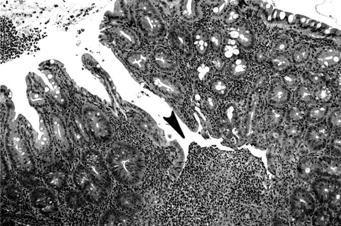
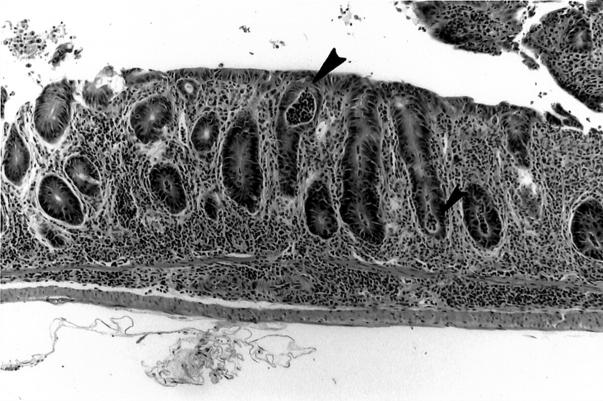
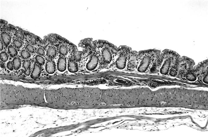
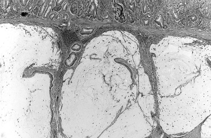
Germ-free interleukin-10 knockout (IL-10 KO) mice developed inflammatory bowel disease (IBD) after they were colonized with a pure culture of Enterococcus faecalis. E. faecalis not only induced IBD (primarily in colon and rectum) but rectal dysplasia and adenocarcinoma was also found in the IL-10 KO mice. Conventional (complex-intestinal flora) IL-10 KO mice developed IBD within 10 to 15 weeks of age and showed more pathology in the cecum (typhlitis) than we observed with E. faecalis-induced IBD in gnotobiotic IL-10 KO mice. Conversely, neither germ-free IL-10 mice nor IL-10 KO mice colonized as adults, with a pure culture of Candida albicans, Escherichia coli, Lactobacillus casei, L. reuteri, L. acidophilus, a Bifidobacterium sp., Lactococcus lactis, or a Bacillus sp. developed IBD during the 25- to 30-week study. E. faecalis is a common intestinal microbe of man and animals that can trigger IBD, dysplasia, and carcinoma in a genetically susceptible murine host.
Figures
References
-
- Karlinger K, Gyorke T, Mako E, Mester A, Tarjan Z: The epidemiology and the pathogenesis of inflammatory bowel disease. Eur J Radiol 2000, 35:154-167 - PubMed
-
- Fiocchi C: Inflammatory bowel disease: etiology and pathogenesis. Gastroenterology 1998, 115:182-205 - PubMed
-
- Prantera C, Scribano ML: Crohn’s disease: the case for bacteria. Ital J Gastroenterol Hepatol 1999, 31:244-246 - PubMed
-
- Boismenu R, Chen Y: Insights from mouse models of colitis. J Leukoc Biol 2000, 67:267-278 - PubMed
-
- MacDonald TT: Cytokine gene deleted mice in the study of gastrointestinal inflammation. Eur J Gastroenterol Hepatol 1997, 9:1051-1055 - PubMed
Publication types
MeSH terms
Substances
Grants and funding
LinkOut - more resources
Full Text Sources
Molecular Biology Databases
Research Materials
