. 2002 Jul;184(13):3530-8.
doi: 10.1128/JB.184.13.3530-3538.2002.

Roles for sigma factors in global circadian regulation of the cyanobacterial genome

Affiliations

Roles for sigma factors in global circadian regulation of the cyanobacterial genome

Usha Nair et al. J Bacteriol. 2002 Jul.

Abstract

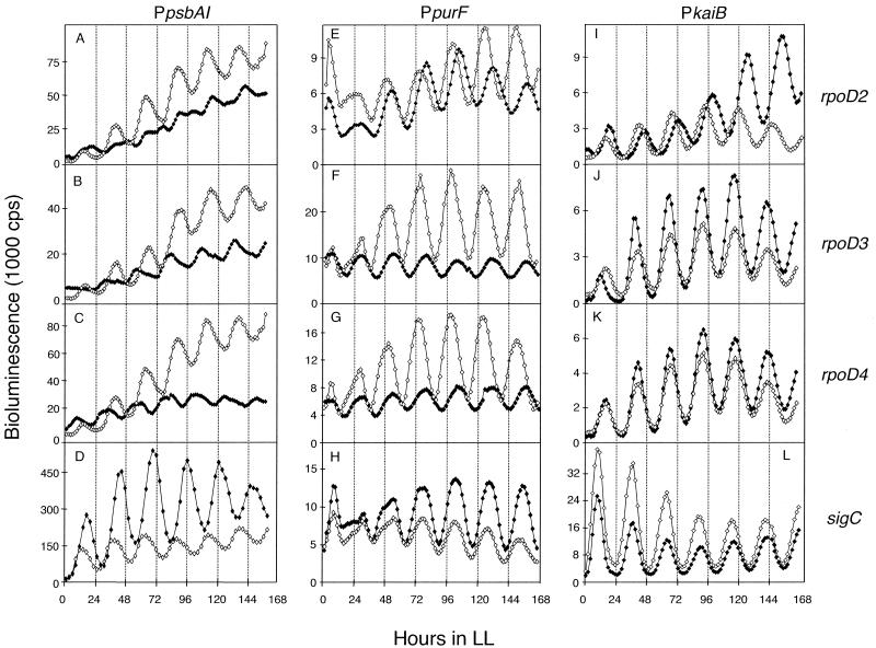
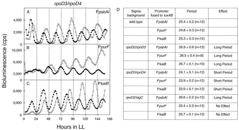
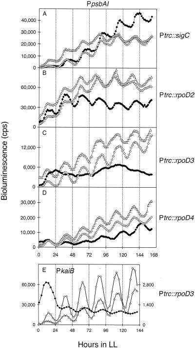
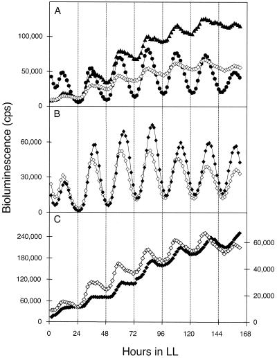
The circadian clock of the unicellular cyanobacterium Synechococcus elongatus PCC 7942 imposes a global rhythm of transcription on promoters throughout the genome. Inactivation of any of the four known group 2 sigma factor genes (rpoD2, rpoD3, rpoD4, and sigC), singly or pairwise, altered circadian expression from the psbAI promoter, changing amplitude, phase angle, waveform, or period. However, only the rpoD2 mutation and the rpoD3 rpoD4 and rpoD2 rpoD3 double mutations affected expression from the kaiB promoter. A striking differential effect was a 2-h lengthening of the circadian period of expression from the promoter of psbAI, but not of those of kaiB or purF, when sigC was inactivated. The data show that separate timing circuits with different periods can coexist in a cell. Overexpression of rpoD2, rpoD3, rpoD4, or sigC also changed the period or abolished the rhythmicity of PpsbAI expression, consistent with a model in which sigma factors work as a consortium to convey circadian information to downstream genes.


Figures

FIG. 1.
FIG. 1.
Circadian expression rhythms of PpsbAI::luxAB, PpurF::luxAB, and PkaiB::luxAB reporter strains in which the rpoD2, rpoD3, rpoD4, or sigC gene has been inactivated. Bioluminescence traces are shown from wild-type (open diamonds) and sigma-inactivated mutant (closed diamonds) strains that carry the reporter gene shown above each column of panels. The gene inactivated in each reporter strain is indicated at the end of each row. The x axis shows time in hours after the cells were released into LL. The y axis indicates bioluminescence (in units of 1,000 cps). Sigma factor gene inactivations are: rpoD2 (A, E, and I), rpoD3 (B, F, and J), rpoD4 (C, G, and K), and sigC (D, H, and L). Traces are representative of bioluminescence measured from at least three replicates of four independent transformants for each sigma factor mutant. Note that the wild-type psbAI::luxAB reporter strain used for inactivation of sigC in panel D is of a different genetic background than that used for inactivation of the other sigma factor genes in panels A, B, and C (see Materials and Methods). The overall expression level is higher for the AMC393 reporter than for AMC669, but the circadian properties of the strains are equivalent.
FIG. 2.
FIG. 2.
Effects of pairwise inactivations of sigma factors on period lengths of PpsbAI, PpurF, and PkaiB expression rhythms. Effect of the rpoD3 rpoD4 double null mutation on PpsbAI::luxAB (A), PpurF::luxAB (B), and PkaiB::luxAB (C) bioluminescence rhythms. Open diamonds and closed diamonds represent wild-type and mutant traces, respectively. The x and y axes are labeled as described for Fig. 1 except that the units for bioluminescence are counts per second. Traces are representative of bioluminescence measured from at least three replicates of four independent transformants. (D) Effects of pairwise inactivations of rpoD2-rpoD3, rpoD3-rpoD4, and rpoD2-sigC on the periods of PpsbAI::luxAB, PpurF::luxAB, and PkaiB::luxAB expression rhythms.
FIG. 3.
FIG. 3.
Differential effects of sigma factor overexpression. The overexpression of sigC (A), rpoD2 (B), rpoD3 (C), or rpoD4 (D) on rhythms from a PpsbAI::luxAB and rpoD3 (E) on a PkaiB::luxAB reporter strain. Overexpression was induced in bioluminescent reporter strains by the addition of 2 mM IPTG immediately before the entraining 12-h dark pulse. Open triangles, bioluminescence rhythms from wild-type reporter strains; open and closed diamonds, rhythms from reporter strains bearing sigma factor overexpression constructs in the absence and presence, respectively, of the inducer. The gene overexpressed from the trc promoter is indicated at the end of each row. The x and y axes are the same as in Fig. 2. Traces are representative of bioluminescence measured from at least six independent samples for each sigma factor overexpression.
FIG. 4.
FIG. 4.
Expression patterns of rpoD2, rpoD3, and sigC and the effect of the loss of sigC and rpoD2 on their respective oscillations. (A) The upstream regions of rpoD2, rpoD3, and sigC were fused to luxAB, and the expression patterns of these sigma factor genes were monitored. Closed triangles, PrpoD3::luxAB; closed circles, PsigC::luxAB; open diamonds, PrpoD2::luxAB. (B) Inactivation of sigC in a PsigC::luxAB reporter strain. Open diamonds, wild-type PsigC::luxAB reporter strain; closed diamonds, sigC mutant PsigC::luxAB reporter strain. (C) Inactivation of rpoD2 in a PrpoD2::luxAB reporter strain. Open diamonds, wild-type PrpoD2::luxAB reporter strain, with y axis scale at right; closed diamonds, PrpoD2::luxAB rpoD2 mutant trace, with y axis scale at left. The x and y axes are the same as for Fig. 2. Traces are representative of bioluminescence measured from at least three replicates of four independent transformants.
FIG. 5.
FIG. 5.
Rhythmic abundance of RpoD4 during the circadian cycle. An entrained wild-type strain was sampled at 4-h intervals between hours 4 and 52 in LL (200 microeinsteins m−2 s−1) and examined by immunoblot analysis with an RpoD4 antiserum (one sample was lost at 40 h). The posttransfer gel was stained with Coomassie blue to verify equal loading of protein samples. White and hatched bars indicate time points corresponding to subjective day and night, respectively.

References

    1. Andersson, C. A., N. F. Tsinoremas, J. Shelton, N. V. Lebedeva, J. Yarrow, H. Min, and S. S. Golden. 2000. Application of bioluminescence to the study of circadian rhythms in cyanobacteria. Methods Enzymol. 305:527-542. - PubMed
    1. Bell-Pedersen, D., J. C. Dunlap, and J. J. Loros. 1996. Distinct cis-acting elements mediate clock, light, and developmental regulation of the Neurospora crassa eas (ccg-2) gene. Mol. Cell. Biol. 16:513-521. - PMC - PubMed
    1. Bustos, S. A., and S. S. Golden. 1991. Expression of the psbDII gene in Synechococcus sp. strain PCC 7942 requires sequences downstream of the transcription start site. J. Bacteriol. 173:7525-7533. - PMC - PubMed
    1. Bustos, S. A., and S. S. Golden. 1992. Light-regulated expression of the psbD gene family in Synechococcus sp. strain PCC 7942: evidence for the role of duplicated psbD genes in cyanobacteria. Mol. Gen. Genet. 232:221-230. - PubMed
    1. Clark, S. R., I. J. Oresnik, and M. F. Hynes. 2001. RpoN of Rhizobium leguminosarum bv. viciae strain VF39SM plays a central role in FnrN-dependent microaerobic regulation of genes involved in nitrogen fixation. Mol. Gen. Genet. 264:623-633. - PubMed

Publication types

MeSH terms

Substances

LinkOut - more resources