. 2002 Jun 10;157(6):909-14.
doi: 10.1083/jcb.200201025. Epub 2002 Jun 10.

Np95 is regulated by E1A during mitotic reactivation of terminally differentiated cells and is essential for S phase entry

Affiliations

Np95 is regulated by E1A during mitotic reactivation of terminally differentiated cells and is essential for S phase entry

Ian Marc Bonapace et al. J Cell Biol. .

Abstract

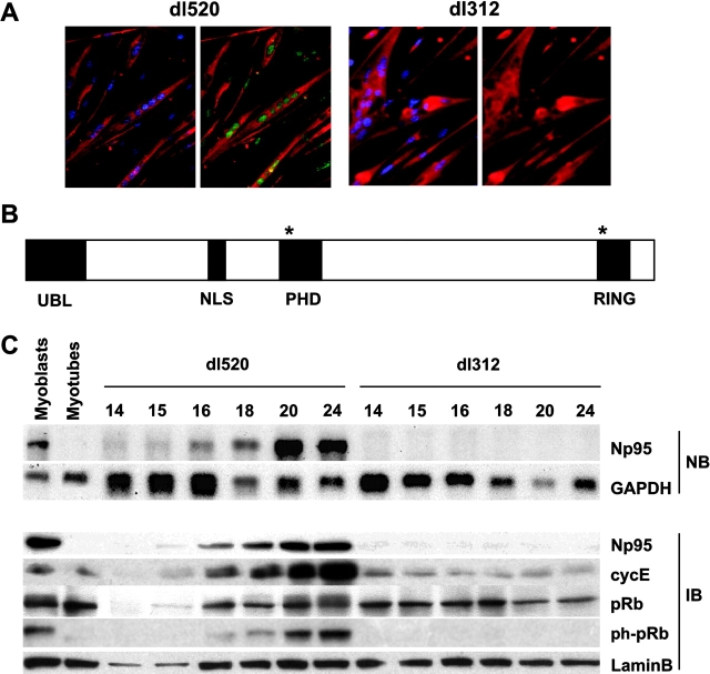
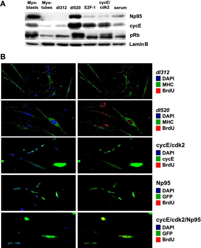
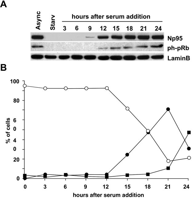
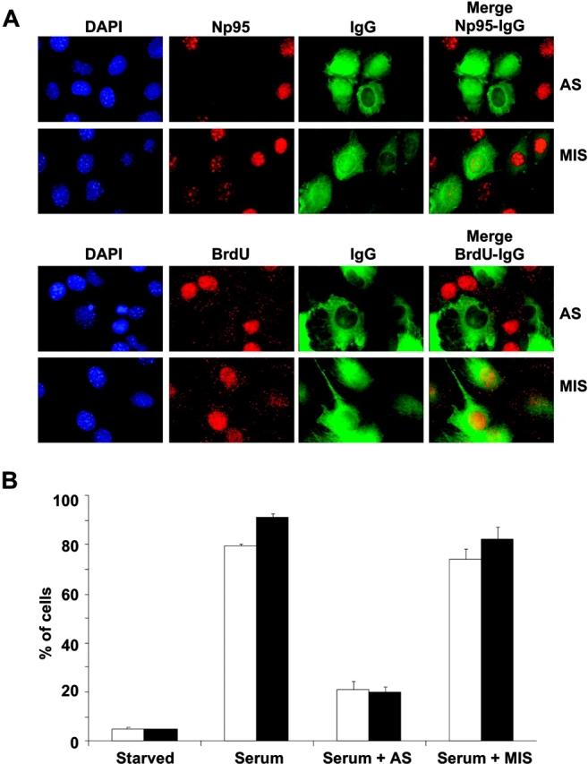
Terminal differentiation exerts a remarkably tight control on cell proliferation. However, the oncogenic products of DNA tumor viruses, such as adenovirus E1A, can force postmitotic cells to proliferate, thus representing a powerful tool to study progression into S phase. In this study, we identified the gene encoding Np95, a murine nuclear phosphoprotein, as an early target of E1A-induced transcriptional events. In terminally differentiated (TD) cells, the activation of Np95 was specifically induced by E1A, but not by overexpression of E2F-1 or of the cyclin E (cycE)-cyclin-dependent kinase 2 (cdk2) complex. In addition, the concomitant expression of Np95 and of cycE-cdk2 was alone sufficient to induce S phase in TD cells. In NIH-3T3 cells, the expression of Np95 was tightly regulated during the cell cycle, and its functional ablation resulted in abrogation of DNA synthesis. Thus, expression of Np95 is essential for S phase entry. Previous evidence suggested that E1A, in addition to its well characterized effects on the pRb/E2F-1 pathway, activates a parallel and complementary pathway that is also required for the reentry in S phase of TD cells (Tiainen, M., D. Spitkousky, P. Jansen-Dürr, A. Sacchi, and M. Crescenzi. 1996. Mol. Cell. Biol. 16:5302-5312). From our results, Np95 appears to possess all the characteristics to represent the first molecular determinant identified in this pathway.


Figures

Figure 1.
Figure 1.
Np95 is an early E1A-induced gene. (A) TD myotubes were infected with either dl520 (left, MOI, 400 pfu/cell) or dl312 as a control (right, MOI, 400 pfu/cell). 24 h later, cells were treated with BrdU. 48 h after infection, cells were fixed and stained with anti–myosin heavy chain (red) to check differentiation and anti-BrdU (green) antibodies. Nuclear counterstaining was performed with DAPI (blue). (B) Schematic of mouse Np95; the various domains of the protein are indicated (Ubl, ubiquitin-like; NLS, nuclear localization signal). Two putative pRb-binding motifs, LxCxE and IxCxE (Dahiya et al., 2000), are indicated by asterisks. (C) TD myotubes were infected with either dl520 (MOI, 400 pfu/cell) or dl312 (MOI, 400 pfu/cell), and mRNA or proteins were harvested at the indicated time points. RNAs (2 μg Poly(A)+, top) were analyzed in Northern blot (NB) with the indicated probes. Proteins (40 μg of total cellular lysates, bottom) were immunoblotted (IB) with the indicated antibodies. pRb and ph-pRb are a pan–anti-pRb and a specific anti-phosphoRb, respectively (see Materials and methods). The levels of Np95 before (myoblasts) and after (myotubes) differentiation are also shown.
Figure 2.
Figure 2.
Role of Np95 in the reentry in the cell cycle of TD myotubes. (A) TD myotubes were infected with dl520 (MOI, 400 pfu/cell), dl312 (MOI, 400 pfu/cell), or adenoviral vectors encoding E2F-1 (MOI, 400 pfu/cell), cycE (MOI, 100 pfu/cell)/cdk2 (MOI, 400 pfu/cell), or stimulated with 5% FCS, as indicated on top. 48 h after infection, total cellular lysates were extracted and immunoblotted (40 μg) with the indicated antibodies. Lanes myoblasts and myotubes are as in Fig. 1 C. (B) TD myotubes were infected with adenoviral vectors, as indicated on the right. MOIs were as follows: cycE, 100 pfu/cell; cdk2, 400 pfu/cell; and Np95, 60 pfu/cell. 24 h later, cells were treated with BrdU. 48 h after infection, cells were fixed and stained with anti-BrdU (red), and either anti–myosin heavy chain (green, top two rows, MHC), or anti-cycE (green, middle row) antibodies, or detected by epifluorescence (green, bottom two rows, to detect GFP-Np95). Nuclear counterstaining was performed with DAPI (blue). For each condition (horizontally paired panels), two pictures of the same microscopic field are shown. All left panels display the blue (DAPI) and green (either MHC, or cycE, or GFP) channels. All right panels display the green (either MHC, or cycE, or GFP) and red (BrdU) channels. Stainings are also indicated by a color code (squares).
Figure 3.
Figure 3.
Cell cycle–dependent regulation of Np95 in NIH-3T3 cells. (A) NIH-3T3 cells were serum-starved for 48 h, and then stimulated with 10% FCS for the indicated time points. (A) Immunoblots (40 μg of total proteins) with the indicated antibodies. The levels of Np95 in serum-starved cells (Starv) or in an asynchronously growing population (Async) are also shown. (B) FACS® profiles: open circles, G0/G1; closed circles, S; squares, G2/M.
Figure 4.
Figure 4.
Functional ablation of Np95 in NIH-3T3 cells. (A) NIH-3T3 were serum starved for 48 h, and then microinjected with the Np95 antisense (AS) oligonucleotide or its mismatched (MIS) control. IgGs were also microinjected to identify microinjected cells. BrdU and 10% FCS were then added, and cells were fixed 24 h later and stained as indicated. Nuclear counterstaining was performed with DAPI. (B) A quantitative assessment was performed on three independent experiments, in which at least 150 microinjected cells per experiment were counted. Results are shown as a percentage of microinjected cells (±SD) that were Np95-positive (solid bars) or BrdU-positive (empty bars).

References

    1. Alevizopoulos, K., B. Catarin, J. Vlach, and B. Amati. 1998. A novel function of adenovirus E1A is required to overcome growth arrest by the CDK2 inhibitor p27(Kip1). EMBO J. 17:5987–5997. - PMC - PubMed
    1. Alevizopoulos, K., B. Sanchez, and B. Amati. 2000. Conserved region 2 of adenovirus E1A has a function distinct from pRb binding required to prevent cell cycle arrest by p16INK4a or p27Kip1. Oncogene. 19:2067–2074. - PubMed
    1. Arany, Z., W.R. Sellers, D.M. Livingston, and R. Eckner. 1994. E1A-associated p300 and CREB-associated CBP belong to a conserved family of coactivators. Cell. 77:799–800. - PubMed
    1. Arany, Z., D. Newsome, E. Oldread, D.M. Livingston, and R. Eckner. 1995. A family of transcriptional adaptor proteins targeted by the E1A oncoprotein. Nature. 374:81–84. - PubMed
    1. Crescenzi, M., S. Soddu, and F. Tatò. 1995. Mitotic cycle reactivation in terminally differentiated cells by adenovirus infection. J. Cell. Physiol. 162:26–35. - PubMed

Publication types

MeSH terms