. 2002 Jun;13(6):1832-45.
doi: 10.1091/mbc.01-10-0504.

Kinesin-like protein CHO1 is required for the formation of midbody matrix and the completion of cytokinesis in mammalian cells

Affiliations

Kinesin-like protein CHO1 is required for the formation of midbody matrix and the completion of cytokinesis in mammalian cells

Jurgita Matuliene et al. Mol Biol Cell. 2002 Jun.

Abstract

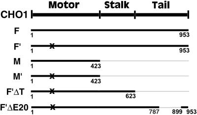
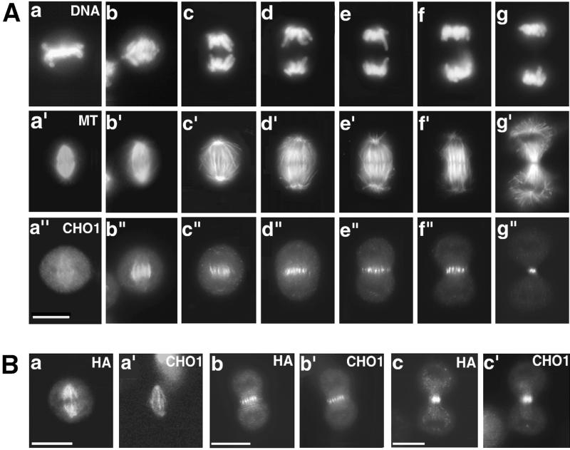
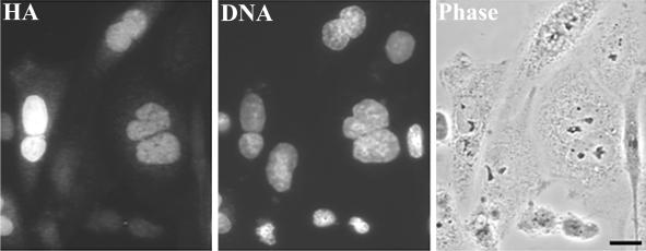
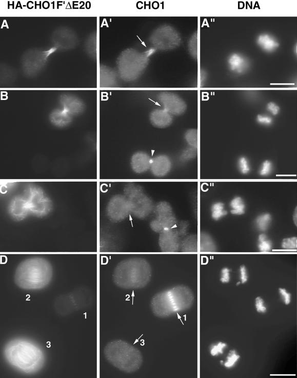
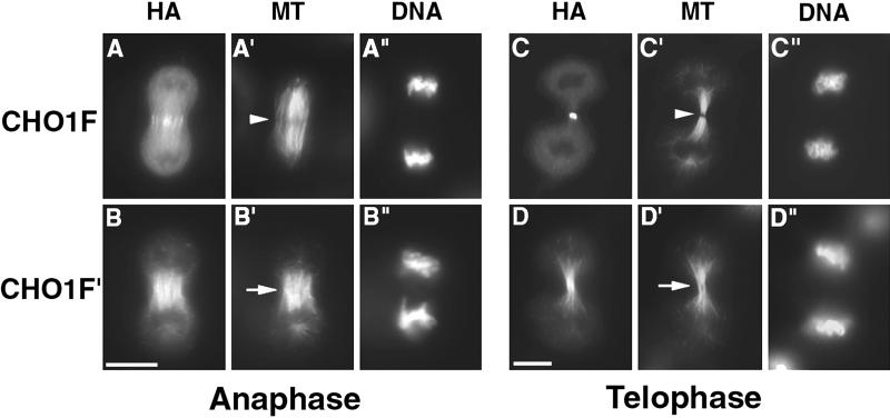
CHO1 is a mammalian kinesin-like motor protein of the MKLP1 subfamily. It associates with the spindle midzone during anaphase and concentrates to a midbody matrix during cytokinesis. CHO1 was originally implicated in karyokinesis, but the invertebrate homologues of CHO1 were shown to function in the midzone formation and cytokinesis. To analyze the role of the protein in mammalian cells, we mutated the ATP-binding site of CHO1 and expressed it in CHO cells. Mutant protein (CHO1F') was able to interact with microtubules via ATP-independent microtubule-binding site(s) but failed to accumulate at the midline of the central spindle and affected the localization of endogenous CHO1. Although the segregation of chromosomes, the bundling of midzone microtubules, and the initiation of cytokinesis proceeded normally in CHO1F'-expressing cells, the completion of cytokinesis was inhibited. Daughter cells were frequently entering interphase while connected by a microtubule-containing cytoplasmic bridge from which the dense midbody matrix was missing. Depletion of endogenous CHO1 via RNA-mediated interference also affected the formation of midbody matrix in dividing cells, caused the disorganization of midzone microtubules, and resulted in abortive cytokinesis. Thus, CHO1 may not be required for karyokinesis, but it is essential for the proper midzone/midbody formation and cytokinesis in mammalian cells.


Figures

Figure 1
Figure 1
A map of CHO1 constructs used for the current study. The total number of amino acids, encoded by each construct, is identified beneath the bold lines, representing the constructs. All constructs are tagged with the HA epitope at the N terminus. X indicates the mutation in the ATP-binding site of CHO1.
Figure 2
Figure 2
(A) Immunolocalization of CHO1 in CHO cells at different stages of mitosis. After synchronization, cells were fixed at the different stages of mitotic progression and stained with DAPI (a–g), antitubulin (a′–g′) and polyclonal anti-CHO1 (a"–g") antibodies. CHO1 changes its distribution from the entire spindle (a") to the midzone region during anaphase (b"–f") and concentrates to a midbody at the end of cytokinesis (g"). (B) Distribution of exogenous CHO1F in transfected cells is identical to the distribution of endogenous CHO1. Cells fixed at metaphase (a, a′), anaphase (b, b′), and telophase (c, c′) were stained with anti-HA (a, b, and c) and polyclonal anti-CHO1 (a′, b′, and c′) antibodies. Bar, 10 μm.
Figure 3
Figure 3
ATP-binding mutant of CHO1 (CHO1F′) interacts with microtubules via ATP-independent microtubule binding site(s) but does not concentrate at the midline of the central spindle during anaphase. (A) CHO1M associates with microtubules when expressed in CHO cells. (B) CHO1 M′ generally does not display microtubule binding (compare HA staining in B and tubulin staining in B′). CHO1F′ interacts with microtubules in both mitotic (C, metaphase; D, anaphase; E, telophase) and interphase cells (F). Although CHO1F′ contains the NLS and is generally seen within the nucleus, in some interphase cells with the high level of expression, CHO1F′ can also be detected in the cytoplasm (F). CHO1F′ΔT strongly binds and bundles microtubules (G), whereas endogenous CHO1 is confined within the nucleus (G′). This indicates that microtubule binding by mutated CHO1F′ and CHO1F′ΔT constructs is not due to the dimerization with endogenous CHO1 but is due to the ATP-independent microtubule binding site(s) present in the motor protein. CHO1F′ fails to concentrate at the midline of the central spindle during anaphase (D) and distributes along the entire intercellular bridge at the end of cytokinesis (E). Staining was performed using monoclonal anti-HA (A and C–E), polyclonal anti-HA (B, F, and G), monoclonal antitubulin (B′ and F′), monoclonal anti-CHO1 antibodies (G′) and DAPI (F" and G"). Bars, 10 μm.
Figure 4
Figure 4
High level of CHO1F′ expression inhibits completion of cytokinesis in CHO cells. Although the midzone bundles are forming in cells expressing CHO1F′ (A′ and A"), the daughter cells remain connected by the cytoplasmic bridges after mitosis (B–F). Cells expressing mutant protein are stained by anti-HA (A′, B, C, D′, E′, and F) and either anti-CHO1 polyclonal (A", B′, D", and F′) or antitubulin (C′ and E") antibodies. DNA is visualized by DAPI staining (A and E). Bars, 10 μm
Figure 5
Figure 5
Expression of CHO1F′ induces binucleation in CHO cells (also see Table 1). Cells were fixed at 48 h after transfection with CHO1F′ construct and stained with anti-HA antibodies and DAPI to visualize the nuclei. Cells were observed by the fluorescence and phase contrast microscopy. Bar, 10 μm.
Figure 6
Figure 6
Mutant CHO1 inhibits the accumulation of endogenous CHO1 at the midzone/midbody region. (A–D) The effect of CHO1F′ΔE20 expression on the localization of endogenous CHO1. When the expression of CHO1F′ΔE20 increases from low (A) to medium (B) and high levels (C), the endogenous CHO1 becomes depleted from midzone/midbody area of the dividing cells (arrows in B′ and C′). (D–D") Three anaphase cells expressing different levels of mutated protein in the same field. The higher the level of CHO1F′ΔE20 expression (D), the weaker the accumulation of endogenous protein at the midzone region (arrows 1 to 3 in D′). Staining was performed using anti-HA (A–D) and anti-CHO1 E20 (A′–D′) antibodies and DAPI (A"–D"). Bars, 10 μm.
Figure 7
Figure 7
Formation of stem body and midbody matrices is inhibited in dividing cells expressing mutated CHO1. CHO cells, transfected with CHO1F (A and C) and CHO1F′ (B and D) constructs were fixed at anaphase (A and B) and telophase (C and D) stages and stained using anti-HA (A–D), antitubulin (A′–D′) antibodies, and DAPI (A"–D"). The stem body and midbody matrix is easily detected in cells expressing wild-type CHO1F (arrowheads in A′ and C′). However, in cells expressing mutant protein, the dense matrix material is missing, based on the absence of dark spots in tubulin staining (arrows in B′ and D′). Bars, 10 μm.
Figure 8
Figure 8
Expression of CHO1F′ inhibits the organization of electron dense midbody matrix. Thin section electron micrographs show a control cell (A and B) and a cell expressing CHO1F′ (C and D) at the late stage of cell division. Both cells are seen at low (A and C) and high (B and D) magnifications. Although the electron dense midbody matrix can be easily detected in the intercellular bridge connecting two control cells (arrows in A and B), the electron-dense structure is completely disorganized in the cell expressing CHO1F′. Bars: A and C, 5 μm; B and D, 0.5 μm.
Figure 9
Figure 9
RNAi directed against endogenous CHO1 interferes with the organization of central spindle and the formation of midbody matrix in CHO cells. Mock- and siRNA-transfected cells were synchronized and fixed at the different stages of mitosis 30 h after transfection. Cells were visualized by staining with antitubulin (A–H), anti-CHO1 polyclonal (A′-H′) antibodies, and DAPI (A"-H"). To correctly represent the amount of endogenous CHO1 in all cells, both mock- and siRNA-transfected cells were fixed and immunostained in an identical manner, and all images were captured and developed using identical exposure and conditions. RNAi directed against endogenous CHO1 affects neither the formation of bipolar spindle (B–B") nor chromosome segregation (D", E", G", and H") in CHO cells. However, the reduced level of CHO1 expression (D′ and G′) diminishes the matrix formation in anaphase (arrowhead in D) and telophase (arrowhead in G) cells in comparison with control (arrows in C and F). Almost complete depletion of CHO1 (E′ and H′) causes the severe disorganization of central spindles (E and H).

Similar articles

Cited by

References

    1. Adams RR, Tavares AAM, Salzberg A, Bellen HJ, Glover DM. pavarotti encodes a kinesin-like protein required to organize the central spindle and contractile ring for cytokinesis. Genes Dev. 1998;12:1483–1494. - PMC - PubMed
    1. Ausubel FM, Brent R, Kingston RE, Moore DD, Seidman JG, Smith JA, Struhl K. Current protocols in molecular biology. New York: John Wiley & Sons, Inc; 1994.
    1. Boman AL, Kuai J, Zhu X, Chen J, Kuriyama R, Kahn RA. Arf proteins bind to mitotic kinesin-like protein 1 (MKLP1) in a GTP-dependent fashion. Cell Motil Cytoskeleton. 1999;44:119–132. - PubMed
    1. Cao LG, Wang YL. Signals from the spindle midzone are required for the stimulation of cytokinesis in cultured epithelial cells. Mol Biol Cell. 1996;7:225–232. - PMC - PubMed
    1. Chen M, Detrich HW., III The kinesin superfamily of zebrafish embryos. Mol Biol Cell. 1996;7:396a.

Publication types

Substances

LinkOut - more resources