. 2002 Jun;13(6):1906-15.
doi: 10.1091/mbc.02-02-0025.

Pancreatic beta-cell protein granuphilin binds Rab3 and Munc-18 and controls exocytosis

Affiliations

Pancreatic beta-cell protein granuphilin binds Rab3 and Munc-18 and controls exocytosis

Thierry Coppola et al. Mol Biol Cell. 2002 Jun.

Abstract

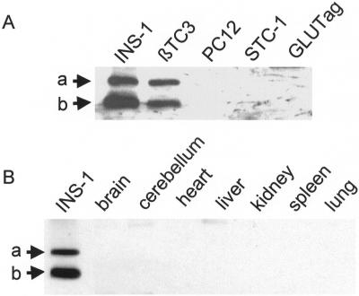
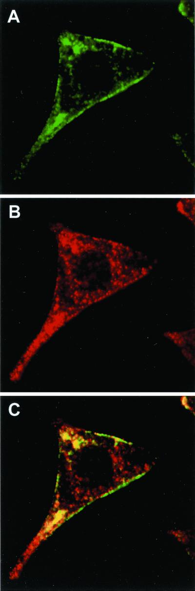
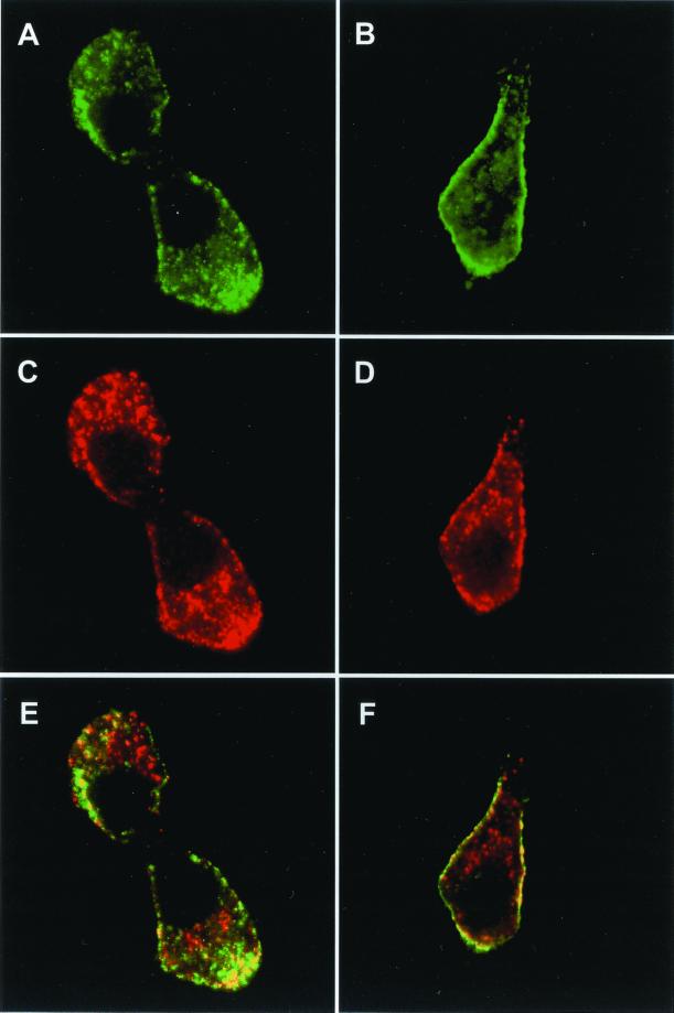
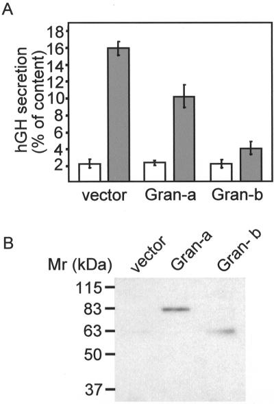
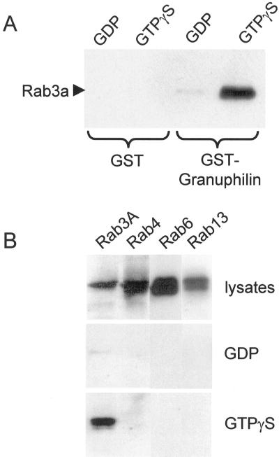
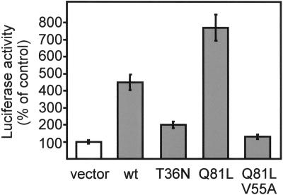
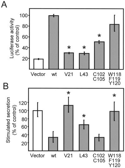
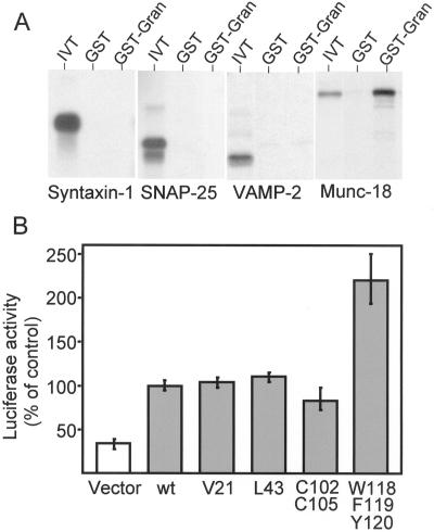
Granuphilin/Slp-4 is a member of the synaptotagmin-like protein family expressed in pancreatic beta-cells and in the pituitary gland. We show by confocal microscopy that both granuphilin-a and -b colocalize with insulin-containing secretory granules positioned at the periphery of pancreatic beta-cells. Overexpression of granuphilins in insulin-secreting cell lines caused a profound inhibition of stimulus-induced exocytosis. Granuphilins were found to bind to two components of the secretory machinery of pancreatic beta-cells, the small GTP-binding protein Rab3 and the soluble N-ethylmaleimide-sensitive factor attachment protein receptor (SNARE)-binding protein Munc-18. The interaction with Rab3 occurred only with the GTP-bound form of the protein and was prevented by a point mutation in the effector domain of the GTPase. Structure-function studies using granuphilin-b mutants revealed that complete loss of Rab3 binding is associated with a reduction in the capacity to inhibit exocytosis. However, the granuphilin/Rab3 complex alone is not sufficient to mediate the decrease of exocytosis, suggesting the existence of additional binding partners. Taken together, our observations indicate that granuphilins play an important role in pancreatic beta-cell exocytosis. In view of the postulated role of Munc-18 in secretory vesicle docking, our data suggest that granuphilins may also be involved in this process.


Figures

Figure 1
Figure 1
Tissue distribution of rat granuphilin-a and -b. (A) Aliquots (30 μg of protein) of homogenates of the indicated cell lines were separated on SDS-PAGE and blotted on nitrocellulose membranes. The proteins were analyzed by Western blotting using a polyclonal antibody directed against the common N-terminal fragment of granuphilins. The arrows indicate the position of granuphilin-a (a) and -b (b). The apparent Mr of bands a and b were 80 and 60 kDa, respectively. (B) Aliquots (30 μg of protein) of homogenates of the indicated rat organs were analyzed by Western blotting using the anti-granuphilin antibody. An INS-1 extract was run in parallel as a positive control.
Figure 2
Figure 2
Subcellular localization of endogenous granuphilins. INS-1 cells were grown on glass coverslips coated with laminin and poly-l-lysine. The cells were fixed with paraformaldehyde and the coverslips incubated with a rabbit polyclonal antibody directed against the N-terminus of granuphilins and an anti-insulin antibody raised in guinea pig. The subcellular distribution of granuphilins was analyzed by confocal microscopy using an Oregon-green–labeled anti-rabbit antibody. The position of secretory granules was visualized with an anti-guinea pig antibody coupled to Cy3. (A) Anti-granuphilins; (B) anti-insulin; (C) overlay between images A and B.
Figure 3
Figure 3
Subcellular localization of granuphilin-a and -b. INS-1 cells transiently transfected with myc-tagged granuphilin-a (A, C, E) or -b (B, D, F) were grown for 2 days on glass coverslips coated with laminin and poly-l-lysine. The cells were fixed and analyzed by confocal microscopy using an anti-myc antibody and an anti-insulin antibody. (A and B) Anti-myc; (C and D) anti-insulin; (E and F) overlay of the signal obtained with the anti-myc and anti-insulin antibodies.
Figure 4
Figure 4
Effect of granuphilin-a and -b on exocytosis. HIT-T15 cells were transiently cotransfected with a plasmid encoding hGH and either an empty vector (vector) or plasmids encoding granuphilin-a (Gran-a) or granuphilin-b (Gran-b). After 3 days in culture, the cells were incubated for 10 min under basal conditions (open bars) or in the presence of stimulatory concentrations of K+ and glucose (filled bars). The total amount of hGH expressed by the cells and the fraction released during the incubation period were determined by ELISA. (A) Percentage of hGH present in the cells that is secreted under basal or stimulatory conditions. (B) Expression level of granuphilin-a and -b assessed by Western blotting using an antimyc antibody.
Figure 5
Figure 5
The N-terminus of granuphilin binds to Rab3. (A) GST or GST-granuphilin 1–300 were immobilized on glutathione-agarose beads and incubated with purified wild-type Rab3A loaded with GDP or with GTPγS. The proteins remaining associated with the affinity columns were visualized by Western blotting using a monoclonal antibody against Rab3A. (B) HIT-T15 cells were transiently transfected with wild-type Rab3A, Rab4, Rab6, or Rab13. Two days later, the cells were homogenized and the cell lysates incubated either with GDP or GTPγS for 15 min at 30°C. They were then loaded onto GST-granuphilin 1–300 affinity columns. The proteins interacting with GST-granuphilin 1–300 were detected by Western blotting using an antibody against the epitope tag.
Figure 6
Figure 6
Granuphilin interacts with the GTP-bound form of Rab3 in living cells. HIT-T15 cells were cotransfected with a fusion protein of VP16 with granuphilin-b, a fusion protein of GAL4 with the indicated mutants of Rab3A, and a plasmid containing five binding sites for GAL4 upstream of the firefly luciferase gene. The interaction between granuphilin-b and the Rab3A mutants was quantified by measurement of firefly luciferase activity. The luciferase activity produced in cells transfected with VP16/granuphilin-b and a GAL4 empty vector was set to 100%. The results are the mean ± SD of three independent experiments performed in duplicate. wt, wild-type.
Figure 7
Figure 7
Effect of different point mutations of granuphilin on Rab3 binding and on exocytosis. (A) HIT-T15 cells were cotransfected with a fusion protein of GAL4 with the GTPase-deficient mutant Rab3AQ81L, a fusion protein of VP16 with the indicated mutants of granuphilin-b and a plasmid containing five binding sites for GAL4 upstream of the firefly luciferase gene. The interaction of the GTP-bound form of Rab3A with the mutants of granuphilin-b was assessed by measuring firefly luciferase activity. The luciferase activity produced in cells expressing the fusion proteins of GAL4 with Rab3AQ81L and VP16 with wild-type (wt) granuphilin-b was set to 100%. The results are the mean ± SD of three independent experiments performed in duplicate. Asterisks indicate the mutants whose interaction with Rab3A is significantly (p < 0.01) different from wild-type granuphilin-b. (B) HIT-T15 cells were transiently transfected with the indicated mutants of granuphilin-b and with a plasmid encoding hGH. After 3 days in culture, the cells were incubated under basal conditions or in the presence of depolarizing concentrations of K+ and glucose. The amount of hGH released into the medium under basal and stimulatory conditions was determined by ELISA. The response of the cells transfected with the hGH plasmid together with an empty vector (vector) was set to 100%. The figure shows the mean ± SD of at least five independent experiments measured in triplicate. Asterisks indicate the mutants whose effect on exocytosis is significantly (p < 0.01) different from that of wild-type granuphilin-b.
Figure 8
Figure 8
Granuphilin interacts with Munc-18 in vitro and in living cells. (A) GST or GST-granuphilin 1–300 (GST-Gran) were immobilized on glutathione agarose beads and incubated with in vitro translated 35S-labeled syntaxin-1, SNAP-25, VAMP-2, and Munc-18 (IVT). The proteins remaining associated with the affinity columns were separated by SDS-PAGE and visualized by autoradiography. (B) HIT-T15 cells were cotransfected with a fusion protein of GAL4 with Munc-18, a fusion protein of VP16 with the indicated mutants of granuphilin-b, and a firefly luciferase reporter gene. The luciferase activity produced in cells expressing the fusion proteins of GAL4 with Munc-18 and VP16 with wild-type (wt) granuphilin-b was set to 100%. The results are the mean ± SD of three independent experiments performed in duplicate.

References

    1. Asfari M, Janjic D, Meda P, Li G, Halban PA, Wollheim CB. Establishment of 2-mercaptoethanol-dependent differentiated insulin-secreting cell lines. Endocrinology. 1992;130:167–178. - PubMed
    1. Brondyk WH, McKiernan CJ, Burstein ES, Macara IG. Mutants of Rab3A analogous to oncogenic Ras mutants. J Biol Chem. 1993;268:9410–9415. - PubMed
    1. Chung SH, Takai Y, Holz RW. Evidence that the Rab3a-binding protein, rabphilin3a, enhances regulated secretion. J Biol Chem. 1995;270:16714–16717. - PubMed
    1. Coppola T, Hirling H, Perret-Menoud V, Gattesco S, Catsicas S, Joberty G, Macara IG, Regazzi R. Rabphilin dissociated from Rab3 promotes endocytosis through interaction with rabaptin-5. J Cell Sci. 2001;114:1757–1764. - PubMed
    1. Coppola T, Perret-Menoud V, Lüthi S, Farnsworth CC, Glomset JA, Regazzi R. Disruption of Rab3-calmodulin interaction, but not other effector interactions, prevents Rab3 inhibition of exocytosis. EMBO J. 1999;18:5885–5891. - PMC - PubMed

Publication types

MeSH terms

LinkOut - more resources