

Effects of subcortical ischemic vascular dementia and AD on entorhinal cortex and hippocampus
- PMID: 12058091
- PMCID: PMC1820858
- DOI: 10.1212/wnl.58.11.1635
Effects of subcortical ischemic vascular dementia and AD on entorhinal cortex and hippocampus
Abstract
Objective: To determine the effects of subcortical ischemic vascular dementia (SIVD) and AD on entorhinal cortex (ERC) and hippocampus.
Methods: Thirty-eight cognitively normal subjects, 18 patients with SIVD, and 22 patients with AD were included. Volumes of ERC and hippocampus were manually measured based on MRI. Global cerebral changes of cortical gray matter, subcortical gray matter, white matter, sulcal CSF, ventricular CSF (vCSF), and white matter signal hyperintensities (WMSH) were assessed.
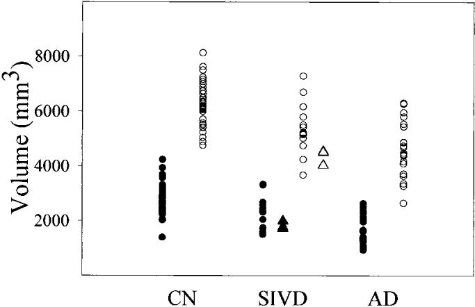
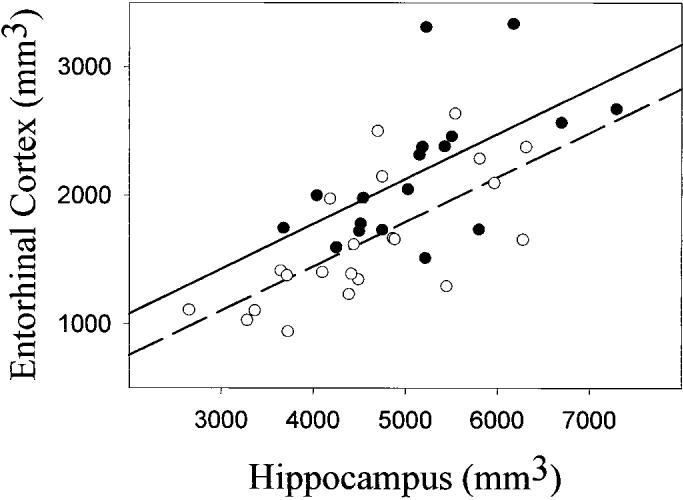
Results: Patients with SIVD had 21.7% (p < 0.01) smaller ERC and 18.2% (p < 0.01) smaller hippocampi than cognitively normal subjects and 24.4% (p < 0.01) larger ERC and 11.1% (p < 0.05) larger hippocampi than patients with AD. In addition, patients with SIVD had less cortical gray matter and white matter and more vCSF and WMSH (all p < 0.01) than cognitively normal subjects and more vCSF and WMSH (p < 0.01) than patients with AD. The volumes of ERC and hippocampus were positively correlated to similar extents (p < 0.01) in SIVD and AD. Cortical gray matter loss was positively correlated (p < 0.01) with hippocampal atrophy, but not with ERC atrophy, in SIVD and AD. Hippocampal volume alone could classify 82% of patients with SIVD from cognitively normal subjects and 63% of patients with SIVD from subjects with AD. Adding global cerebral changes to hippocampus substantially improved the classification to 96% between patients with SIVD and cognitively normal subjects and 83% between subjects with SIVD and those with AD, whereas adding ERC change to hippocampus did not significantly improve the discrimination.
Conclusions: The entorhinal cortex and hippocampus are less affected by subcortical ischemic vascular dementia than by AD.
Figures
References
-
- Jellinger K, Danielczyk W, Fischer P, Gabriel E. Clinicopathological analysis of dementia disorders in the elderly. J Neurol Sci. 1990;95:239–258. - PubMed
-
- Wade JP, Mirsen TR, Hachinski VC, Fisman M, Lau C, Merskey H. The clinical diagnosis of Alzheimer's disease. Arch Neurol. 1987;44:24–29. - PubMed
-
- Seab JP, Jagust WJ, Wong ST, Roos MS, Reed BR, Budinger TF. Quantitative NMR measurements of hippocampal atrophy in Alzheimer's disease. Magn Reson Med. 1988;8:200–208. - PubMed
-
- Kesslak JP, Nalcioglu O, Cotman CW. Quantification of magnetic resonance scans for hippocampal and parahippocampal atrophy in Alzheimer's disease. Neurology. 1991;41:51–54. - PubMed
-
- Jack CRJ, Petersen RC, O'Brien PC, Tangalos EG. MR-based hippocampal volumetry in the diagnosis of Alzheimer's disease. Neurology. 1992;42:183–188. - PubMed
Publication types
MeSH terms
Grants and funding
LinkOut - more resources
Full Text Sources
Medical