

Delayed toxicity of cyclophosphamide on the bladder of DBA/2 and C57BL/6 female mouse
- PMID: 12059909
- PMCID: PMC2517663
- DOI: 10.1046/j.1365-2613.2002.00208.x
Delayed toxicity of cyclophosphamide on the bladder of DBA/2 and C57BL/6 female mouse
Abstract
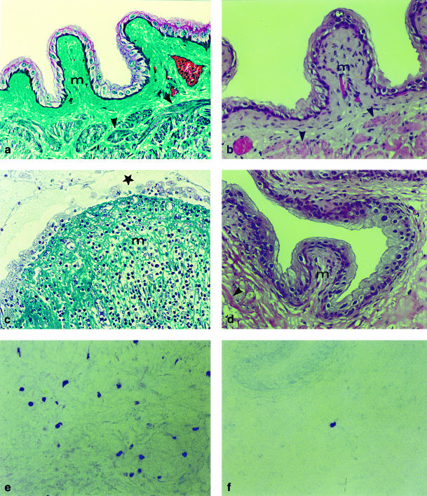
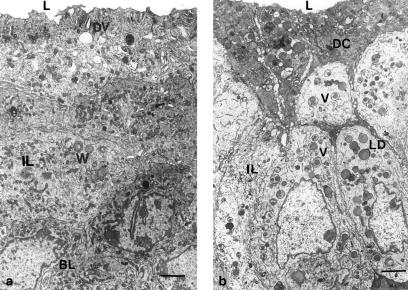
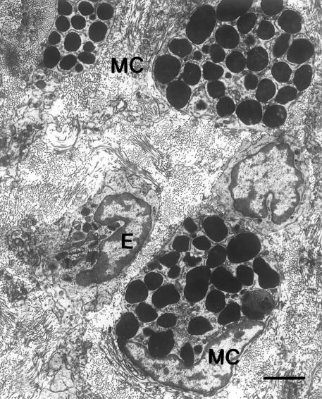
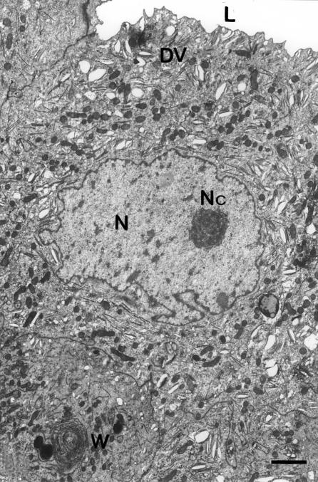
The present study describes the delayed development of a severe bladder pathology in a susceptible strain of mice (DBA/2) but not in a resistant strain (C57BL/6) when both were treated with a single 300 mg/kg dose of cyclophosphamide (CY). Inbred DBA/2 and C57BL/6 female mice were injected with CY, and the effect of the drug on the bladder was assessed during 100 days by light microscopy using different staining procedures, and after 30 days by conventional electron microscopy. Early CY toxicity caused a typical haemorrhagic cystitis in both strains that was completely repaired in about 7-10 days. After 30 days of CY injection ulcerous and non-ulcerous forms of chronic cystitis appeared in 86% of DBA/2 mice but only in 4% of C57BL/6 mice. Delayed cystitis was characterized by infiltration and transepithelial passage into the lumen of inflammatory cells and by frequent exfoliation of the urothelium. Mast cells appeared in the connective and muscular layers of the bladder at a much higher number in DBA/2 mice than in C57BL/6 mice or untreated controls. Electron microscopy disclosed the absence of the typical discoidal vesicles normally present in the cytoplasm of surface cells. Instead, numerous abnormal vesicles containing one or several dark granules were observed in the cytoplasm of cells from all the epithelial layers. Delayed cystitis still persisted in DBA/2 mice 100 days after treatment. These results indicate that delayed toxicity of CY in female DBA/2 mice causes a bladder pathology that is not observed in C57BL/6 mice. This pathology resembles interstitial cystitis in humans and could perhaps be used as an animal model for studies on the disease.
Figures
Similar articles
-
Ultrastructural study of the effect of cyclophosphamide on the growth area of incisor teeth of DBA/2 and C57BL/6 mice.Int J Exp Pathol. 1996 Apr;77(2):83-8. doi: 10.1046/j.1365-2613.1996.00967.x. Int J Exp Pathol. 1996. PMID: 8762867 Free PMC article.
-
Increased sensitivity of glutathione S-transferase P-null mice to cyclophosphamide-induced urinary bladder toxicity.J Pharmacol Exp Ther. 2009 Nov;331(2):456-69. doi: 10.1124/jpet.109.156513. Epub 2009 Aug 20. J Pharmacol Exp Ther. 2009. PMID: 19696094 Free PMC article.
-
[Experimental studies on microcirculatory inflammatory reactions of the urinary bladder].Magy Seb. 2012 Aug;65(4):184-90. doi: 10.1556/MaSeb.65.2012.4.3. Magy Seb. 2012. PMID: 22940386 Hungarian.
-
Differential sensitivity of DBA/2 and C57BL/6 mice to cyclophosphamide.J Appl Toxicol. 1993 Nov-Dec;13(6):423-7. doi: 10.1002/jat.2550130609. J Appl Toxicol. 1993. PMID: 8288846
-
[Reconstitution of cyclophosphamide-induced, impaired function of the immune system in animal models].Postepy Hig Med Dosw. 2003;57(1):55-66. Postepy Hig Med Dosw. 2003. PMID: 12765123 Review. Polish.
Cited by
-
Immune Relevant and Immune Deficient Mice: Options and Opportunities in Translational Research.ILAR J. 2018 Dec 31;59(3):211-246. doi: 10.1093/ilar/ily026. ILAR J. 2018. PMID: 31197363 Free PMC article. Review.
-
Cyclophosphamide leads to persistent deficits in physical performance and in vivo mitochondria function in a mouse model of chemotherapy late effects.PLoS One. 2017 Jul 10;12(7):e0181086. doi: 10.1371/journal.pone.0181086. eCollection 2017. PLoS One. 2017. PMID: 28700655 Free PMC article.
-
Normalization of proliferation and tight junction formation in bladder epithelial cells from patients with interstitial cystitis/painful bladder syndrome by d-proline and d-pipecolic acid derivatives of antiproliferative factor.Chem Biol Drug Des. 2011 Jun;77(6):421-30. doi: 10.1111/j.1747-0285.2011.01108.x. Epub 2011 Apr 27. Chem Biol Drug Des. 2011. PMID: 21352500 Free PMC article.
-
NLRP3 Inflammasome: A Promising Therapeutic Target for Drug-Induced Toxicity.Front Cell Dev Biol. 2021 Apr 12;9:634607. doi: 10.3389/fcell.2021.634607. eCollection 2021. Front Cell Dev Biol. 2021. PMID: 33912556 Free PMC article. Review.
-
Keratinocyte Growth Factor Reduces Injury and Leads to Early Recovery from Cyclophosphamide Bladder Injury.Am J Pathol. 2020 Jan;190(1):108-124. doi: 10.1016/j.ajpath.2019.09.015. Epub 2019 Oct 22. Am J Pathol. 2020. PMID: 31654636 Free PMC article.
References
-
- Anton E. Differential sensitivity of DBA/2 and C57BL/6 mice to cyclophosphamide. J.Appl.Toxicol. 1993;13:423–427. - PubMed
-
- Anton E. Detection of apoptosis by a modified trichrome technique. J.Histotechn. 1999;22:301–304.
-
- Anton E, Brandes D. Lysosomes in mice mammary tumors treated with cyclophosphamide. Cancer. 1968;21:483–500. - PubMed
-
- Cox PJ. Cyclophosphamide cystitis. Identification of acrolein as the causative agent. Biochem.Pharmacol. 1979;28:2045–2049. - PubMed
Publication types
MeSH terms
Substances
LinkOut - more resources
Full Text Sources
Other Literature Sources
Medical
Molecular Biology Databases
