. 2002 Jun 11;99(12):7980-5.
doi: 10.1073/pnas.132241399.

Crystal structure of conserved hypothetical protein Aq1575 from Aquifex aeolicus

Affiliations

Crystal structure of conserved hypothetical protein Aq1575 from Aquifex aeolicus

Dong Hae Shin et al. Proc Natl Acad Sci U S A. .

Abstract

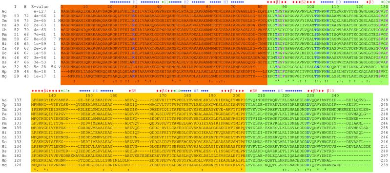
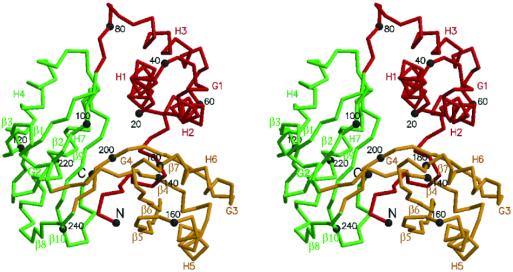
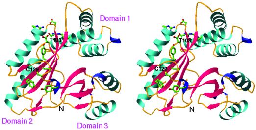
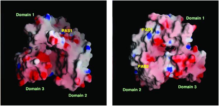
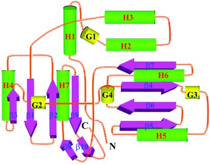
The crystal structure of a conserved hypothetical protein, Aq1575, from Aquifex aeolicus has been determined by using x-ray crystallography. The protein belongs to the domain of unknown function DUF28 in the Pfam and PALI databases for which there was no structural information available until now. A structural homology search with the DALI algorithm indicates that this protein has a new fold with no obvious similarity to those of other proteins of known three-dimensional structure. The protein reveals a monomer consisting of three domains arranged along a pseudo threefold symmetry axis. There is a large cleft with approximate dimensions of 10 A x 10 A x 20 A in the center of the three domains along the symmetry axis. Two possible active sites are suggested based on the structure and multiple sequence alignment. There are several highly conserved residues in these putative active sites. The structure based molecular properties and thermostability of the protein are discussed.


Figures

Figure 1
Figure 1
Sequence comparison between Aq1575 and its homologs. Aq, A. aeolicus; Tp, Treponema pallidum; Tm, Thermotoga maritima; Pa, Pseudomonas aeruginosa; Ch, Clostridium histolyticum; Pm, Pasteurella multocida; Bm, Brucella melitensis; Hi, Haemophilus influenzae; Ca, Clostridium acetobutylicum; Ec, E. coli; Mt, Mycobacterium tuberculosis; Bs, Bacillus subtilis; Ho, Homo sapiens; Mp, Mycoplasma pneumoniae; Mg, Mycoplasma genitalium. “I” represents % identity, “H” represents % homology, and E values are from the result of PSI-blast. The domains are represented as follows: scarlet for domain 1; green for domain 2; and yellow for domain 3. Blue characters represent the residues at putative active site 1 (PAS1), and pink residues at PAS2 (see text). The “−”s represent gaps; “*” for conserved residues; “:” for homologous residues having hydrophobic side chain; and “.” for homologue residues having polar side chains.
Figure 2
Figure 2
A stereo drawing of a Cα trace of Aq1575. Domain 1 (scarlet), domain 2 (green), and domain 3 (yellow) of Aq1575 are represented by a thick line. Every twentieth residue is numbered and represented by a dot. The N (residue Ser-5) and C termini (residue Lys-247) and the secondary elements are labeled. The figure was generated by molscript (38).
Figure 3
Figure 3
A ribbon diagram of Aq1575. The figure was generated by using the program ribbons (39). α-helices are colored in cyan, β-strands are colored red, and 310-helix are colored blue. The highly conserved residues around putative active site are represented by a ball-and-stick model (blue for nitrogen atoms, red for oxygen, yellow for sulfur, and green for carbon). Each domain, N termini, Thr-103 (located at the center of the first putative active site, PAS1) and Cys-129 (the second putative active site, PAS2) are labeled.
Figure 4
Figure 4
The electrostatic surface potential of Aq1575. (Left) A molecular surface is created by the program GRASP (red, negative; blue, positive; white, uncharged) (40). The residues, 5–17 and 243–247, covering a cleft were deleted to show a clear view of the inside of the protein in both Left and Right. The putative active site (PAS1) is indicated. (Right) The figure was drawn after 180° rotation of Left versus y axis. The putative active sites (PAS1 and PAS2) are indicated.
Figure 5
Figure 5
A topology diagram of Aq1575. α-helices are represented by green cylinders, β-strands by pink thick arrows, and 310-helices by gold cylinders. Secondary structure elements of helices and strands are labeled.

References

    1. Altschul S F, Madden T L, Schaffer A A, Zhang J, Zhang Z, Miller W, Lipman D J. Nucleic Acids Res. 1997;25:3389–3402. - PMC - PubMed
    1. Murzin A G, Patty L. Curr Opin Struct Biol. 1999;9:359–361.
    1. Marcotte E M, Pellegrini M, Thompson M J, Yeates T O, Eisenberg D. Nature (London) 1999;402:83–86. - PubMed
    1. Zarembinski T I, Hung L-W, Mueller-Dieckmann H -J, Kim K-K, Yokota H, Kim R, Kim S-H. Proc Natl Acad Sci USA. 1998;95:15189–15193. - PMC - PubMed
    1. Hwang K Y, Chung J H, Kim S-H, Han Y S, Cho Y. Nat Struct Biol. 1999;6:691–696. - PubMed

Publication types

Substances

LinkOut - more resources