. 2002 Jun 11;99(12):8048-53.
doi: 10.1073/pnas.112664499.

Integration of Runx and Smad regulatory signals at transcriptionally active subnuclear sites

Affiliations

Integration of Runx and Smad regulatory signals at transcriptionally active subnuclear sites

Sayyed K Zaidi et al. Proc Natl Acad Sci U S A. .

Abstract

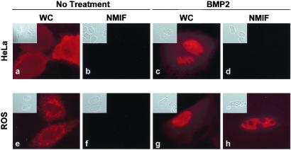
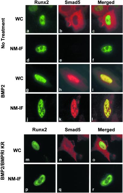
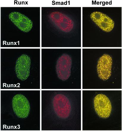
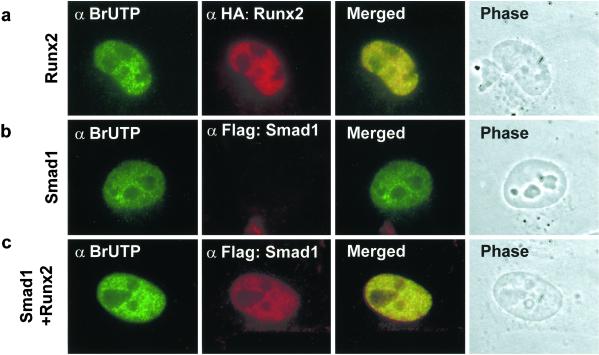
Runx factors control lineage commitment and are transcriptional effectors of Smad signaling. Genetic defects in these pathways interfere with normal development. The in situ localization of Runx and Smad proteins must impact the mechanisms by which these proteins function together in gene regulation. We show that the integration of Runx and Smad signals is mediated by in situ interactions at specific foci within the nucleus. Activated Smads are directed to these subnuclear foci only in the presence of Runx proteins. Smad-Runx complexes are associated in situ with the nuclear matrix, and this association requires the intranuclear targeting signal of Runx factors. The convergence of Smad and Runx proteins at these sites supports transcription as reflected by BrUTP labeling and functional cooperativity between the proteins. Thus, Runx-mediated intranuclear targeting of Smads is critical for the integration of two distinct pathways essential for fetal development.


Figures

Figure 1
Figure 1
Differential subnuclear organization of Smad5 in nonosseous and osseous cells. Human cervical carcinoma HeLa cells (ad) or rat osteosarcoma ROS 17/2.8 cells (eh) were grown on gelatin-coated coverslips and transfected with 0.5 μg of expression construct coding for Flag Smad5. After 24 h of transfection, cells were processed in situ for the WC and the NM-IF preparation and immunofluorescence microscopy. A mouse mAb against Flag tag (dilution: 1:1000) was used to detect Smad5.
Figure 2
Figure 2
Runx2 transcription factor targets Smads to subnuclear sites. HeLa cells, which lack endogenous Runx proteins, were transfected with 0.5 μg of expression construct for Flag Smad5 together with HA-Runx2 expression vector. BMP2-treated or untreated cells were processed for WC (ac, untreated cells; gi, BMP2-treated cells) or NM-IF (df, untreated cells; jl, BMP2-treated cells) preparation and in situ immunofluorescence 24 h after transfection. Smad5 was detected with mouse mAb against Flag tag whereas Runx2 was detected by a rabbit polyclonal Ab against HA tag. To confirm that the activation of Smad5 by BMP2 is required for nuclear accumulation and consequent subnuclear targeting of Smad5, a dominant negative inhibitor of BMP signaling, BMPRI (KR), was expressed along with expression constructs for HA-Runx2 and Flag-Smad5. Cells were treated with BMP2 and were processed for WC (mo) or NM-IF (pr) preparation and in situ immunofluorescence as described.
Figure 3
Figure 3
Runx factors specify subnuclear localization of Smads. HeLa cells were transfected with 0.5 μg of expression construct for Flag Smad1 along with Runx1, Runx2, or Runx3 expression vectors. Cells were treated with BMP2, and Smad1 was examined for association with Runx factors in the nuclear matrix (NM-IF preparation) by in situ immunofluorescence 24 h after transfection. Smad1 was detected with mouse mAb against Flag tag. Runx proteins were detected with rabbit polyclonal Abs at a dilution of 1:200. Images were taken by a Zeiss Axioplan microscope coupled with a charge-coupled device (CCD) camera and were processed for deconvulation microscopy with METAMORPH bioimaging software.
Figure 4
Figure 4
Runx2 is required for subnuclear targeting of Smad5. (a) A schematic of Runx2 protein. The region where Smads interact is shown. The Y428A mutation, which disrupts association of Runx2 with the nuclear matrix, was introduced by PCR-based site-directed mutagenesis. QA, poly glutamine-poly alanine stretch; RHD, runt homology domain; NMTS, nuclear matrix-targeting signal; VWRPY, a highly conserved motif that mediate interactions of Runx2 with groucho/TLE proteins. (b) ROS17/2.8 cells grown in 100-mm plates were transfected with 10 μg each of expression constructs for Flag-Smad1 and wild-type HA-Runx2 or HA-Runx2 Y428A mutant. Cells were treated with 300 ng/ml of BMP2 for 24 h and processed for immunoprecipitation. One microgram Ab against Flag tag was used for immunoprecipitation. The immunoprecipitated complex was resolved by 8% SDS/PAGE. Wild-type or mutant Runx2 proteins were detected with mouse mAb against HA tag (dilution 1:2000). HeLa cells were transfected with 0.5 μg of expression constructs for Flag-Smad1 along with HA-tagged wild-type or Runx2 Y428A mutant proteins. Cells were treated with BMP2 and subjected to WC (ce) or NM-IF (fh) preparation and double-labeled in situ immunofluorescence with mouse mAb against Flag tag and rabbit polyclonal Ab against HA tag.
Figure 5
Figure 5
Runx2 recruits Smad1 to subnuclear sites. HeLa cells were transfected with 0.5 μg of indicated expression constructs. BMP2-treated cells were labeled with BrUTP and subjected to NM-IF preparation and in situ immunofluorescence. A rat mAb (dilution 1:20) was used to detect BrUTP. Flag-Smad was detected with a mouse mAb against Flag tag.
Figure 6
Figure 6
Recruitment of Smads to sites of active transcription is coupled with the regulation of gene expression. HeLa cells were transfected with 0.45 95 g of pTβRE-Luc (Upper) along with 250 ng of pcDNA 3.1 (EV), Runx2 (wild-type or Y428A mutant), and/or Smad3 or 5. A promoterless luciferase gene was used as internal control for the transfection efficiency. Cells were treated with TGF-β (10 ng/ml) or BMP2 (300 ng/ml) for 6 h and harvested in passive lysis buffer 24 h after transfection. Ten microliters of the cell lysate was used for dual luciferase assay. The activity of the firefly luciferase was normalized with that of Renilla luciferase. The graphs represent three independent experiments with n = 6. TβRE, TGF-β-responsive element.

References

    1. Massague J. Annu Rev Biochem. 1998;67:753–791. - PubMed
    1. Finidori J. Vitam Horm. 2000;59:71–97. - PubMed
    1. Ihle J N. Curr Opin Cell Biol. 2001;13:211–217. - PubMed
    1. Akiyama T. Cytokine Growth Factor Rev. 2000;11:273–282. - PubMed
    1. Merriman H L, van Wijnen A J, Hiebert S, Bidwell J P, Fey E, Lian J, Stein J, Stein G S. Biochemistry. 1995;34:13125–13132. - PubMed

Publication types

MeSH terms

LinkOut - more resources